El uso de cerdos en la investigación ha aumentado en los últimos años. Sin embargo, los cerdos se caracterizan por una anatomía difícil de las vías respiratorias. Al demostrar cómo realizar la intubación endotraqueal guiada endoscópicamente, el presente protocolo tiene como objetivo aumentar aún más la seguridad de los animales de laboratorio para evitar el sufrimiento de los animales y la muerte innecesaria.
Se requiere una suscripción a JoVE para ver este contenido. Inicie sesión o comience su prueba gratuita.
Artículo de método
Intubación endotraqueal con un endoscopio de intubación flexible como modelo estandarizado para el manejo seguro de las vías respiratorias en cerdos
En este artículo
Aviso de erratum
Important: There has been an erratum issued for this article. View Erratum Notice
Resumen
Resumen
La intubación endotraqueal es a menudo un requisito básico para la investigación traslacional en modelos porcinos para diversas intervenciones que requieren una vía aérea segura o altas presiones de ventilación. La intubación endotraqueal es una habilidad desafiante, que requiere un número mínimo de intubaciones endotraqueales exitosas para lograr una alta tasa de éxito en condiciones óptimas, lo que a menudo es inalcanzable para los investigadores no anestesiólogos. Debido a la anatomía específica de las vías respiratorias porcinas, generalmente se puede asumir una vía aérea difícil. La imposibilidad de establecer una vía aérea segura puede provocar lesiones, eventos adversos o la muerte del animal de laboratorio. Utilizando un enfoque de evaluación prospectiva, aleatorizada y controlada, se ha demostrado que la intubación endotraqueal asistida por fibra óptica lleva más tiempo, pero tiene una tasa de éxito de primer paso más alta que la intubación convencional sin causar caídas clínicamente relevantes en la saturación de oxígeno. Este modelo presenta un régimen estandarizado para la intubación endotraqueal guiada endoscópicamente, proporcionando una vía aérea segura, especialmente para los investigadores que no tienen experiencia en la técnica de intubación endotraqueal mediante laringoscopia directa. Se espera que este procedimiento minimice el sufrimiento de los animales y las pérdidas innecesarias de animales.
Introducción
La intubación endotraqueal es a menudo un requisito básico para la investigación traslacional en modelos porcinos para diversas intervenciones que requieren una vía aérea segura o altas presiones de ventilación (como la ventilación durante la reanimación cardiopulmonar1 o el síndrome de dificultad respiratoria aguda2) o requieren que el flujo sanguíneo cerebral no se vea comprometido a través de la compresión interna por dispositivos supraglóticos de las vías respiratorias3 , que ocasionalmente se propagan como alternativas en el contexto de una vía aérea difícil prevista en cerdos
Acceso restringido. Inicie sesión o comience una prueba gratuita para ver este contenido.
Protocolo
Los experimentos en este protocolo fueron aprobados por el Comité Estatal e Institucional de Cuidado de los Animales (Landesuntersuchungsamt Rheinland-Pfalz, Koblenz, Alemania; aprobación no. G20-1-135). Los experimentos se llevaron a cabo siguiendo las directrices de ARRIVE. En total, se utilizaron para el presente estudio 10 cerdos machos anestesiados (Sus scrofa domestica) con un peso medio de 30 kg ± 2 kg y 12-16 semanas de edad.
1. Preparación de animales
- Mantenga un ambiente normal para los animales para minimizar el estrés. Retenga los alimentos 6 h antes del experimento programado para reducir el ....
Acceso restringido. Inicie sesión o comience una prueba gratuita para ver este contenido.
Resultados
La intubación endotraqueal se realizó en 10 cerdos machos (edad 12-16 semanas, peso 30 kg ± 3 kg) en un entorno de estudio prospectivo, aleatorizado y controlado. Los cerdos fueron aleatorizados en dos grupos: uno fue intubado convencionalmente laringoscópicamente (grupo IC), y el otro grupo fue intubado asistido a través de un endoscopio de intubación flexible como se describe en el protocolo (grupo FIE). La asignación grupal se realizó tirando de sobres sellados. El investigador fue asignado al azar diariament.......
Acceso restringido. Inicie sesión o comience una prueba gratuita para ver este contenido.
Discusión
En estudios previos, nuestro grupo de investigación ya ha descrito detalles específicos sobre los beneficios traslacionales del modelo porcino 2,27,32,33. En general, reducir el nivel de estrés del animal y el dolor innecesario debe ser una parte integral de cualquier protocolo de estudio y es primordial para generar datos reproducibles de manera confiable. Por lo tanto, la intubación guiada en.......
Acceso restringido. Inicie sesión o comience una prueba gratuita para ver este contenido.
Divulgaciones
El endoscopio de intubación flexible y sus accesorios han sido proporcionados incondicionalmente por el fabricante solo con fines de investigación. Los autores declaran no tener más conflictos de intereses financieros o de otro tipo.
Agradecimientos
Los autores quieren agradecer a Dagmar Dirvonskis por su excelente soporte técnico.
....Acceso restringido. Inicie sesión o comience una prueba gratuita para ver este contenido.
Materiales
| Nombre | Empresa | Número de catálogo | Comentarios |
|---|---|---|---|
| Ambu aScope Regular | Ambu GmbH, Medizinprodukte, Bad Nauheim, Alemania | Diámetro exterior de fibra óptica desechable 5 mm | |
| Ambu aView Monitor | Ambu GmbH, Medizinprodukte, Bad Nauheim, Alemania | monitor | |
| Atracurio Hikma 50 mg/5mL | Hikma Pharma GmbH, Martinsried | atracurio | |
| Azaperona (Stresnil) 40mg/mL | Lilly Deutschland GmbH, Bad Homburg, Alemania | azaperona | |
| BD Discardit II Spritze 2, 5, 10, 20 mL | Becton Dickinson S.A. Carretera, Mequinenza Fraga, España | jeringa | |
| BD Luer Connecta | Becton Dickinson Infusion Therapy, AB Helsingborg, Schweden | Llave de paso de 3 vías | |
| BD Microlance 3 20 G | Becton Dickinson S.A. Carretera, Mequinenza Fraga, España | cánula | |
| Curafix i.v. classics | Lohmann & Rauscher International GmbH & Co. KG, Rengsdorf, Alemania | Apósito de retención de cánulas | |
| Engströ m Carestation | GE Heathcare, Madison USA | ventilador | |
| Fentanilo-Janssen 0,05 mg/mL | Janssen-Cilag GmbH, Neuss | fentanilo | |
| Fü hrungsstab, Durchmesser 4.3 | Rü | Introductor de tubo endotraqueal | |
| IBM SPSS Statistics for Windows, Versión 20 | IBM SPSS Statistics for Windows, Version 20.0. Armonk, NY: IBM Corp.) | Software estadístico | |
| Incetomat-line 150 cm | Fresenius, Kabi Deutschland, GmbH | línea de perfusores | |
| Intrafix Primeline | B. Braun Melsungen AG, Melsungen, Alemania | Línea de infusión | |
| JOZA Einmal Nitril Untersuchungshandschuhe | JOZA, Mü nchen, Alemania | guantes desechables | |
| Laringoscopio, 45.48.50, KL 2000 | Medicon | Mango de laringoscopio | |
| Littmann Classic III Estetoscopio | 3M Deutschland GmbH, Neuss, Alemania | estetoscopio | |
| Luer Lock | B.Braun Melsungen AG, Alemania | ||
| Mmutilado Vlieskompresse | Marmed GmbH, Neuenkirchen, Alemania | Compresa de vellón para fijar la lengüeta | |
| Masimo LNCS Adtx SpO2 sensor | Masimo Corporation Irvine, Ca 92618 USA | clip de saturación para la cola | |
| Masimo LNCS TC-I SpO2 sensor de pinza para la oreja | Masimo Corporation Irvine, Ca 92618 USA | Pinza de saturación para la oreja | |
| Masimo Radical 7 | Masimo Corporation Irvine, Ca 92618 EE. UU | Saturación de oxígeno periférico | |
| Midazolam 15 mg/3 mL | Hameln Pharma GmbH, Hameln, Alemania | midazolam | |
| Midmark Mascarilla canina de plástico pequeño con diafragma FRSCM-0005 | Midmark Corp., Dayton, Ohio, EE. UU | mascarilla de ventilación para perros | |
| Octeniderm farblos | Schü LKE & Mayr GmbH, Nordenstedt, Alemania | Desinfectante alcohólico | |
| Original Jeringa Perfusor 50 mL | B.Braun Melsungen AG, Alemania | jeringa perfusora | |
| Perfusor FM Braun | B.Braun Melsungen AG, Alemania | bomba de jeringa | |
| Propofol 2% 20 mg/mL (matraces de 50 mL) | Fresenius, Kabi Deutschland, GmbH | propofol | |
| Rü SCH Fü hrungsstab fü r Tubo endotraqueal (ID 5,6 mm) | Teleflex Medical Sdn. Bhd, Malasia Alambre guía de | tubo recubierto | |
| Rü schelit Super Safety Clear >ID 6/6.5 /7.0 mm | Teleflex Medical Sdn. Bhd, Malasia | tubo endotraqueal | |
| Inoxidable Macintosh Grö ß e 4 | Welch Allyn69604 | cuchilla para laringoscopio | |
| Sterofundin | B.Braun Melsungen AG, Melsungen, Alemania | Solución electrolítica equilibrada | |
| Ultrastop Antibeschlagmittel frasco con gotero 25 mL | Sigmapharm Arzneimittel GmbH, Wien, Austria | Agente antivaho | |
| Vasofix Safety 22 G-16 G | B.Braun Melsungen AG, Alemania | catéter | |
| venosoManómetro de manguito VBM | Medizintechnik GmbH, Sulz a.N., Alemania | manómetro de manguito | |
| Zelette | Lohmann & Rauscher International GmbH & Co. KG, Rengsdorf, Alemania | Hisopo de tejido |
Referencias
- Kleinman, M. E., Oh, W., Stonestreet, B. S. Comparison of intravenous and endotracheal epinephrine during cardiopulmonary resuscitation in newborn piglets. Critical Care Medicine. 27 (12), 2748-2754 (1999).
- Rissel, R., et al.
Acceso restringido. Inicie sesión o comience una prueba gratuita para ver este contenido.
Reimpresiones y permisos
Erratum
Formal Correction: Erratum: Endotracheal Intubation Using a Flexible Intubation Endoscope As a Standardized Model for Safe Airway Management in Swine
Posted by JoVE Editors on 4/03/2023. Citeable Link.
An erratum was issued for: Endotracheal Intubation Using a Flexible Intubation Endoscope As a Standardized Model for Safe Airway Management in Swine. The Protocol, Representative Results, and Discussion sections were updated.
In the Protocol, step 1.5 was updated from:
Disinfect the skin with a disinfectant (alcoholic) before inserting a peripheral vein cannula (22 G) into an ear vein. Spray the area, wipe once, then spray again, and allow the disinfectant to dry.
to:
Disinfect the skin with a disinfectant (alcoholic) before inserting a peripheral vein cannula (22 G) into an ear vein. Spray the area, wipe once, then spray again, and allow the disinfectant to dry. Secure the ear cannula with a band-aid (See Table of Materials).
In the Protocol, step 3.7 was updated from:
While maintaining the position of the endoscope, advance the endotracheal tube until it becomes visible in the camera image.
NOTE: If the endotracheal tube cannot be advanced through the glottic plane, there is a possibility that it has become caught on the arytenoid cartilage. In this case, the endotracheal tube must be withdrawn 1 cm and rotated by 90° before gently advancing again. If necessary, this maneuver can be repeated. Similar calibers of flexible intubation endoscope and endotracheal tube can minimize the risk of this issue occurring. If the endotracheal tube cannot be advanced despite this maneuver, it is likely that the subglottic narrowness-the narrowest part of the porcine larynx-cannot be passed. In this case, a smaller endotracheal tube size needs to be selected. Regular commercially available endotracheal tubes in sizes 6.5 cm or 7.0 cm ID should be able to pass the glottis as long as no anatomic abnormalities are present.
to:
While maintaining the position of the endoscope, advance the endotracheal tube until it becomes visible in the camera image.
NOTE: If the endotracheal tube cannot be advanced through the glottic plane, there is a possibility that it has become caught on the arytenoid cartilage. In this case, the endotracheal tube must be withdrawn 1 cm and rotated by 90° before gently advancing again. If necessary, this maneuver can be repeated. Similar calibers of flexible intubation endoscope and endotracheal tube can minimize the risk of this issue occurring. If the endotracheal tube cannot be advanced despite this maneuver, it is likely that the subglottic narrowness-the narrowest part of the porcine larynx-cannot be passed. In this case, a smaller endotracheal tube size needs to be selected. Regular commercially available endotracheal tubes in sizes 6.5 cm or 7.0 cm ID should be able to pass the glottis as long as no anatomic abnormalities are present. Endotracheal tube size requirements vary depending on the piglet size and breed.
In the Representative Results, the sixth paragraph was updated from:
Statistical analyses were performed using commercially available software (see Table of Materials). Normal distribution was examined using the Kolmogorov-Smirnoff test28. If a normal distribution was determined, group differences were analyzed using t-tests of independent samples29 or the Mann-Whitney U test30 for the non-parametric version. Data are presented as mean (± standard deviation). Correlations of ordinal-scale data were examined using Spearman's correlation coefficient31. A significance level of p < 0.05 was assumed.
to:
Statistical analyses were performed using commercially available software (see Table of Materials). Normal distribution was examined using the Kolmogorov-Smirnoff test28. If a normal distribution was determined, group differences were analyzed using t-tests of independent samples29 or the Mann-Whitney U test30 for the non-parametric version. Data are presented as mean (± standard deviation). Correlations of ordinal-scale data were examined using Spearman's correlation coefficient31. A significance level of p < 0.05 was assumed. All tests were performed with exploratory intention; therefore p-values are descriptive. Nevertheless, p < 0.05 was accepted as indicative of statistical significance.
In the Representative Results, the legend for figure 1 was updated from:
Figure 1: Number of intubation attempts in group comparison. For the group that was intubated using a flexible intubation endoscope, every intubation attempt was successful; in the group that was conventionally intubated, it took an average of 1.4 attempts before the endotracheal tube could be placed correctly. Error bars show the standard deviation. Please click here to view a larger version of this figure.
to:
Figure 1: Number of intubation attempts in group comparison. For the group that was intubated using a flexible intubation endoscope, every intubation attempt was successful; in the group that was conventionally intubated, it took an average of 1.4 attempts before the endotracheal tube could be placed correctly. Error bars show the standard deviation. n = 5 (for each group). Please click here to view a larger version of this figure.
In the Representative Results, figure 2 was updated from:
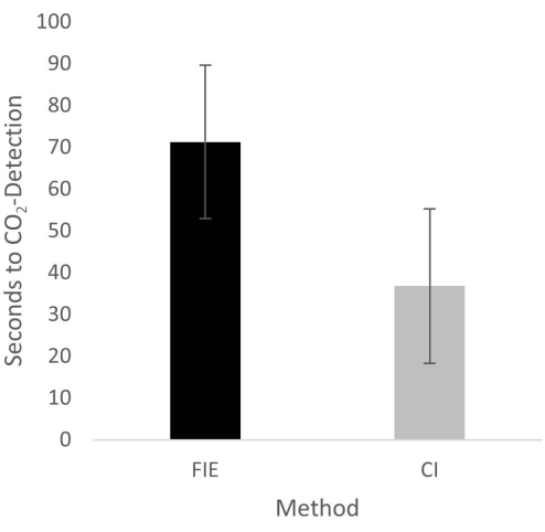
Figure 2: Time until CO2 detection in group comparison. For the group that was intubated using a flexible intubation endoscope, it took significantly longer until end-tidal CO2 could be detected, depicted as mean and standard deviation. Please click here to view a larger version of this figure.
to:
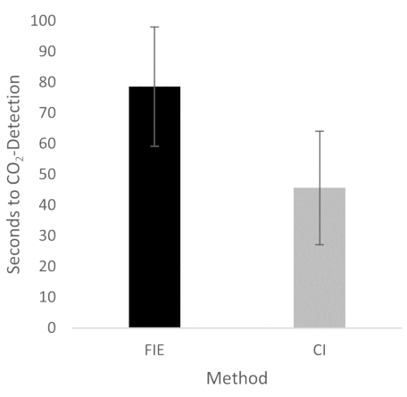
Figure 2: Time until CO2 detection in group comparison. For the group that was intubated using a flexible intubation endoscope, it took significantly longer until end-tidal CO2 could be detected, depicted as mean and standard deviation. n = 5 (for each group). Please click here to view a larger version of this figure.
In the Discussion, the fifth paragraph was updated from:
The increased duration had no clinical significance in this cohort. At no time was the termination criterion-a saturation of less than 93%-reached. This is shown in the results because a procedure change was unnecessary at any time. Prior adequate mask ventilation is a critical step to allow sufficient time for fiberoptic endotracheal tube placement to avoid rapid desaturation34. These results are consistent with previous studies comparing conventional intubation and endoscopically assisted intubations with inexperienced providers35.
to:
The increased duration had no clinical significance in this cohort. At no time was the termination criterion-a saturation of less than 93%-reached. This is shown in the results because a procedure change was unnecessary at any time. Prior adequate mask ventilation is a critical step to allow sufficient time for fiberoptic endotracheal tube placement to avoid rapid desaturation34. These results are consistent with previous studies comparing conventional intubation and endoscopically assisted intubations with inexperienced providers35. We attribute the prolonged duration of fiberoptic intubation to the fact that one must first reorient again after insertion, whereas with conventional intubation, one retains a view of the glottis. It is also important to avoid contact with the mucosa with the flexible intubation endoscope during advancement. This requires occasional corrective maneuvers. Last but not least, after successful placement, retraction of the relatively long endoscope is required, which increases the time to CO2 detection slightly.
