Este protocolo describe enfoques in vivo e in vitro para investigar cómo el N-lactoil-fenilalanina afecta la cardiomiopatía diabética, el metabolismo lipídico cardíaco y la síntesis de colesterol, con especial atención a la vía de la proteína quinasa activada por AMP alfa 1/reductasa de la hidroximetilglutaril-coenzima A.
Se requiere una suscripción a JoVE para ver este contenido. Inicie sesión o comience su prueba gratuita.
Artículo de investigación
La N-lactoil-fenilalanina mejora la función cardíaca en un modelo de ratón de miocardiopatía diabética mediante la regulación del metabolismo lipídico
En este artículo
Resumen
Resumen
La cardiomiopatía diabética (DCM) es una de las complicaciones más graves de la diabetes. La N-lactoil-fenilalanina (Lac-Phe), un metabolito endógeno, puede promover la pérdida de peso y mejorar la homeostasis glucídica. Este estudio tuvo como objetivo investigar el efecto terapéutico de Lac-Phe sobre la DCM y explorar además el mecanismo subyacente. Se administró Lac-Phe a ratones con diabetes tipo 2 que presentaban DCM. Lac-Phe mejoró la función contráctil cardíaca, redujo el remodelado patológico y disminuyó la acumulación de lípidos en el corazón. In vitro, se estudiaron los efectos de Lac-Phe en células H9c2 estimuladas con glucosa alta y ácido palmítico. Lac-Phe redujo la acumulación de lípidos al disminuir la síntesis de colesterol en las células H9c2. Mecanísticamente, Lac-Phe promovió la fosforilación y activación de la proteína quinasa activada por monofosfato de adenosina alfa 1 (AMPKα1). Posteriormente, la AMPKα1 activada fosforiló e inhibió su diana descendente, la 3-hidroxi-3-metilglutárico-coenzima A reductasa (HMGCR), lo que condujo a una reducción en la síntesis de colesterol y, en última instancia, a la mejora del desequilibrio del metabolismo lipídico en las células H9c2. Estos hallazgos indican que Lac-Phe mejora el metabolismo lipídico en células H9c2 en el contexto de la DCM a través de la vía AMPKα1/HMGCR, mejorando así la función contráctil cardíaca y reduciendo el remodelado patológico. Lac-Phe podría representar un enfoque terapéutico potencial para la cardiomiopatía diabética.
Introducción
La diabetes es una enfermedad metabólica caracterizada por hiperglucemia persistente1. En las últimas dos décadas, la diabetes se ha convertido en una de las diez principales causas de muerte en todo el mundo, con más de seis millones de personas fallecidas cada año debido a la diabetes y sus complicaciones.2. La miocardiopatía diabética (DCM), una afección cardíaca patológica resultante de la diabetes, representa una de las complicaciones más graves asociadas con la diabetes3,4. Actúa como un factor de riesgo independiente para la insuficiencia cardíaca y se caracteriza por alteraciones estructurales y funcionales del miocardio5,6, incluyendo fibrosis cardíaca e hipertrofia de los miocitos cardíacos, que finalmente conducen a disfunción diastólica y sistólica. Aunque varias terapias cardiovasculares convencionales, como β-bloqueantes, inhibidores de la enzima convertidora de angiotensina e inhibidores del receptor de tipo 1 de angiotensina II, se utilizan actualmente en el manejo de la miocardiopatía dilatada7,8, su eficacia sigue siendo subóptima en entornos clínicos. Dado que los mecanismos subyacentes a la miocardiopatía diabética no se han estudiado exhaustivamente9, actualmente no existe una estrategia de tratamiento clara dirigida a esta enfermedad10,11Las manifestaciones clínicas de la miocardiopatía dilatada progresan desde una disfunción diastólica asintomática en la etapa inicial hasta una disfunción sistólica e insuficiencia cardíaca clínica en la etapa avanzada, e incluso la muerte12,13Por lo tanto, existe una necesidad urgente de explorar y desarrollar opciones de tratamiento eficaces para esta afección.
El metabolismo lipídico desequilibrado es una característica clave de la miocardiopatía diabética (DCM)14. En la DCM, los niveles elevados de ácidos grasos libres (AGL) en sangre pueden provocar una captación excesiva de ácidos grasos por los cardiomiocitos15,16. El metabolismo de ácidos grasos en los cardiomiocitos depende principalmente de la oxidación de ácidos grasos. Con el tiempo, la oxidación de ácidos grasos en los cardiomiocitos es insuficiente para metabolizar completamente todos los ácidos grasos ingeridos, y se acumulan excesos de ácidos grasos y sus metabolitos intermedios en los cardiomiocitos17. Esta sobrecarga lipídica excesiva induce lipotoxicidad, altera el suministro energético miocárdico y promueve el estrés oxidativo cardíaco y las respuestas inflamatorias, causando finalmente un daño severo a los cardiomiocitos18,19. Por lo tanto, regular activamente el metabolismo lipídico cardíaco podría representar un objetivo terapéutico potencial para la DCM20.
La N-lactoil-fenilalanina (Lac-Phe) es un metabolito endógeno cuya concentración aumenta rápidamente después del ejercicio21. Mecánicamente, la Lac-Phe es sintetizada por la enzima citosólica carnosina dipeptidasa 2 (CNDP2) mediante la condensación de lactato y fenilalanina. Esta vía biosintética está ampliamente distribuida en diversos tipos celulares, incluidos macrófagos, células epiteliales y células madre mesenquimales, que secretan Lac-Phe a la circulación tras la actividad física22. Se ha demostrado que la administración crónica de Lac-Phe reduce eficazmente la ingesta de alimentos, disminuye la obesidad y el peso corporal, y mejora la homeostasis glucosa21. Un estudio reciente ha mostrado que la Lac-Phe puede mejorar la capacidad del metabolismo lipídico de las microglías/macrófagos tras una lesión de la médula espinal23. Curiosamente, estudios indican que tanto la administración aguda como crónica de metformina, un fármaco de primera línea para la diabetes tipo 2, puede elevar los niveles de Lac-Phe y que esta última media los efectos antiobesidad de la metformina22. Además, Sha et al. recientemente demostraron que la Lac-Phe mejora la muerte celular miocárdica aliviar la disfunción mitocondrial, mejorando así la insuficiencia cardíaca inducida por la constricción aórtica transversal (TAC)24. La hiperglucemia prolongada y la obesidad son factores importantes que contribuyen a la desregulación del metabolismo lipídico en la miocardiopatía diabética (DCM)25. En conjunto, esta evidencia sugiere que la Lac-Phe podría tener un efecto terapéutico potencial sobre la DCM. Sin embargo, hasta la fecha, no se ha investigado el papel específico de la Lac-Phe en la DCM.
En este estudio, descubrimos que la administración exógena de Lac-Phe a ratones con cardiomiopatía diabética mejoró eficazmente la función contráctil del corazón y atenuó la remodelación patológica cardíaca. Encontramos que Lac-Phe mejoró la función cardíaca en ratones con MCD mediante la regulación del desequilibrio del metabolismo lipídico. Lac-Phe redujo la acumulación de gotas lipídicas (GL) en cardiomiocitos de ratones con MCD y disminuyó el contenido de colesterol en el corazón. Mecánicamente, Lac-Phe promovió la fosforilación y activación de la proteína quinasa activada por adenosina monofosfato alfa 1 (AMPKα1), promoviendo así la fosforilación de la 3-hidroxi-3-metilglutaril-coenzima A reductasa (HMGCR). La fosforilación de HMGCR reduce su actividad, lo que resulta en una menor producción de colesterol y, por ende, mejora el desequilibrio del metabolismo lipídico en los cardiomiocitos. Estos resultados proporcionan una base teórica para el posible tratamiento de la MCD con Lac-Phe.
Acceso restringido. Inicie sesión o comience una prueba gratuita para ver este contenido.
Protocolo
The animal study protocol was approved by the Institutional Ethics Committee of Nanjing Drum Tower Hospital (Approval Number: 2025AE1065, 26 September 2025) and was conducted in accordance with the guidelines outlined in the Guidelines for the Care and Use of Laboratory Animals (8th Edition), published by the National Institutes of Health of the United States.
Animals
Thirty 5-week-old male C57BL/6J mice were used in this study. After 1 week of adaptive feeding, all mice were housed with access to food and water under an alternating 12 h day-night cycle at a temperature of 22°C ± 2°C and humidity of 40%–60%. An HFD combined with low-dose STZ was used to establish the DCM mouse model (Figure 1A). Four weeks after initiation of high-fat diet (HFD; 60% fat, 20% protein, and 20% carbohydrate) feeding, mice were fasted overnight and subsequently administered streptozotocin (STZ; 8 mg/mL dissolved in citrate buffer, pH 4.5) via intraperitoneal injection at a dosage of 35 mg/kg/day for three consecutive days. Control group mice were fed a control diet (10% fat, 20% protein, and 70% carbohydrate) and received equivalent volumes of citrate buffer. Following 15 additional weeks of high-fat diet maintenance, mice meeting both criteria, random blood glucose of ≥13.3 mmol/L and left ventricular ejection fraction (LVEF) of <55%, were defined as successfully established DCM models. These validated DCM mice were numbered and randomly assigned to the DCM and DCM + Lac-Phe groups using a random number table. Approximately half of the mice failed to develop the model; these animals were euthanized in accordance with animal ethics requirements. In brief, mice were divided into three groups: Control, DCM, and DCM + Lac-Phe (n = 10 for each group). All 10 mice per group underwent echocardiography, body weight measurement, and OGTT. Following the 28-day treatment period, hearts were collected for subsequent analyses. There were 10 mice in each group. Of these, three mice per group were used for transcriptome sequencing, three were used for transmission electron microscopy and histological staining, and three were used for protein expression analysis. The remaining mouse in each group was reserved as a backup. After successful establishment of the DCM model, Lac-Phe was dissolved in normal saline to a final concentration of 10 mg/mL. Mice in the DCM + Lac-Phe group received intraperitoneal Lac-Phe at 50 mg/kg/day in an injection volume of 5 mL/kg, once daily for 28 consecutive days. Mice in the Control and DCM groups were administered an equivalent volume of normal saline as vehicle control.
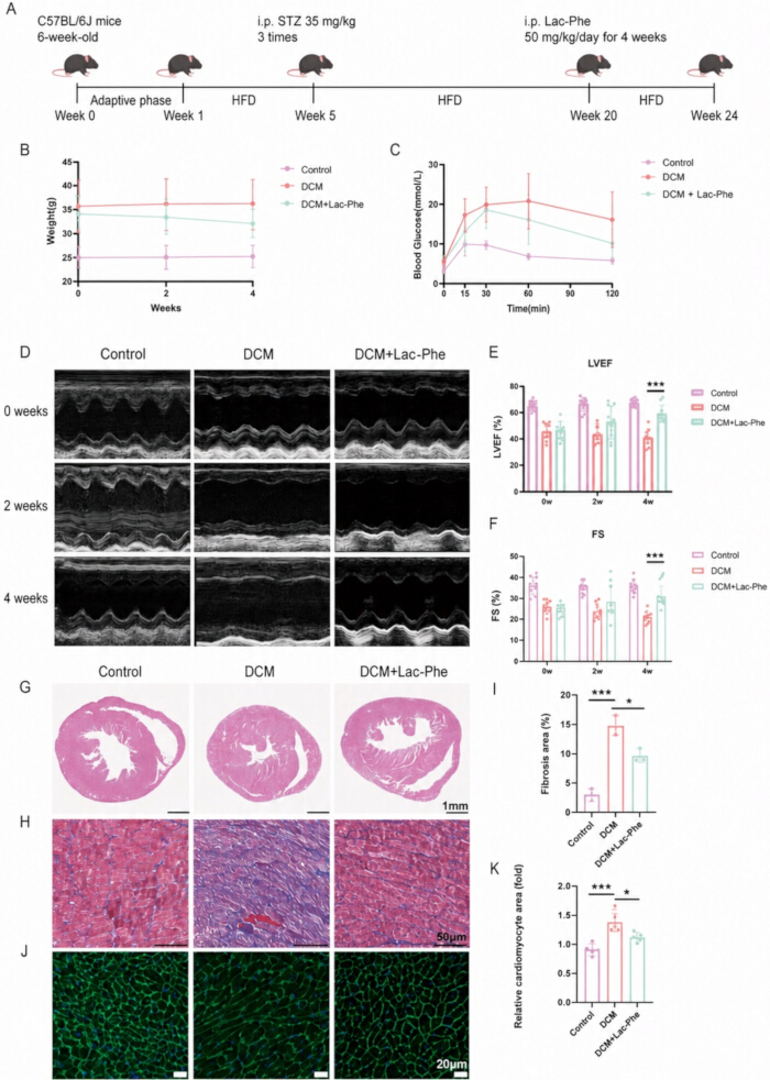
Figure 1. Lac-Phe improves cardiac function and pathological remodeling in mice with diabetic cardiomyopathy. (A) Schematic of the HFD/STZ-induced DCM mouse model and Lac-Phe treatment protocol. (B) Body weights of mice in the Control, DCM, and DCM + Lac-Phe groups during the 4-week treatment period; n = 10 per group. (C) Oral glucose tolerance test (OGTT) in the indicated groups; n = 10 per group. (D) Representative echocardiographic images at 0, 2, and 4 weeks of treatment. (E,F) Quantification of left ventricular ejection fraction (LVEF) (E) and fractional shortening (FS) (F); n = 10 per group. (G) Representative hematoxylin and eosin (H&E)-stained heart sections. Scale bar = 1 mm. (H,I) Representative Masson’s trichrome staining of heart sections (H) and quantification of fibrotic area (I); n = 3 per group. Scale bar = 50 μm. (J,K) Representative wheat germ agglutinin (WGA) staining of heart sections (J) and quantification of relative cardiomyocyte area (K); n = 3 per group. Scale bar = 20 μm. Data are presented as the mean ± standard deviation. P < 0.05, **P < 0.001. Please click here to view a larger version of this figure.
Cardiac function
Following anesthesia with isoflurane inhalation, mice were secured in a supine position on a temperature-controlled platform (37°C). Left ventricular dimensions and mitral valve blood flow velocity during diastole and systole were measured using an echocardiography machine equipped with a 40 MHz transducer, from which LVEF and shortening fraction (FS) were calculated. Three consecutive cardiac cycles were collected from each mouse.
Cell treatment
H9c2 cells were cultured in Dulbecco’s modified Eagle’s medium supplemented with 10% fetal bovine serum and 1% penicillin–streptomycin at 37°C in a humidified atmosphere containing 5% CO₂. Cells were passaged every 2–3 days upon reaching 70%–80% confluence. For experiments, cells were seeded in 10 cm dishes and then divided into three groups: Control, high glucose/palmitic acid (HG/PA), and HG/PA + Lac-Phe. Cells in the HG/PA and HG/PA + Lac-Phe groups were first exposed to 0.25 mM palmitic acid and 50 mM glucose for 24 h. Thereafter, cells in the HG/PA + Lac-Phe group were treated with 10 μg/mL Lac-Phe for an additional 24 h while HG/PA exposure was maintained; cells in the HG/PA group received an equivalent volume of PBS under the same HG/PA conditions. In the p-AMPK inhibition experiments, 10 μM AMPK-IN-3 (dissolved in DMSO) was added simultaneously with Lac-Phe for 24 h, and the corresponding control groups received an equal volume of DMSO. For the atorvastatin experiments, H9c2 cells under HG/PA conditions were treated with 10 μM atorvastatin for 24 h, either alone or in combination with 10 μg/mL Lac-Phe.
Histological staining
After 28 days of Lac-Phe administration, mouse hearts were harvested and immersion-fixed in 4% paraformaldehyde (pH 7.4) for 24 h. Fixed cardiac tissues were paraffin-embedded and sectioned into 6-μm-thick slices. Tissue sections were stained with hematoxylin and eosin, wheat germ agglutinin, and Masson’s trichrome. All staining procedures were performed strictly following the manufacturers’ protocols. Whole-slide imaging was performed at 20× magnification using a high-resolution brightfield microscope equipped with a digital camera system.
Western blot analysis
Protein concentration was determined using the Bradford assay. Tissue lysates were resolved by SDS-PAGE on 10% (w/v) acrylamide gels and electrophoretically transferred to polyvinylidene difluoride membranes. Nonspecific binding sites were blocked with 5% bovine serum albumin for 2 h at room temperature. Membranes were then incubated overnight at 4°C with primary antibodies (diluted 1:1,000), followed by six washes with TBST (5 min each) on a shaker. The membranes were then incubated with horseradish peroxidase-conjugated secondary antibodies (diluted 1:5,000) for 2 h at room temperature, followed by another six washes with TBST (5 min each). Protein bands were visualized using an enhanced chemiluminescence reagent kit. Quantification of western blot results was performed by densitometric analysis using ImageJ software. All antibodies used in this study are listed in the Table of Materials.
Transcriptomics
Total RNA was isolated from cardiac tissue of C57BL/6J mice using TRIzol Reagent and RNeasy mini kits. RNA quality was evaluated using a 2100 bioanalyzer, demonstrating high RNA purity and integrity. RNA-seq libraries were prepared according to the VAHTS Universal V10 RNA-seq Library Prep Kit and sequenced using an Illumina NovaSeq 6000 System (single-read, 150 bp). Differentially expressed genes were identified using DESeq2, with an adjusted P value < 0.05 and an absolute log₂ fold change > 1 considered statistically significant. Gene Ontology (GO) enrichment analysis was conducted on differentially expressed genes, and the significance of enrichment of differentially expressed genes in GO terms was calculated using the hypergeometric distribution algorithm.
Oil Red O staining and LD540 lipid staining
Cardiac tissue was embedded in OCT compound and then cut into 10-μm-thick sections. The frozen sections were equilibrated at room temperature for 30 min and fixed with 4% paraformaldehyde for 10 min. After washing with PBS, the sections were placed in 60% isopropyl alcohol for 15 s and covered with Oil Red O working solution for 30 min. The sections were then treated with 60% isopropyl alcohol for 15 s and washed three times with PBS. For LD540 lipid staining, H9c2 cells were washed with PBS and fixed with 4% paraformaldehyde for 10 min at room temperature. The cells were then incubated with 10 μM LD540 for 30 min at room temperature.
Transmission electron microscopy (TEM)
Fresh mouse heart tissues were cut into 1–2 mm3 pieces, fixed in electron microscope fixative at 4°C for 4 h, then washed with double-distilled water and immersed in ethanol solutions of progressively increasing concentrations. The samples were embedded, sectioned, and then observed under a transmission electron microscope.
Total cholesterol (TC) content
Total cholesterol (TC) levels in serum and H9c2 cell samples were measured using the Amplex Red Cholesterol and Cholesteryl Ester Assay Kit according to the manufacturer’s instructions.
Oral Glucose Tolerance Test (OGTT)
Mice were fasted for 14 h (from 19:00 to 9:00) and gavaged with glucose (20% stock, 2 g/kg). Blood glucose levels were determined using blood collected from the tail vein at 0, 15, 30, 60, and 120 min after gavage.
Cell counting kit-8 (CCK-8) measurement
CCK-8 was used to measure cell viability. H9c2 cells were seeded into 96-well plates at an initial density of 3 × 10⁴ cells/well. After treatment with different concentrations of Lac-Phe under HG/PA conditions for 24 h, 10 μL of the kit reagent was added to 100 μL of culture medium per well, followed by incubation at 37°C for 4 h. The absorbance was measured at 450 nm using a multimode microplate reader to obtain the optical density (OD) value.
Molecular docking
The crystal structures of Lac-Phe and AMPKα1 were downloaded from PubChem26. The molecular docking process was carried out using CB-DOCK227.
Statistical analysis
All echocardiographic measurements, histological staining, and image quantifications were performed by investigators blinded to group assignment. All data analyses were performed using Prism and SPSS 20.0. Each experiment was repeated at least three times, and the data were presented as the mean ± standard deviation. Homogeneity of variance was assessed using Levene’s test. Normality was confirmed by graphical inspection of residuals. Comparisons among multiple groups were conducted using one-way analysis of variance, followed by Tukey's post hoc test for pairwise comparisons. For OGTT data, two-way repeated-measures ANOVA was used, with group as the between-subjects factor and time as the within-subjects factor, followed by Bonferroni's post hoc test. P < 0.05 was considered statistically significant.
Acceso restringido. Inicie sesión o comience una prueba gratuita para ver este contenido.
Resultados
Lac-Phe improves cardiac function in diabetic cardiomyopathy mice
We induced DCM in C57BL/6J mice using an HFD combined with STZ injections (Figure 1A) and randomly assigned the successfully established DCM mice to an untreated group (DCM group) and a Lac-Phe treatment group, with control C57BL/6J mice serving as the negative control. According to the study by Li et al.21, we selected 50 mg/kg as the therapeutic dose of Lac...
Acceso restringido. Inicie sesión o comience una prueba gratuita para ver este contenido.
Discusión
En este estudio, presentamos evidencia que respalda un efecto terapéutico de Lac-Phe en la miocardiopatía dilatada (DCM). Lac-Phe mejoró el remodelado cardíaco y la disfunción cardíaca en la DCM, al mismo tiempo que redujo la producción de colesterol en los cardiomiocitos y mejoró el desequilibrio del metabolismo lipídico. Mecánicamente, Lac-Phe incrementó la fosforilación de AMPKα1. Posteriormente, el aumento en la fosforilación de AMPKα1 se asoció con un incremento en la fosforilación y la inhibición de su diana descen...
Acceso restringido. Inicie sesión o comience una prueba gratuita para ver este contenido.
Divulgaciones
Conflicto de intereses:
Los autores no tienen nada que declarar.
Agradecimientos
Queremos expresar nuestro sincero agradecimiento a los investigadores y al personal del Hospital Clínico Colegio Nanjing Drum Tower y del Hospital Afiliado de la Facultad de Medicina de la Universidad de Nanjing por su apoyo técnico. Esta investigación fue financiada por subvenciones de la Fundación Nacional de Ciencias Naturales de China (números de subvención 81870291 y 82300384), la Fundación China para la Ciencia Postdoctoral (número de subvención 2023M731625) y la Fundación de Ciencias Naturales de la Provincia de Jiangsu (número de subvención BK20220175). También agradecemos los recursos y el apoyo colaborativo proporcionados por el Instituto de Síntesis Avanzada de la Universidad de Nanjing Tech.
Acceso restringido. Inicie sesión o comience una prueba gratuita para ver este contenido.
Materiales
| Nombre | Empresa | Número de catálogo | Comentarios |
|---|---|---|---|
| AMPK-IN-3 (inhibidor de p-AMPK) | MedChemExpress (MCE) | HY-132921 | Utilizado en 10 μM durante experimentos de inhibición de AMPK para inhibir la señalización de AMPK en células tratadas con Lac-Phe. |
| Kit de ensayo Amplex Red para colesterol y esterol de colesterol | Beyotime | S0211S | Utilizado para determinar los niveles totales de colesterol en suero y en muestras de células H9c2. |
| Anti-AMPKα1 | Cell Signaling Technology | 2532; RRID: AB_330331 | Anticuerpo primario utilizado para el análisis por inmunotransferencia de AMPKα1. |
| Anti-GAPDH | Cell Signaling Technology | 5174; RRID: AB_10622025 | Anticuerpo primario utilizado para el análisis por inmunotransferencia de GAPDH. |
| Anti-HMGCR | Abcam | ab174830; RRID: AB_2749818 | Anticuerpo primario utilizado para el análisis por inmunotransferencia de HMGCR. |
| Anti-p-AMPKα1 (Thr172) | Cell Signaling Technology | 2535; RRID: AB_331250 | Anticuerpo primario utilizado para el análisis por inmunotransferencia de AMPK fosforiladaα1. |
| Anti-p-HMGCR (Ser872) | Abcam | ab124350 | Anticuerpo primario utilizado para el análisis por inmunotransferencia de HMGCR fosforilada. |
| anticuerpo secundario anti-IgG de conejo, conjugado con HRP | Cell Signaling Technology | 7074; RRID: AB_2099233 | Utilizado a una dilución 1:5.000 para la detección en inmunotransferencia western. |
| Anti-β-tubulina | Cell Signaling Technology | 2128; RRID: AB_823664 | Anticuerpo primario utilizado para el análisis por inmunotransferencia de β-tubulina. |
| Atorvastatina | MedChemExpress (MCE) | HY-17379 | Utilizado solo o en combinación con Lac-Phe para evaluar los efectos sobre la acumulación de lípidos, los niveles totales de colesterol y la fosforilación de HMGCR en células H9c2 tratadas con HG/PA. |
| Albúmina sérica bovina | Sigma-Aldrich | A7906 | Utilizado al 5 % para bloquear la unión inespecífica durante el análisis de western blot. |
| reactivo/kit de ensayo de Bradford | Thermo Fisher Scientific | 23200 | Utilizado para determinar la concentración de proteína antes del análisis mediante western blot. |
| C57BL/6J ratones (machos, de 5 semanas de edad) | Centro de Investigación de Animales Modelo de la Universidad de Nanjing | N/A | Utilizado para establecer el modelo murino de cardiomiopatía diabética inducida por dieta rica en grasas (HFD) y estreptozotocina (STZ). |
| Kit de conteo celular-8 (CCK-8) | Sharebio | SB-CCK8S | Utilizado para medir la viabilidad de células H9c2 tras el tratamiento. |
| Tampón de citrato | Preparado internamente | N/A | Preparado a pH 4,5 y utilizado para disolver la STZ y como control vehicular correspondiente. |
| Dieta control (10 % grasa, 20 % proteína, 70 % carbohidrato) | Research Diets, Inc. | D12450J | Utilizado como dieta para los ratones del grupo control. |
| D-(+)-Glucosa | Sigma-Aldrich | N/A | Utilizado para el tratamiento de células HG/PA y para la prueba de tolerancia oral a la glucosa. |
| DMSO (dimetilsulfóxido) | Sigma-Aldrich | D2650 | Utilizado como disolvente para AMPK-IN-3 y como control vehicular correspondiente. |
| Dulbecco’s modificado de Eagle’medio s | Gibco, Thermo Fisher Scientific | 11965092 | Utilizado como medio de cultivo basal para células H9c2. |
| Equipo de ecocardiografía con transductor de 40 MHz | FUJIFILM VisualSonics | Vevo 3100 LT; transductor de 40 MHz | Utilizado para medir las dimensiones del ventrículo izquierdo y la velocidad del flujo sanguíneo en la válvula mitral, y para derivar la FEVI y la FS. |
| Fijador para microscopía electrónica | ServiceBio | G1102 | Utilizado para fijar tejido cardíaco fresco de ratón para microscopía electrónica de transmisión. |
| Etanol | Sigma-Aldrich | E7023 | Utilizado en concentraciones progresivamente crecientes para la deshidratación durante la preparación de muestras para microscopía electrónica de transmisión. |
| Suero bovino fetal | Gibco, Thermo Fisher Scientific | 10099141 | Utilizado al 10% para suplementar el medio de cultivo celular H9c2. |
| Línea celular H9c2 | Sunncell | RRID: CVCL_0286 | Utilizado como modelo celular cardíaco in vitro para experimentos de tratamiento con HG/PA y Lac-Phe. |
| Tinción con hematoxilina y eosina | ServiceBio | G1005-1/2 | Utilizado para la evaluación histológica del tejido cardíaco. |
| Dieta alta en grasas (60 % kcal de grasas) | Research Diets, Inc. | D12492 | Utilizado con STZ de baja dosis para establecer el modelo de ratón de cardiomiopatía diabética. |
| Microscopio de campo claro de alta resolución | Leica | DM2500 | Utilizado para la obtención de imágenes completas de cortes de tejido cardíaco teñidos a 20× aumento |
| ImageJ | Institutos Nacionales de la Salud | N/A | Utilizado para la cuantificación densitométrica de los resultados de inmunotransferencia en Western blot. |
| Isoflurano | Sigma-Aldrich | 792632 | Utilizado para anestesia durante la ecocardiografía en ratones. |
| Alcohol isopropílico | Sigma-Aldrich | I9516 | Utilizado al 60 % durante la tinción con Oil Red O de secciones cardíacas congeladas. |
| tinción lipídica LD540 | Beyotime | C2050S-1 | Utilizado en 10 μM para teñir lípidos intracelulares. |
| Masson’tinción con tricrómico s | ServiceBio | G1006 | Utilizado para la tinción histológica de tejido cardíaco. |
| N-lactoil-fenilalanina (Lac-Phe) | MedChemExpress (MCE) | HY-146098 | Administrado a ratones con miocardiopatía dilatada y utilizado para tratar células H9c2 expuestas a glucosa alta/ácido palmítico. |
| Solución de trabajo de Oil Red O | ServiceBio | G1015-100ML | Utilizado para teñir lípidos en cortes de tejido cardíaco congelado. |
| Kit Omni-ECL de Quimioluminiscencia Eficiente | EpiZyme | SQ203L | Utilizado para la visualización quimioluminiscente de bandas proteicas tras el western blot. |
| Ácido palmítico | Sigma-Aldrich | P0500 | Utilizado a 0,25 mM con glucosa alta para establecer el modelo de células H9c2 tratadas con HG/PA. |
| Paraformaldehído | Sigma-Aldrich | P6148 | Preparado como una solución al 4 % y utilizado para fijar tejido cardíaco y células antes de la tinción histológica o de lípidos. |
| Penicilina-estreptomicina | Gibco, Thermo Fisher Scientific | 15140122 | Utilizado al 1% en medio de cultivo celular H9c2. |
| Solución salina tamponada con fosfato (PBS) | Gibco, Thermo Fisher Scientific | 10010023 | Utilizado para lavados durante los procedimientos de tinción y como vehículo correspondiente en experimentos de tratamiento celular. |
| Prism | GraphPad Software | Versión 8.0 | Utilizado para el análisis estadístico de datos experimentales. |
| membrana de transferencia de PVDF, 0.45 μm | Thermo Fisher Scientific | 88518 | Utilizado para la transferencia electroforética de proteínas durante el análisis de western blot. |
| RNeasy Mini Kit | QIAGEN | 74104 | Utilizado para el aislamiento/purificación de ARN total a partir de tejido cardíaco de ratón. |
| Solución de cloruro de sodio, 0,9 % (suero fisiológico) | Sigma-Aldrich | S8776 | Utilizado para disolver Lac-Phe y como control vehicular para los ratones Control y DCM. |
| SPSS | IBM | Versión 20.0 | Utilizado para el análisis estadístico de datos experimentales. |
| Estreptozotocina (STZ) | Sigma-Aldrich | S0130 | Administrado junto con dieta rica en grasas (HFD) para establecer el modelo de ratón de cardiomiopatía diabética. |
| Tampón TBS Tween-20 (20×) | Thermo Fisher Scientific | 28360 | Utilizado para lavar membranas tras la incubación con anticuerpos primarios y secundarios durante el análisis de western blot. |
| Tissue-Tek O.C.T. Compound | Sakura Finetek | 4583 | Empleada para incluir tejido cardíaco antes de la preparación de cortes congelados para la tinción con Oil Red O. |
| Microscopio electrónico de transmisión | HITACHI | HT7800 | Utilizado para examinar cambios ultraestructurales en el tejido cardíaco de ratón. |
| TRIzol Reagente | Invitrogen | 15596026 | Utilizado para el aislamiento de ARN total a partir de tejido cardíaco de ratón. |
| Kit de preparación de bibliotecas VAHTS Universal V10 para RNA-seq | Vazyme | NR616-01 | Utilizado para preparar bibliotecas de RNA-seq para análisis transcriptómico. |
| Aglutinina de germen de trigo (WGA) | ServiceBio | W7024 | Utilizado para la evaluación histológica del tejido cardíaco. |
Referencias
- Pan KL, Hsu YC, Chang ST, Chung CM, Lin CL. The role of cardiac fibrosis in diabetic cardiomyopathy: from pathophysiology to clinical diagnostic tools. Int J Mol Sci. 2023;24(10):8604.
- Saeedi P, et al. Global and regional diabetes prevalence estimates for 2019 and projections for 2030 and 2045: results from the International Diabetes Federation Diabetes Atlas, 9th edition. Diabetes Res Clin Pract. 2019;157:107843.
- Zou R, et al. PGAM5-mediated PHB2 dephosphorylation contributes to diabetic cardiomyopathy by disrupting mitochondrial quality surveillance. Research. 2022;2022:0001.
- Jia G, DeMarco VG, Sowers JR. Insulin resistance and hyperinsulinaemia in diabetic cardiomyopathy. Nat Rev Endocrinol. 2016;12(3):144–53.
- Low Wang CC, Hess CN, Hiatt WR, Goldfine AB. Clinical update: cardiovascular disease in diabetes mellitus: atherosclerotic cardiovascular disease and heart failure in type 2 diabetes mellitus—mechanisms, management, and clinical considerations. Circulation. 2016;133(24):2459–502.
- Liu P, Zhang Z, Chen H, Chen Q. Pyroptosis: mechanisms and links with diabetic cardiomyopathy. Ageing Res Rev. 2024;94:102182.
- Park JJ. Epidemiology, pathophysiology, diagnosis and treatment of heart failure in diabetes. Diabetes Metab J. 2021;45(2):146–57.
- Gilca GE, et al. Diabetic cardiomyopathy: current approach and potential diagnostic and therapeutic targets. J Diabetes Res. 2017;2017:1310265.
- Nakamura K, et al. Pathophysiology and treatment of diabetic cardiomyopathy and heart failure in patients with diabetes mellitus. Int J Mol Sci. 2022;23(7):3587.
- Han X, et al. Cardiomyocyte OTUD1 drives diabetic cardiomyopathy via directly deubiquitinating AMPKα2 and inducing mitochondrial dysfunction. Nat Commun. 2025;16(1):666.
- Jia G, Hill MA, Sowers JR. Diabetic cardiomyopathy: an update of mechanisms contributing to this clinical entity. Circ Res. 2018;122(4):624–38.
- Yao Y, Yang B, Shi J. TiO(x)(OH)(4-2x) nanosheets with catalytic antioxidative activity alleviate oxidative injury in diabetic cardiomyopathy. J Am Chem Soc. 2025;147(4):3885–95.
- Seferovic PM, et al. Diabetic myocardial disorder: a clinical consensus statement of the Heart Failure Association of the ESC and the ESC Working Group on Myocardial & Pericardial Diseases. Eur J Heart Fail. 2024;26(9):1893–903.
- Wang H, et al. Inhibition of fatty acid uptake by TGR5 prevents diabetic cardiomyopathy. Nat Metab. 2024;6(6):1161–77.
- Zhao Y, et al. Ferroptosis: roles and molecular mechanisms in diabetic cardiomyopathy. Front Endocrinol. 2023;14:1140644.
- Nakamura M. Lipotoxicity as a therapeutic target in obesity and diabetic cardiomyopathy. J Pharm Pharm Sci. 2024;27:12568.
- Nakamura M, et al. Glycogen synthase kinase-3α promotes fatty acid uptake and lipotoxic cardiomyopathy. Cell Metab. 2019;29(5):1119–34.e12.
- Ritchie RH, Abel ED. Basic mechanisms of diabetic heart disease. Circ Res. 2020;126(11):1501–25.
- Thakur M, Tupe RS. Lipoxin and glycation in SREBP signaling: insight into diabetic cardiomyopathy and associated lipotoxicity. Prostaglandins Other Lipid Mediat. 2023;164:106698.
- Dhar A, et al. A comprehensive review of the novel therapeutic targets for the treatment of diabetic cardiomyopathy. Ther Adv Cardiovasc Dis. 2023;17:17539447231210170.
- Li VL, et al. An exercise-inducible metabolite that suppresses feeding and obesity. Nature. 2022;606(7915):785–90.
- Xiao S, et al. Lac-Phe mediates the effects of metformin on food intake and body weight. Nat Metab. 2024;6(4):659–69.
- Ying W, et al. N-lactoyl-phenylalanine modulates lipid metabolism in microglia/macrophage via the AMPK-PGC1α-PPARγ pathway to promote recovery in mice with spinal cord injury. J Neuroinflammation. 2025;22(1):167.
- Sha L, et al. N-Lac-Phe ameliorates myocardial cell death by improving mitochondrial dysfunction, ultimately improving TAC-induced heart failure. Cardiovasc Drugs Ther. 2026;40:1391–401.
- Ke J, Pan J, Lin H, Gu J. Diabetic cardiomyopathy: a brief summary on lipid toxicity. ESC Heart Fail. 2023;10(2):776–90.
- PubChem. PubChem. Available from: https://pubchem.ncbi.nlm.nih.gov/. Accessed August 15, 2026.
- Liu Y, Yang X, Gan J, et al. CB-Dock2: improved protein-ligand blind docking by integrating cavity detection, docking and homologous template fitting. Nucleic Acids Res. 2022;50(W1):W159–W164.
- GeneCards. GeneCards: the Human Gene Database. Version 6.1, June 17, 2026.
- Daina A, Michielin O, Zoete V. SwissTargetPrediction: updated data and new features for efficient prediction of protein targets of small molecules. Nucleic Acids Res. 2019;47(W1):W357–W364.
- Jo Y, DeBose-Boyd RA. Post-translational regulation of HMG CoA reductase. Cold Spring Harb Perspect Biol. 2022;14(12):a041253.
- Steinberg GR, Hardie DG. New insights into activation and function of the AMPK. Nat Rev Mol Cell Biol. 2023;24(4):255–72.
- Istvan ES, Deisenhofer J. Structural mechanism for statin inhibition of HMG-CoA reductase. Science. 2001;292(5519):1160–4.
- Beaudry D, et al. Statin therapy in cardiovascular diseases other than atherosclerosis. Curr Atheroscler Rep. 2007;9(1):25–32.
- Koch CG. Statin therapy. Curr Pharm Des. 2012;18(38):6284–90.
- Dewanjee S, et al. Autophagy in the diabetic heart: a potential pharmacotherapeutic target in diabetic cardiomyopathy. Ageing Res Rev. 2021;68:101338.
- Madonna R, et al. Empagliflozin inhibits excessive autophagy through the AMPK/GSK3β signalling pathway in diabetic cardiomyopathy. Cardiovasc Res. 2023;119(5):1175–89.
- Madhavi YV, et al. Targeting AMPK in diabetes and diabetic complications: energy homeostasis, autophagy and mitochondrial health. Curr Med Chem. 2019;26(27):5207–29.
- Fang C, et al. The AMPK pathway in fatty liver disease. Front Physiol. 2022;13:970292.
- Liu S, et al. AICAR-induced activation of AMPK inhibits TSH/SREBP-2/HMGCR pathway in liver. PLoS One. 2015;10(5):e0124951.
- Zhou H, et al. Empagliflozin rescues diabetic myocardial microvascular injury via AMPK-mediated inhibition of mitochondrial fission. Redox Biol. 2018;15:335–46.
- Lu QB, et al. METRNL ameliorates diabetic cardiomyopathy via inactivation of cGAS/STING signaling dependent on LKB1/AMPK/ULK1-mediated autophagy. J Adv Res. 2023;51:161–79.
- Wang D, et al. FGF1(ΔHBS) prevents diabetic cardiomyopathy by maintaining mitochondrial homeostasis and reducing oxidative stress via AMPK/Nur77 suppression. Signal Transduct Target Ther. 2021;6(1):133.
- He L. Metformin and systemic metabolism. Trends Pharmacol Sci. 2020;41(11):868–81.
- Yang F, et al. Metformin inhibits the NLRP3 inflammasome via AMPK/mTOR-dependent effects in diabetic cardiomyopathy. Int J Biol Sci. 2019;15(5):1010–9.
- Regitz-Zagrosek V, Gebhard C. Gender medicine: effects of sex and gender on cardiovascular disease manifestation and outcomes. Nat Rev Cardiol. 2023;20(4):236–47.
- Evrony GD, Hinch AG, Luo C. Applications of single-cell DNA sequencing. Annu Rev Genomics Hum Genet. 2021;22:171–97.
- Verboom K, et al. Smarter single cell total RNA sequencing. Nucleic Acids Res. 2019;47(16):e93.
Acceso restringido. Inicie sesión o comience una prueba gratuita para ver este contenido.
Reimpresiones y permisos
Este artículo ha sido publicado
Video próximamente