This hypothesis-generating narrative review evaluates ARC/NOL3 as a potential apoptosis-regulatory node in uncommon EGFR-mutant lung adenocarcinoma while explicitly distinguishing disease-specific evidence from mechanistic extrapolation and unverified hypotheses.
Artículo de revisión
Apoptosis Repressor with Caspase Recruitment Domain in Uncommon Epidermal Growth Factor Receptor-Mutant Lung Adenocarcinoma: An Aging Hypothesis
* These authors contributed equally
En este artículo
Resumen
Resumen
Uncommon epidermal growth factor receptor (EGFR)-mutant lung adenocarcinoma comprises clinically and biologically heterogeneous subgroups with variable sensitivity to targeted therapy. Incomplete responses and eventual progression suggest that factors beyond receptor inhibition influence disease persistence during therapy. This hypothesis-generating narrative review examines whether apoptosis repressor with caspase recruitment domain (ARC), encoded by nucleolar protein 3 (NOL3), warrants investigation as a downstream node in the regulation of apoptosis within an aging-informed framework. The available evidence is considered across three levels: direct evidence from uncommon EGFR-mutant lung adenocarcinoma; evidence from broader EGFR-mutant lung adenocarcinoma or lung cancer; and evidence extrapolated from other cancers or nonmalignant tissues. Within the literature evaluated, no primary study directly demonstrated mutation-specific ARC/NOL3 enrichment, induction after EGFR inhibition, functional dependence, or an association with post-treatment tumor cell survival or clinical outcomes in uncommon EGFR-mutant lung adenocarcinoma. Accordingly, established ARC functions in caspase regulation, mitochondrial protection, and stress tolerance are not interpreted as evidence of disease-specific causality. Instead, they provide a biological rationale for testing whether uncommon EGFR signaling, regulation of apoptosis, and aging-associated stress responses intersect in selected tumor contexts. We outline the experimental and clinical evidence required to evaluate this hypothesis, including mutation-specific expression analyses, functional perturbation studies, longitudinal sampling, and biomarker validation. The framework identifies experimental and clinical priorities for evaluating the potential role of ARC/NOL3 in this disease setting.
Introducción
Epidermal growth factor receptor (EGFR)-directed therapy has transformed the treatment of lung adenocarcinoma, particularly for tumors carrying exon 19 deletions or L858R substitutions1. However, uncommon epidermal growth factor receptor mutations comprise structurally and clinically heterogeneous subgroups with variable sensitivity to available therapies2. Although several targeted treatments produce meaningful responses, incomplete responses and subsequent progression remain common clinical problems3. This raises a fundamental question: does treatment persistence result solely from incomplete receptor inhibition, or can failure to execute apoptosis also permit tumor-cell survival after oncogenic signaling is suppressed?
This question is particularly relevant when an uncommon epidermal growth factor receptor-mutant lung adenocarcinoma is viewed through the lens of aging biology. Lung cancer is common in older adults, and the aged tissue environment is shaped by genomic instability, mitochondrial stress, altered proteostasis, chronic low-grade inflammation, and changes in immune surveillance4,5. These processes do not merely accompany cancer; they may influence how cancer cells tolerate therapeutic stress. Aging can modify the threshold for programmed cell death, weaken the clearance of damaged cells, and promote adaptation to oxidative, metabolic, and hypoxic stress4,5. In this setting, a tumor cell may become less dependent on rapid proliferation and more reliant on survival mechanisms that protect mitochondrial integrity and restrain caspase activation. Whether uncommon epidermal growth factor receptor variants generate an ARC-dependent survival state remains unknown. These variants can produce signaling and therapeutic response patterns that differ from those associated with classical exon 19 deletions or L858R substitutions2. However, current evidence does not establish that such differences involve ARC/NOL3 or create a specific dependency on this protein. The possibility that incomplete treatment responses reflect both imperfect receptor inhibition and downstream resistance to apoptosis, therefore, represents a hypothesis that requires direct experimental evaluation.
Apoptosis repressor with caspase recruitment domain (ARC), encoded by nucleolar protein 3, provides a candidate mechanistic lens through which this possibility may be examined, although the supporting evidence currently derives largely from general apoptosis biology, other malignancies, and nonmalignant tissues. This protein was first recognized in long-lived tissues, including myocardium, skeletal muscle, and neurons, where excessive apoptosis would compromise tissue integrity6,7. Such a physiological role is closely connected to aging because aging tissues must continually balance the removal of damaged cells with the preservation of cells with limited regenerative capacity. Apoptosis repressor with caspase recruitment domain is structurally equipped to regulate this balance. Its caspase recruitment domain enables interactions with initiator caspases and death receptor components, while additional regions support interactions with mitochondrial regulators, including BAX. Through these interactions, the protein can restrain both extrinsic and intrinsic apoptosis, limit mitochondrial outer membrane permeabilization, and influence other regulated death programs8. In cancer, however, this protective function may be redirected toward malignant persistence. Increased expression of nucleolar protein 3 or apoptosis repressor with caspase recruitment domain has been linked to tumor survival, treatment resistance, hypoxia adaptation, metabolic remodeling, and immune escape in several cancer types9,10,11. These features overlap substantially with major themes in aging-related cancer biology, particularly mitochondrial dysfunction, inflammaging, impaired immunogenic cell death, and stress tolerance.
The present hypothesis-generating narrative review asks whether ARC/NOL3 warrants investigation in uncommon EGFR-mutant lung adenocarcinoma within an aging-informed framework. We first summarize the molecular and clinical heterogeneity of uncommon EGFR variants and then review established ARC biology according to the experimental systems in which it was demonstrated. Evidence is considered according to its disease specificity, ranging from findings in uncommon EGFR-mutant lung adenocarcinoma to evidence from broader lung cancer settings, other malignancies, and nonmalignant tissues. Because direct evidence linking ARC/NOL3 to uncommon EGFR-mutant lung adenocarcinoma is currently absent or extremely limited, the proposed intersection among uncommon EGFR signaling, ARC-mediated regulation of apoptosis, and aging-associated stress biology is presented as a testable conceptual model rather than an established mechanism. The review concludes by defining the evidence required to evaluate this model and determine whether ARC is a functional dependency, a correlated stress marker, or neither in this disease setting.
In this article, “aging-informed” refers to an analytical framework that considers, but does not equate, several distinct dimensions: chronological age; biological aging and frailty; age-related changes in normal lung tissue and systemic immunity; tumor-intrinsic adaptation to hypoxia, metabolic stress, or therapy; and the effects of host functional reserve on treatment selection and tolerance. Chronological age is treated as a demographic variable rather than a surrogate for frailty, biological aging, tumor evolutionary history, or apoptotic state. Likewise, hypoxia and metabolic stress within a tumor are not assumed to result from the patient’s age. The framework is used to generate testable questions about how host context and tumor-cell stress biology might separately modify apoptotic competence. It does not assert that aging causes ARC/NOL3 expression or dependency in uncommon epidermal growth factor receptor-mutant lung adenocarcinoma.
Revisión y perspectiva
Review methodology
This article was designed as a hypothesis-generating narrative review rather than a systematic or scoping review. PubMed/MEDLINE, Embase, Web of Science Core Collection, and Scopus were searched from database inception through June 20, 2026. Four complementary search concepts were used: (1) uncommon EGFR-mutant lung cancer: (“epidermal growth factor receptor” OR EGFR) AND (“lung adenocarcinoma” OR “non-small cell lung cancer” OR NSCLC) AND (uncommon OR rare OR G719X OR L861Q OR S768I OR “exon 20 insertion*” OR “compound mutation*”); (2) ARC/NOL3 biology: (“apoptosis repressor with caspase recruitment domain” OR “ARC protein” OR NOL3 OR “nucleolar protein 3”) AND (apoptosis OR caspase* OR BAX OR mitochondri* OR cancer OR carcinoma); (3) treatment persistence: (EGFR OR “epidermal growth factor receptor”) AND (“drug-tolerant persister*” OR “residual disease” OR apoptosis OR resistance OR caspase* OR mitochondri*); and (4) aging-related context: (aging OR ageing OR senescence OR inflammaging OR immunosenescence) AND (cancer OR “lung cancer”) AND (apoptosis OR mitochondri* OR “cell death” OR stress OR hypoxia). Database-specific syntax and controlled vocabulary were adapted as required. The reference lists of eligible primary studies and relevant reviews were also examined to identify additional publications.
English-language, peer-reviewed full-text articles were eligible if they reported clinical or preclinical evidence concerning uncommon EGFR mutant lung cancer, broader EGFR-mutant or lung cancer biology, ARC/NOL3-mediated regulation of cell death, treatment-persistent residual disease, or aging-associated modulation of apoptosis and tumor stress responses. Clinical trials, prospective and retrospective clinical studies, and primary mechanistic studies were prioritized. Reviews and clinical guidelines were used to establish background, terminology, and broader biological context and to identify primary studies, but they were not treated as substitutes for primary evidence when relevant original studies were available. Conference abstracts without a corresponding full report, editorials, commentaries, non-peer-reviewed reports, and articles without direct relevance to the review questions were excluded.
Duplicate records were removed before screening. Two authors independently screened titles and abstracts, followed by full-text assessment of potentially eligible articles; disagreements were resolved by discussion and consensus with a senior author. Evidence was evaluated qualitatively according to its directness to uncommon EGFR-mutant lung adenocarcinoma, study design, use of functional perturbation, relevance of the measured outcomes, and consistency with other studies. Disease-specific primary studies were assigned the greatest interpretive weight, whereas evidence from other disease settings and nonmalignant systems was used primarily to assess biological plausibility. As a hypothesis-generating narrative synthesis, the review did not include a formal risk-of-bias assessment, a meta-analysis, or pooled effect estimates, and was not designed to provide an exhaustive systematic assessment of the literature.
Throughout this review, evidence levels are designated as follows: “direct disease-specific evidence” refers to findings demonstrated in human tumors, clinically annotated specimens, or experimental models carrying uncommon EGFR variants; “established outside the target disease” refers to findings demonstrated in broader EGFR-mutant lung cancer, other malignancies, or nonmalignant systems; “indirectly suggested” refers to relationships inferred by integrating separate bodies of evidence that have not been tested together; and “unverified hypothesis” refers to disease-specific relationships proposed in this review without direct experimental support. The proposed connection among uncommon EGFR signaling, ARC/NOL3 activity, aging-associated stress biology, and treatment persistence is therefore classified as an unverified hypothesis rather than a demonstrated disease-specific causal pathway.
Molecular and clinical heterogeneity of uncommon epidermal growth factor receptor mutations in lung adenocarcinoma
In this review, “uncommon EGFR mutations” is used as an umbrella term for pathogenic or likely pathogenic activating EGFR variants other than the two common sensitizing genotypes, exon 19 deletions and exon 21 L858R. This umbrella category is subdivided into four clinically non-equivalent groups: major uncommon substitutions, represented by G719X, S768I, and L861Q; exon 20 insertion variants; compound EGFR genotypes containing two or more EGFR variants; and other individually low-frequency activating variants. Acquired resistance mutations, such as T790M and C797S, are not included within the primary scope of this review. Exon 20 insertions and compound genotypes are analyzed separately rather than pooled with G719X, S768I, and L861Q. Because several pivotal studies enrolled patients with non-small cell lung cancer rather than exclusively lung adenocarcinoma, their populations are reported according to the original eligibility criteria and are not assumed to be histologically identical to the population specified in the title.
Clinical interventions are classified in this review by their current status within the specified disease setting. “Approved treatment” denotes a therapy with current regulatory authorization for the stated genotype and line of therapy; “guideline-supported treatment” denotes an option recommended for routine clinical consideration but not necessarily supported by a mutation-specific regulatory indication; “investigational” or “trial-only treatment” denotes an intervention under clinical evaluation without established routine use; “preclinical” denotes an intervention evaluated only in experimental models; “historical treatment” denotes a previously available intervention whose relevant approval has been withdrawn; and “hypothetical strategy” denotes an author-proposed approach without direct disease-specific testing. Regulatory status is reported according to the United States context unless otherwise specified and may differ across jurisdictions.
These categories differ structurally as well as clinically2. G719X substitutions occur within the phosphate-binding loop of exon 18, S768I lies in exon 20 near the αC-helix region, and L861Q affects the activation-loop region of exon 21. Exon 20 insertions comprise multiple in-frame insertion or duplication variants located within or immediately after the αC-helix and should not be considered a single molecular entity, because insertion position and sequence can influence kinase conformation and inhibitor sensitivity. Compound genotypes are similarly context-dependent: the therapeutic behavior of an uncommon variant may differ depending on whether its partner is another uncommon mutation, a common sensitizing mutation, or a resistance-associated alteration2.
Regulatory status and clinical efficacy should be considered separately. In the United States, afatinib is approved for the first-line treatment of metastatic non-small cell lung cancer harboring non-resistant epidermal growth factor receptor mutations, specifically including G719X, L861Q, and S768I12. In the pooled post hoc analysis of LUX-Lung 2, 3, and 6, 75 tyrosine kinase inhibitor-naive patients receiving afatinib had uncommon epidermal growth factor receptor mutations13. Mutation-specific objective response rates were 77.8% among 18 patients with G719X, 56.3% among 16 patients with L861Q, and 100% among 8 patients with S768I; however, the small subgroup sizes and inclusion of compound mutations limit comparisons among variants13. Osimertinib has prospective phase 2 evidence in these uncommon-mutation populations but does not carry a mutation-specific United States regulatory indication for G719X, L861Q, or S768I3,14,15. In KCSG-LU15-09, which excluded exon 20 insertions and included both first- and later-line treatment, osimertinib produced an objective response rate of 50% among 36 evaluable patients, with a median progression-free survival of 8.2 months and a median duration of response of 11.2 months14. In the first-line UNICORN study, which also excluded exon 20 insertions, 40 eligible patients had an objective response rate of 55.0%, a median progression-free survival of 9.4 months, and a median duration of response of 22.7 months3. Thus, afatinib has an established mutation-specific regulatory status for G719X, L861Q, and S768I in the United States, whereas osimertinib for these variants is supported principally by prospective clinical evidence rather than a mutation-specific regulatory indication. These estimates describe heterogeneous, uncommon-mutation cohorts and should not be interpreted as equivalent mutation-specific effects.
Exon 20 insertion-positive disease has a distinct clinical and regulatory evidence base. In the first-line PAPILLON trial, 308 patients were randomly assigned to amivantamab plus carboplatin and pemetrexed or chemotherapy alone. Median progression-free survival was 11.4 versus 6.7 months, respectively, with a hazard ratio of 0.40 (95% confidence interval, 0.30–0.53); objective response rates were 73% and 47%, and median durations of response were 9.7 and 4.4 months16. On the basis of these findings, amivantamab plus carboplatin and pemetrexed is approved in the United States for the first-line treatment of locally advanced or metastatic non-small cell lung cancer with epidermal growth factor receptor exon 20 insertion mutations17. In the post-platinum CHRYSALIS cohort, amivantamab monotherapy achieved an objective response rate of 40% among 81 evaluable patients, with a median progression-free survival of 8.3 months18. Amivantamab monotherapy has received traditional approval in the United States for locally advanced or metastatic disease that has progressed after platinum-based chemotherapy17. Sunvozertinib has received accelerated approval in the United States for adults with locally advanced or metastatic exon 20 insertion-positive non-small cell lung cancer whose disease has progressed on or after platinum-based chemotherapy; in the supporting cohort, the objective response rate was 46%, and the median duration of response was 11.1 months19. Continued approval for this indication may be contingent on verification and description of clinical benefit in a confirmatory trial. Zipalertinib has demonstrated activity in platinum-pretreated disease but remains investigational and should not be presented as an approved or standard treatment20. Mobocertinib should be described only as a historical treatment. Although its phase 1/2 study reported an independently assessed objective response rate of 28% and a median progression-free survival of 7.3 months among 114 platinum-pretreated patients, the first-line phase 3 EXCLAIM-2 confirmatory trial did not meet its primary endpoint21,22. The manufacturer subsequently initiated a voluntary global withdrawal23, and withdrawal of the United States accelerated approval became effective on July 15, 202424. Mobocertinib should therefore not be presented as a currently available treatment option, and its efficacy data are retained here only as historical evidence. Cross-trial comparisons remain inappropriate because treatment line, eligibility criteria, insertion-variant composition, response assessment, regulatory basis, and follow-up differed. Table 1 summarizes the definitions, structural context, quantitative clinical outcomes, treatment settings, and principal limitations of these distinct mutation categories. This clinical heterogeneity also argues against applying a single apoptosis repressor with caspase recruitment domain-centered interpretation to all uncommon epidermal growth factor receptor variants. Figure 1, therefore, presents uncommon epidermal growth factor receptor signaling, apoptosis-regulatory mechanisms, and aging-associated host biology as separate evidence domains. Their proposed convergence through ARC represents a testable hypothesis whose relevance may differ among uncommon EGFR mutation subtypes.
From receptor inhibition to cell death execution
The success of EGFR tyrosine kinase inhibitors has reinforced a receptor-centered model of treatment. In this model, the receptor acts as the dominant oncogenic driver; an inhibitor suppresses receptor activity; downstream signaling collapses; and tumor cells die. This sequence is often effective in classical EGFR-mutant lung adenocarcinoma, but it does not fully explain rare EGFR-mutant disease1,2. In many patients with rare variants, the tumor may show partial shrinkage while leaving behind a population of viable cells. This clinical pattern suggests that receptor inhibition and execution of cell death are linked but separable events.
Apoptosis is the primary programmed cell death pathway by which many targeted and cytotoxic therapies eliminate cancer cells25. It depends on the coordination among extrinsic death receptor signaling, intrinsic mitochondrial signaling, and caspase activation25. When these pathways are sufficiently engaged, the cell passes a point of no return and is eliminated. When the apoptotic threshold is high, however, therapy may suppress growth without fully clearing malignant cells. This difference between growth arrest and true cell death is central to understanding residual disease. Potential explanations for incomplete tumor clearance differ in evidential support. Reduced inhibitor binding is supported for selected variants, particularly some exon 20 insertions2,26. By contrast, the proposed contributions of adaptive survival signaling and altered mitochondrial apoptotic priming are extrapolated from broader EGFR-mutant lung cancer and general cancer biology. Neither mechanism has been shown to depend on ARC/NOL3 in uncommon EGFR-mutant lung adenocarcinoma. These mechanisms are not mutually exclusive. A tumor cell can receive a weakened receptor signal, activate compensatory survival pathways, and simultaneously maintain antiapoptotic protection at the mitochondrial level.
An aging-informed interpretation requires separation of host aging from tumor-intrinsic stress adaptation. Chronological age does not establish biological aging, frailty, an aged tumor microenvironment, or a specific apoptotic state. Biological aging may involve changes in mitochondrial function, inflammation, immune surveillance, vascular integrity, and tissue repair, but the direction and magnitude of their effects on apoptotic priming can vary by tissue, genotype, treatment, and disease state4,5. Tumor hypoxia, metabolic stress, and adaptation to therapy are tumor-level processes that may occur in patients of any age and should not be inferred from chronological age alone. No study identified in this review directly tested whether an aged host environment or age-associated biological phenotype increases ARC/NOL3 expression or dependency in uncommon epidermal growth factor receptor-mutant lung adenocarcinoma. The relevance of aging biology to ARC/NOL3, therefore, remains a testable contextual hypothesis rather than an established component of the disease mechanism.
Apoptosis repressor with caspase recruitment domain as a multi-level regulator of programmed cell death
Apoptosis repressor with caspase recruitment domain (ARC), encoded by nucleolar protein 3 (NOL3), is a small survival protein with a broad functional range. It was initially identified as an apoptosis inhibitor enriched in skeletal muscle and cardiac tissue, where it interacts with selected caspases and limits inappropriate cell death6. Later work expanded this view, showing that ARC is not restricted to a single apoptosis pathway but can influence several points in the programmed cell death network7,8. This makes it particularly relevant to cancer cells exposed to therapeutic stress. Structurally, ARC contains a caspase recruitment domain that mediates interactions with initiator caspases and death-related signaling components6,7,8. It can interfere with extrinsic apoptosis by limiting death receptor-mediated caspase activation7,8. It can also restrain intrinsic apoptosis by protecting mitochondria, limiting BAX-associated mitochondrial permeabilization, and dampening caspase-9-associated signaling7,8. These functions place ARC close to the execution phase of cell death, where it can determine whether upstream stress becomes lethal.
ARC acts near the execution phase of cell death, distinguishing its experimentally established functions from receptor-level resistance mechanisms. In general, apoptosis models, nonmalignant tissues, and malignancies outside of uncommon EGFR-mutant lung adenocarcinoma, ARC has been reported to restrain caspase activation and mitochondrial apoptosiss6,7,8. These findings support the biological possibility of broad stress tolerance but do not demonstrate protection from EGFR inhibition in the target disease. ARC’s physiological functions in long-lived tissues provide a conceptual link to the biology of aging, in which limiting unnecessary apoptosis may preserve tissue integrity6,7. Its proposed contribution to malignant persistence in an aging host, however, is an extrapolation. Reviews have also described ARC as a potential modulator of non-apoptotic regulated cell death8, but the relevance of these effects to uncommon EGFR-mutant lung adenocarcinoma remains unknown. Accordingly, Figure 2 separates canonical apoptotic signaling and reported ARC-associated interactions from the unverified disease-specific link to treatment persistence, whereas Table 2 identifies the evidence source underlying each component of the proposed model.
Table 2 classifies the available literature as direct evidence in uncommon EGFR-mutant lung adenocarcinoma; evidence from broader EGFR-mutant lung adenocarcinoma or lung cancer; evidence extrapolated from other malignancies or nonmalignant systems; evidence from aging biology and host context; and the integrative hypothesis proposed in this review.
Potential convergence of uncommon EGFR signaling and ARC/NOL3: a testable hypothesis
No primary study identified in the literature evaluated here directly linked uncommon EGFR variants to ARC/NOL3 expression, regulation, or functional dependency. Specifically, current evidence does not establish that ARC/NOL3 is enriched in uncommon EGFR-mutant tumors, induced by EGFR inhibition, differentially regulated across mutation subtypes, or required for treatment persistence. The relationships discussed below should therefore be interpreted as mechanistic extrapolations that generate testable hypotheses rather than as evidence of an uncommon EGFR-ARC signaling axis. A hypothetical relationship between uncommon EGFR signaling and ARC/NOL3 can be constructed from several indirect lines of evidence. First, persistent EGFR signaling can maintain downstream survival pathways even when receptor inhibition is incomplete. This is especially relevant for variants with reduced inhibitor accessibility, such as exon 20 insertions2,26. Second, some rare variants may produce signaling outputs that differ from those of classical exon 19 deletions or L858R2. Rather than acting only through proliferative signaling, they may sustain pathways involved in cell survival, stress tolerance, and mitochondrial protection. The phosphoinositide 3-kinase-Akt pathway is one of the most relevant links27. Akt signaling can promote survival through effects on metabolism, protein synthesis, transcriptional regulation, and mitochondrial apoptosis27,28. In a tumor cell treated with an EGFR inhibitor, residual Akt activity may help preserve antiapoptotic proteins and prevent full mitochondrial priming25. If ARC expression or function is sustained by this signaling environment, the cell may resist apoptosis even after receptor activity is reduced.
Evidence from broader lung cancer and general stress response biology indicates that mitogen-activated protein kinase signaling and hypoxia can support adaptive cell survival28,29. Their proposed connection to ARC/NOL3 in uncommon EGFR-mutant lung adenocarcinoma is indirectly suggested rather than directly demonstrated. Mitogen-activated protein kinase signaling may provide another layer of support. Although this pathway is often associated with proliferation, it also participates in adaptive stress responses and protein regulation. Persistent or rebound activity of this pathway after EGFR inhibition may help maintain survival proteins, support cellular repair, and delay apoptotic commitment. In this context, ARC could become part of an adaptive network that converts drug-induced stress into reversible injury rather than committed death. Hypoxia may further strengthen this survival state. Solid tumors often contain poorly perfused regions with low oxygen tension and metabolic stress29. These regions are clinically important because they can foster drug resistance, immune exclusion, and selection of aggressive clones. Aging tissues also exhibit altered vascular function, mitochondrial dysfunction, and impaired stress handling4. If hypoxia-associated signaling increases NOL3 or ARC activity, then hypoxic tumor niches may become reservoirs of apoptosis-resistant cells. Whether hypoxia-associated signaling regulates ARC/NOL3 or creates ARC-dependent residual disease in this setting remains an unverified hypothesis requiring direct experimental testing.
General tumor immunology studies indicate that the mode and extent of cancer cell death can influence antigen release, danger signaling, and subsequent immune activation30. However, no disease-specific study has shown that ARC/NOL3 suppresses immunogenic cell death or immune-mediated clearance in uncommon EGFR-mutant lung adenocarcinoma. The proposed relationship among ARC activity, reduced immunogenic cell death, and immune escape is therefore an extrapolation from broader apoptosis and tumor-immunity literature rather than an established feature of this disease.
Apoptosis repressor with caspase recruitment domain and the persistence of residual disease
Residual disease is a major clinical problem in targeted therapy, and drug-tolerant persister states may permit tumor cell survival before stable genetic resistance mechanisms emerge31,32. Separately, studies in malignancies outside uncommon EGFR-mutant lung adenocarcinoma have associated ARC with apoptosis suppression or treatment resistance9,10,11. No study, however, has demonstrated that ARC maintains residual disease in uncommon EGFR-mutant lung adenocarcinoma. The proposed sequence EGFR inhibition, persistent ARC activity, incomplete mitochondrial or caspase activation, survival of residual cells, and subsequent progression is therefore an unverified hypothesis. Demonstrating this sequence will require showing that ARC perturbation increases apoptosis and reduces the number of residual viable cells in genotype-defined models following EGFR inhibition. More generally, the distinction between cytostasis and committed apoptosis provides a rationale for studying the depth of response, but it does not establish ARC as the determinant of that response.
Residual disease should not be assumed to be more difficult to eliminate solely because a patient is older. In particular, chronological age does not indicate that a tumor evolved under chronic stress or acquired an ARC/NOL3-dependent phenotype. Apparent associations among age, treatment response, and residual disease may be influenced by stage, smoking history, sex, comorbidities, performance status, frailty, molecular co-alterations, treatment line, dose intensity, and treatment selection. Host vulnerability and tumor-cell apoptotic resistance must therefore be evaluated as separate dimensions. Geriatric assessment may help characterize functional reserve and treatment tolerance in older adults, but it does not provide evidence that ARC/NOL3 mediates residual disease33,34.
Therapeutic strategies for weakening the proposed ARC-related survival model
The strategies discussed in this section are hypothesis-driven research directions contingent on first demonstrating ARC/NOL3 dependency in relevant disease models and establishing their preclinical feasibility and safety. If future studies demonstrate that ARC contributes to treatment persistence, direct and indirect strategies could then be evaluated preclinically. Direct inhibition of ARC would be conceptually attractive because it could lower the apoptotic threshold near the point of death execution6,7,8. Possible approaches include blocking ARC-caspase interactions, disrupting ARC-BAX interactions, altering mitochondrial localization, or promoting ARC degradation. At present, these approaches remain mostly preclinical or conceptual, and no established ARC-targeted clinical drug is available. Nevertheless, the concept is valuable because it identifies a defined survival node for future drug discovery.
Indirect strategies may be more immediately feasible. Because ARC is embedded in broader survival networks, targeting upstream pathways that maintain ARC expression or activity may reduce its protective effect. In preclinical studies, EGFR inhibition could be examined together with phosphoinositide 3-kinase-Akt pathway modulation to determine whether simultaneous suppression of receptor signaling and downstream survival pathways increases apoptosis27,28,35. Similarly, EGFR inhibition combined with modulation of the mitogen-activated protein kinase pathway might reduce rebound survival signaling28,35. These strategies must be approached cautiously, particularly in older patients, because combined pathway inhibition can increase the risk of rash, diarrhea, fatigue, metabolic toxicity, and other adverse effects27,35,36. Another approach is to combine EGFR-targeted therapy with apoptosis-sensitizing agents. BCL-2 family inhibitors, inhibitors of apoptosis protein antagonists, and related strategies may lower the mitochondrial threshold for cell death36. If ARC-high tumors depend on parallel anti-apoptotic mechanisms, then partial weakening of one survival layer may expose dependence on another. Whether such a synthetic vulnerability exists remains to be established in genotype-defined uncommon EGFR-mutant models.
General cancer biology indicates that immunogenic forms of tumor-cell death can promote antigen release and danger signaling30. Whether ARC limits these processes in uncommon EGFR-mutant lung adenocarcinoma has not been investigated. Consequently, the possibility that ARC inhibition could enhance the immunogenic consequences of EGFR-directed therapy remains an unverified therapeutic hypothesis. This may be particularly relevant in EGFR-mutant disease, where immune checkpoint blockade alone has limited efficacy in many patients37. Future combinations should therefore assess not only tumor shrinkage but also changes in immune infiltration, antigen release, inflammatory signaling, and residual disease biology. A practical translational framework would not replace EGFR genotyping. Instead, it would add a second layer of assessment focused on apoptotic competence, ARC/NOL3 expression, mitochondrial vulnerability, and host aging-related risk. This proposed strategy is summarized in Table 3 and Figure 3.
Biomarker development and patient selection
ARC/NOL3 biomarker development should proceed through prespecified analytical, clinical, and utility stages. Analytical validity should first be established for a defined assay, such as immunohistochemistry for ARC protein abundance and subcellular localization, while NOL3 RNA expression should be evaluated as a separate measurement. Tissue requirements, preanalytical conditions, positive and negative controls, scoring criteria, interobserver and interlaboratory reproducibility, and assay failure criteria should be specified. Candidate cutoffs should be derived in a training cohort and locked before independent validation. Clinical validity should then be tested in genotype-stratified cohorts, adjusting for disease stage, co-alterations, treatment exposure, and other relevant confounders. An association with outcome independent of treatment would support a prognostic role, whereas predictive validity would require a prespecified biomarker-by-treatment interaction in a prospective randomized or appropriately controlled intervention study. Clinical utility should be claimed only if biomarker-guided management improves patient benefit or treatment safety. Until these requirements are met, “ARC-high” should remain an exploratory research classification rather than a clinically actionable biomarker.
Age-related variables should be recorded as distinct, prespecified measures rather than combined into a single “aging” category. Chronological age should be analyzed separately from frailty, functional status, cognition, nutrition, comorbidity burden, polypharmacy, social support, and other domains captured by geriatric assessment33,34. Candidate biological-aging measures, including inflammatory, immune, metabolic, and mitochondrial markers, should also be distinguished from clinical frailty and from tumor-intrinsic hypoxia or stress-response signatures. The uncommon epidermal growth factor receptor clinical studies summarized in this review included older adults, but they were not designed to establish age-by-genotype or age-by-ARC/NOL3 interactions. No study identified in this review evaluated ARC/NOL3 expression, dependency, or clinical associations by chronological age, frailty, geriatric assessment findings, or biological aging markers. Therefore, age-stratified clinical outcomes cannot currently support the proposed ARC/NOL3 mechanism. Geriatric oncology evidence supports assessment of vulnerabilities and treatment tolerance, but it should inform clinical feasibility rather than be interpreted as evidence of tumor ARC/NOL3 biology33,34. ARC/NOL3 is currently neither a validated prognostic nor a predictive biomarker in uncommon epidermal growth factor receptor mutant lung adenocarcinoma. The term “ARC-high” should remain an assay-defined research category until measurement thresholds, biological relevance, and clinical associations have been validated.
Experimental priorities for mechanistic and translational validation
Because direct disease-specific evidence is currently lacking, the immediate priority is to determine whether ARC/NOL3 is relevant to uncommon EGFR-mutant lung adenocarcinoma before considering therapeutic implications. Studies should assess whether ARC/NOL3 expression differs among uncommon EGFR-mutant, classical EGFR-mutant, and EGFR-wild-type tumors; whether ARC/NOL3 changes after EGFR inhibition; and whether its expression or activity differs across G719X, L861Q, S768I, exon 20 insertion, and compound-mutation models. Functional experiments should determine whether ARC knockdown, knockout, degradation, or blockade increases apoptosis after EGFR inhibition. Clinical cohorts should then evaluate whether ARC/NOL3 is associated with response depth, residual disease, progression-free survival, or acquired resistance. These studies must distinguish causal dependency from a correlation with a marker of cellular stress.
Model selection will be crucial. Standard two-dimensional cell lines may not capture the aged tissue environment or the spatial heterogeneity of solid tumors. Patient-derived organoids, isogenic cell systems, xenografts, co-culture platforms, and spatial profiling may provide more informative data38,39. Models that incorporate stromal cells, immune components, hypoxic gradients, or repeated drug exposure may be especially useful. Because resistance evolves over time, longitudinal sampling before treatment, during early response, at residual disease, and at progression will be more informative than a single time-point analysis.
Clinical studies should embed translational endpoints from the beginning. Tumor biopsies and liquid biopsies should be used not only to identify epidermal growth factor receptor mutations but also to measure ARC/NOL3 expression, apoptotic competence, mitochondrial stress, and immune contexture. Studies evaluating an aging-related hypothesis should prespecify chronological-age strata and independently collect data on frailty, geriatric assessment domains, comorbidities, performance status, smoking history, sex, disease stage, molecular co-alterations, treatment selection, dose intensity, and treatment exposure. Analyses should test whether any association between age-related variables and ARC/NOL3 persists after adjustment for these factors. Mechanistic studies should compare genotype-defined tumor models under controlled young and aged host or stromal conditions rather than treating patient age as a proxy for tumor stress biology. At present, no aged animal model, aged stroma model, or age-stratified human analysis has demonstrated an ARC/NOL3 dependency in uncommon epidermal growth factor receptor mutant lung adenocarcinoma.
Limitations and competing mechanisms
The central relationship proposed in this review has several important limitations. No direct clinical or experimental evidence currently establishes ARC/NOL3 as a mediator of treatment persistence in uncommon EGFR mutant lung adenocarcinoma, and much of the supporting rationale is extrapolated from general apoptosis biology, other malignancies, or nonmalignant tissues. Although the literature search was structured, this hypothesis-generating narrative review was not designed as an exhaustive systematic review or to estimate pooled treatment effects. Furthermore, no ARC-directed intervention has established efficacy or safety in this molecular subgroup. Incomplete response and resistance may instead arise from incomplete target inhibition, pharmacokinetic variability, sanctuary sites, bypass signaling, secondary EGFR alterations, histologic transformation, cellular plasticity, co-mutations, tumor heterogeneity, immune context, or preexisting drug-tolerant states26,32. These mechanisms may coexist with, operate independently of, or ultimately prove more important than the proposed ARC/NOL3 mechanism.
Future perspective: testing an aging-informed framework
Future research in uncommon EGFR-mutant lung adenocarcinoma should determine whether suppression of receptor signaling and execution of cell death become uncoupled in residual tumor cells. Although EGFR inhibition remains the primary therapeutic strategy, incomplete tumor clearance may also reflect downstream mechanisms that preserve mitochondrial integrity or limit caspase activation. ARC is a candidate regulator of this process because of its established roles in apoptosis control and stress tolerance; however, its contribution to uncommon EGFR-mutant disease has not been directly demonstrated. A key priority is therefore to determine whether ARC/NOL3 is merely associated with cellular stress or represents a functional dependency in selected mutation-defined contexts. This will require genotype-specific models and longitudinal analyses that assess ARC/NOL3 expression before and after EGFR inhibition, along with functional perturbation and measurements of apoptosis, residual viability, and treatment response. Mutation subtype, co-alterations, treatment history, tumor microenvironment, and host factors should be considered because they may influence both apoptotic competence and treatment persistence. The aging-informed component of this framework should also be tested rather than assumed. Chronological age, frailty, biological aging, and tumor-intrinsic stress are distinct variables and should be evaluated independently. Studies incorporating age-related host or stromal conditions may help determine whether these factors modify ARC/NOL3-related apoptotic responses, without relying on patient age as a surrogate for tumor biology. Ultimately, the value of ARC/NOL3 will depend on whether it provides information beyond established mechanisms of EGFR resistance. Demonstration of mutation-specific expression or induction, functional dependence after EGFR inhibition, and association with residual disease or clinical outcomes would provide the basis for subsequent biomarker and therapeutic development.
Conclusiones
Current evidence does not establish ARC/NOL3 as a mediator of treatment persistence in uncommon epidermal growth factor receptor-mutant lung adenocarcinoma. Rather, the available literature supports three separate observations: uncommon epidermal growth factor receptor variants show heterogeneous signaling and therapeutic responses; ARC regulates apoptosis and stress tolerance in other biological contexts; and aging can alter mitochondrial, inflammatory, immune, and treatment-tolerance states. The intersection of these observations constitutes a hypothesis rather than a demonstrated disease mechanism. No study identified in this review used an aged disease model or evaluated ARC/NOL3 by chronological age, frailty, geriatric assessment findings, or biological aging markers in uncommon epidermal growth factor receptor-mutant lung adenocarcinoma. Future studies must determine whether ARC/NOL3 is differentially expressed or induced in uncommon epidermal growth factor receptor-mutant tumors, whether its perturbation restores apoptosis after receptor inhibition, and whether it is associated with residual disease or clinical outcomes. Until such evidence is available, ARC/NOL3 should not be considered a validated biomarker or therapeutic target in this population. The principal value of the proposed framework is therefore to define testable questions and the evidence required to determine whether ARC is a functional dependency, a stress-associated marker, or neither in uncommon epidermal growth factor receptor-mutant lung adenocarcinoma.
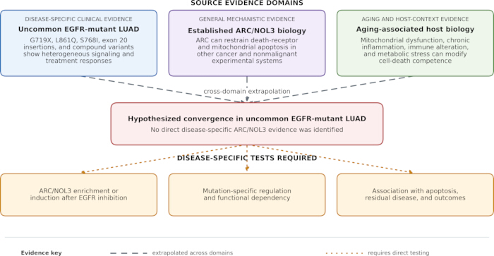
Figure 1: Evidence-stratified framework for the proposed ARC/NOL3 hypothesis in uncommon epidermal growth factor receptor-mutant lung adenocarcinoma. Three independent evidence domains are shown: disease-specific clinical evidence on uncommon epidermal growth factor receptor variants, general mechanistic evidence on ARC/NOL3-mediated regulation of apoptosis, and evidence from aging and host-context biology. Dashed gray arrows indicate cross-domain extrapolation, whereas dotted orange arrows indicate relationships requiring direct disease-specific testing. No primary study identified in this review directly demonstrated the proposed convergence. Please click here to view a larger version of this figure.
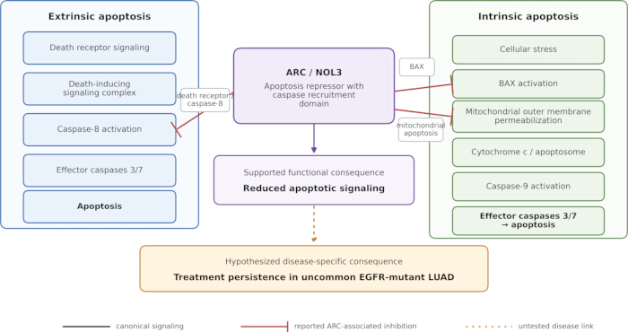
Figure 2: Established ARC/NOL3 functions in apoptotic signaling and their hypothesized disease-specific relevance. The left and right panels depict canonical extrinsic and intrinsic apoptotic signaling, respectively. Red inhibitory lines show ARC-associated interactions reported in general apoptosis models, other malignancies, or nonmalignant systems. The dotted orange arrow indicates the unverified hypothesis that ARC-associated suppression of apoptosis contributes to treatment persistence in uncommon epidermal growth factor receptor-mutant lung adenocarcinoma. This disease-specific relationship has not been directly demonstrated. Please click here to view a larger version of this figure.
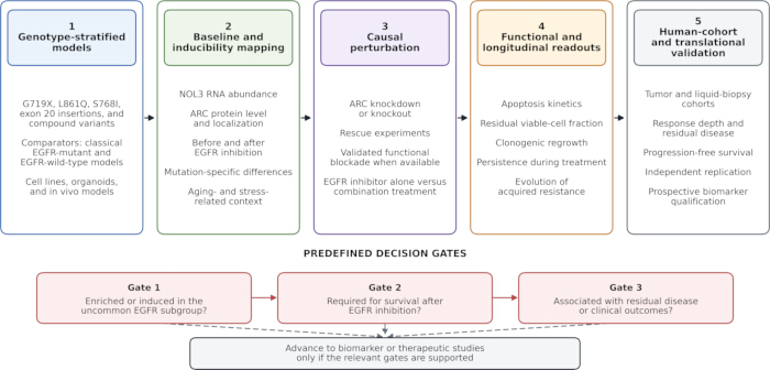
Figure 3: Proposed validation roadmap for the ARC/NOL3 hypothesis. This research framework outlines genotype-stratified model selection, baseline and treatment-induced ARC/NOL3 assessment, causal perturbation, functional and longitudinal readouts, and validation in human cohorts. Progression through the three decision gates requires evidence of ARC/NOL3 enrichment or induction, functional dependency after epidermal growth factor receptor inhibition, and association with residual disease or clinical outcomes before biomarker or therapeutic development is considered. The figure presents a research validation roadmap that progresses from mechanistic assessment to biomarker and translational evaluation. Please click here to view a larger version of this figure.
| Mutation category | Representative variants and approximate frequency | Structural context | Therapy and study | Treatment setting | Efficacy cohort | Principal outcomes | Key limitations |
| G719X | G719A/C/S/D; approximately 2–4% of EGFR-mutant NSCLC | Exon 18; phosphate-binding loop (P-loop); P-loop and αC-helix-compressing class | Afatinib; pooled LUX-Lung 2, 3, and 6 (Ref. 13) | TKI-naive, advanced disease | n = 18 | ORR 77.8% (95% CI, 52.4–93.6) | Post hoc pooled analysis; small cohort; solitary and compound genotypes were not uniformly separated. |
| S768I | S768I; approximately 0.6–2% of EGFR-mutant NSCLC; often occurs in compound genotypes | Exon 20; adjacent to the αC-helix; P-loop and αC-helix-compressing class | Afatinib; pooled LUX-Lung 2, 3, and 6 (Ref. 13) | TKI-naive, advanced disease | n = 8 | ORR 100% (95% CI, 63.1–100.0) | Extremely small cohort; frequent compound mutations; the point estimate is imprecise. |
| L861Q | L861Q; approximately 1–2% of EGFR-mutant NSCLC | Exon 21; activation loop; classical-like structural class | Afatinib; pooled LUX-Lung 2, 3, and 6 (Ref. 13) | TKI-naive, advanced disease | n = 16 | ORR 56.3% (95% CI, 29.9–80.2) | Post hoc pooled analysis; small cohort and wide confidence interval. |
| Major uncommon mutations, excluding exon 20 insertions | Predominantly G719X, L861Q, and S768I; solitary or compound genotypes | Multiple structural classes; should not be treated as biologically uniform | Osimertinib; KCSG-LU15-09 (Ref. 14) | First or later line; 61% received first-line therapy | n = 36 | ORR 50% (95% CI, 33–67); median PFS 8.2 months (95% CI, 5.9–10.5); median DoR 11.2 months (95% CI, 7.7–14.7) | Single-arm trial; mixed treatment lines and genotypes; exon 20 insertions excluded; mutation-specific subgroups were small. |
| Solitary versus compound uncommon mutations, excluding exon 20 insertions | Compound genotype = at least two EGFR mutations in the same tumor; frequency is assay- and definition-dependent | Structural behavior depends on the component variants and their allelic context | Osimertinib; UNICORN phase 2 trial (Ref. 3) | First-line, metastatic disease | n = 22 solitary; n = 18 compound | ORR 45.5% (90% CI, 26.9–65.3) versus 66.7% (90% CI, 43.7–83.7); median PFS 5.4 versus 9.8 months; median OS 23.0 months versus not reached | Nonrandomized; small subgroups; heterogeneous partner variants; between-group differences are descriptive, not proof of superiority. |
| EGFR exon 20 insertions | Heterogeneous in-frame insertions or duplications; approximately 4–12% of EGFR-mutant NSCLC | Within or immediately after the exon 20 αC-helix; position and inserted sequence influence drug sensitivity | Amivantamab plus carboplatin-pemetrexed; PAPILLON phase 3 trial (Ref. 16) | First-line, advanced disease | n = 153 versus n = 155 | ORR 73% versus 47%; median PFS 11.4 versus 6.7 months; HR for progression or death 0.40 (95% CI, 0.30–0.53); median DoR 9.7 versus 4.4 months | Open-label regimen-level comparison; not powered for individual insertion variants; overall-survival data were immature at the primary analysis. |
| EGFR exon 20 insertions | Multiple insertion variants; variant distribution differs across assays and populations | Insertion-specific conformational effects; not one uniform molecular entity | Amivantamab monotherapy; CHRYSALIS phase 1 trial (Ref. 18) | After progression on platinum-based chemotherapy | n = 81 | ORR 40% (95% CI, 29–51); median DoR 11.1 months; median PFS 8.3 months (95% CI, 6.5–10.9); median OS 22.8 months | Single-arm early-phase study; selected population; limited power for variant-level comparisons. |
| EGFR exon 20 insertions | Multiple insertion variants | Insertion-specific conformational effects | Mobocertinib; phase 1/2 platinum-pretreated cohort and EXCLAIM extension (Ref. 21) | After platinum-based chemotherapy | n = 114 | ORR 28% (95% CI, 20–37); median DoR 17.5 months (95% CI, 7.4–20.3); median PFS 7.3 months (95% CI, 5.5–9.2); median OS 24.0 months (95% CI, 14.6–28.8) | Single-arm phase 1/2 evidence; no concurrent comparator; subsequent market withdrawal limits present-day clinical applicability. |
| EGFR exon 20 insertions | Multiple insertion variants | Insertion-specific conformational effects | Sunvozertinib; WU-KONG1B (Ref. 19) | After platinum-based chemotherapy | n = 85 in the primary efficacy population receiving 200 mg once daily | Confirmed ORR 46% (95% CI, 35–57); median DoR 11.1 months (95% CI, 8.2–NE) | Open-label study without a concurrent comparator; accelerated approval was based on ORR and DoR, with confirmatory evidence required. |
| EGFR exon 20 insertions | Multiple insertion variants | Insertion-specific conformational effects | Zipalertinib; REZILIENT1 phase 1/2b study (Ref. 20) | After platinum-based chemotherapy, with or without prior amivantamab | n = 176 in the primary efficacy population; n = 244 treated overall | Confirmed ORR 35.2% (95% CI, 28.2–42.8); median DoR 8.8 months (95% CI, 8.3–12.7) | Single-arm study; heterogeneous prior exon 20-directed treatment; response varied according to prior therapy. |
Table 1: Definitions, structural context, and representative clinical evidence for uncommon epidermal growth factor receptor mutations. Representative uncommon epidermal growth factor receptor mutation categories are presented separately because they differ in structural context, drug sensitivity, and clinical evidence. Frequencies are approximate proportions among epidermal growth factor receptor-mutant non-small cell lung cancers and may vary according to population, testing platform, variant-reporting criteria, and the classification of compound mutations. Clinical outcomes are reported at the study level and should not be interpreted as evidence of therapeutic equivalence across mutation categories or individual exon 20 insertion variants. Most cited trials enrolled patients with non-small cell lung cancer; therefore, extrapolation specifically to lung adenocarcinoma should be made cautiously. Acquired resistance mutations, including T790M and C797S, are outside the scope of this table. CI, confidence interval; DoR, duration of response; EGFR, epidermal growth factor receptor; HR, hazard ratio; NSCLC, non-small cell lung cancer; ORR, objective response rate; OS, overall survival; PFS, progression-free survival; TKI, tyrosine kinase inhibitor.
| Evidence category | Available evidence | Supported interpretation | Relevance to the proposed framework | Key limitation or required test | Representative references |
| Direct evidence in uncommon EGFR-mutant LUAD | No primary study identified in the reviewed literature directly evaluated ARC/NOL3 expression, regulation, functional dependency, or clinical relevance in uncommon EGFR-mutant LUAD. | No disease-specific conclusion can currently be drawn. | Defines the central knowledge gap addressed by this review. | Expression enrichment, induction after EGFR inhibition, mutation-specific regulation, functional dependency, and associations with treatment response or outcomes remain untested. | No eligible direct study identified in the reviewed literature |
| Evidence in broader EGFR-mutant LUAD or lung cancer | Clinical and mechanistic studies demonstrate mutation heterogeneity, variable therapeutic response, prosurvival signaling, and therapy-persistent cell states; these studies did not specifically evaluate ARC/NOL3. | Apoptosis competence and non-genetic persistence may influence response beyond receptor inhibition. | Provides a disease-context rationale for testing ARC/NOL3. | Does not establish an EGFR-to-ARC signaling relationship or ARC dependency. ARC/NOL3 must be measured and perturbed in genotype-defined models and longitudinal clinical samples. | Refs. 1–3, 13–16, 18, 20–22, 26–28, 31,32, 35 |
| Evidence from other malignancies | ARC expression or function has been associated with tumor-cell survival and treatment resistance in selected breast, colorectal, and hematologic cancer models. | ARC can function as a context-dependent cancer-associated survival regulator. | Supports biological plausibility for evaluating ARC in lung cancer. | Cross-cancer findings cannot be assumed to apply to uncommon EGFR-mutant LUAD; disease-specific expression, perturbation, and outcome studies are required. | Refs. 9–11 |
| Evidence from general ARC biology and nonmalignant systems | Mechanistic studies show that ARC can interact with selected caspases and BAX-related mitochondrial death signaling and can limit stress-induced apoptosis in specific experimental systems. | Establishes the molecular capacity of ARC to regulate apoptotic commitment. | Provides the mechanistic basis for a testable apoptosis-resistance hypothesis. | Does not demonstrate tumor-specific, EGFR-specific, mutation-specific, or treatment-specific dependency. | Refs. 6–8 |
| Evidence from aging biology and host context | Aging is associated with mitochondrial dysfunction, oxidative stress, chronic inflammation, immune alterations, frailty, and reduced tolerance of systemic therapy; no age-stratified ARC/NOL3 analysis in uncommon EGFR-mutant LUAD was identified. | Aging-related host and tissue factors may modify treatment response and therapeutic tolerance. | Supports inclusion of aging-related variables in future ARC/NOL3 studies. | Does not establish that aging induces ARC/NOL3 or creates ARC dependency in uncommon EGFR-mutant tumors. | Refs. 4,5,33,34 |
| Integrative hypothesis proposed in this review | The review integrates uncommon EGFR signaling, apoptosis regulation, and aging-associated stress biology into a candidate ARC-related survival model. | Generates experimentally testable questions rather than a causal conclusion. | Prioritizes mutation-specific expression analysis, longitudinal assessment after EGFR inhibition, functional perturbation, and clinical validation. | The proposed intersection remains unverified and should not be interpreted as a validated biomarker, therapeutic target, or clinical algorithm. | Based on the evidence cited above |
Table 2: Evidence map for the proposed relevance of ARC/NOL3 to uncommon epidermal growth factor receptor-mutant lung adenocarcinoma. Evidence is classified as direct disease-specific evidence, evidence established outside the target disease, indirectly suggested relationships, or unverified hypotheses. References in the extrapolated categories support only the individual biological component described and do not demonstrate the complete relationship between uncommon epidermal growth factor receptor–ARC/NOL3–treatment persistence.
| Translational strategy | Rationale | Potential benefit | Key limitation | Most suitable future evidence | References |
| Direct ARC inhibition | Block ARC interactions with caspases or mitochondrial death regulators. | Lower the apoptotic threshold near the execution phase of cell death. | No established ARC-directed clinical inhibitor is available. | Structural biology, target engagement assays, and drug-screening studies. | Refs. 6–11 |
| ARC degradation or destabilization | Reduce ARC abundance or alter its localization to weaken downstream survival protection. | May sensitize tumor cells to EGFR inhibition or cytotoxic therapy. | Selectivity and safety are uncertain, particularly in tissues where ARC may have physiological protective roles. | Preclinical degrader feasibility studies and normal-tissue toxicity assessment. | Refs. 6–11 |
| EGFR plus PI3K-Akt pathway modulation | Suppress the oncogenic receptor while weakening downstream survival signaling. | May reduce adaptive survival and improve apoptosis after EGFR inhibition. | Combined pathway inhibition may increase toxicity, especially in older patients. | Biomarker-selected preclinical models and carefully designed early-phase trials. | Refs. 27,28,35 |
| EGFR plus MAPK-pathway modulation | Limit rebound or persistent adaptive signaling after EGFR inhibition. | May reduce survival signaling that supports residual disease. | Rash, diarrhea, fatigue, and cumulative toxicity may limit long-term use. | Intermittent or short-course combination studies with pharmacodynamic endpoints. | Refs. 28,35 |
| EGFR therapy plus apoptosis sensitizers | Pair receptor inhibition with agents that lower mitochondrial apoptotic thresholds, such as BH3 mimetics or related strategies. | May convert cytostatic stress into committed tumor cell death. | Therapeutic window may be narrow and requires careful patient selection. | Patient-derived models testing apoptosis restoration and combination tolerability. | Refs. 25,36 |
| Immunogenic cell death-oriented combinations | Increase antigen release and danger signaling by improving tumor cell death execution. | May enhance immune-mediated tumor clearance after targeted, cytotoxic, or radiation therapy. | EGFR-mutant tumors often show limited response to immune checkpoint blockade, and aging may reduce immune competence. | Immune profiling, spatial analysis, and trials with cell death and immune endpoints. | Refs. 5,30,37–39 |
| Aging-informed biomarker strategy | Integrate tumor genotype, ARC/NOL3 status, mitochondrial vulnerability, frailty, immune status, and treatment tolerance. | May individualize combination selection for older or biologically vulnerable patients. | Requires standardized assays and prospective validation. | Prospective translational cohorts with geriatric assessment and molecular endpoints. | Refs. 4,5, 33,34, 38,39 |
Table 3: Hypothesis-driven strategies for evaluating the proposed ARC/NOL3-related survival model. The listed approaches are conceptual or preclinical research directions that remain contingent on demonstrating ARC/NOL3 expression, inducibility, and functional dependence in uncommon epidermal growth factor receptor-mutant lung adenocarcinoma. They should not be interpreted as established treatments or clinical recommendations.
Divulgaciones
The authors declare that they have no conflicts of interest.
Author Contributions:
Conceptualization, Fei He and Hong Wei Li; writing and original draft preparation, Jun Ling Wang, Cai E Hou and Kun Rui Chen; writing and review and editing, Han Min Wang, Cheng Yun Zhang, Xiu Ping Li, Jian Peng Hu, Hong Xia Liu, Zhao Min Chen and Chang Wen; Graphical representation, Jun Ling Wang, Cai E Hou, Kun Rui Chen, Yin Yu Zhou, Jian Mei Song, Min Fang Wu, Min Chen, Fan Zhang, Shu Min Li, Ruo Shui Kong and Ying Yu Yang; funding acquisition, Jun Ling Wang, Hong Wei Li, Jian Peng Hu, Ying Yu Yang and Fei He. All authors have read and agreed to the published version of the manuscript.
Agradecimientos
This work was supported financially by grants from the Scientific Research Fund of Yunnan Provincial Department of Science and Technology and Kunming Medical University Joint Project (202301AY070001-071; 202201AY070001-216); Yunnan health training project of high level talents (H-2024062); Qujing University of Medicine and Health Sciences 2025 Annual University-level Scientific Research Fund Project (2025ZD06); Yunnan Provincial Department of Education Engineering Research Center for Precision Medical Testing; The Scientific Research Fund of Yunnan Provincial Department of Education (2024J0152); The Open project of Yunnan Key Laboratory of Public Health and Biosafety (KLPHB_2023_07); The 2024 Key Medical (Reserve) Talent Plan of Qujing Municipal Health Commission; The major science and technology special plan of Yunnan Province (202402AD080006); Qujing University of Medicine and Health Sciences 2025 Annual University-level Scientific Research Fund Project (2025ZD08); Open Project of Yunnan Provincial Clinical Laboratory Medicine Research Center (2017DG005-2024-03).
Referencias
- Soria JC et al. Osimertinib in untreated EGFR-mutated advanced non-small-cell lung cancer. N Engl J Med. 2018;378(2):113-25. doi:10.1056/NEJMoa1713137.
- Harrison PT, Vyse S, Huang PH. Rare epidermal growth factor receptor mutations in non-small cell lung cancer. Semin Cancer Biol. 2020;61:167-79. doi:10.1016/j.semcancer.2019.09.015.
- Okuma Y et al. First-line osimertinib for previously untreated patients with NSCLC and uncommon EGFR mutations: the UNICORN phase 2 nonrandomized clinical trial. JAMA Oncol. 2024;10(1):43-51. doi:10.1001/jamaoncol.2023.5013.
- López-Otín C et al. Hallmarks of aging: an expanding universe. Cell. 2023;186(2):243-78. doi:10.1016/j.cell.2022.11.001.
- Liu Z et al. Immunosenescence: molecular mechanisms and diseases. Signal Transduct Target Ther. 2023;8(1):200. doi:10.1038/s41392-023-01451-2.
- Koseki T, Inohara N, Chen S, Núñez G. ARC, an inhibitor of apoptosis expressed in skeletal muscle and heart that interacts selectively with caspases. Proc Natl Acad Sci U S A. 1998;95(9):5156-60. doi:10.1073/pnas.95.9.5156.
- Ludwig-Galezowska AH, Flanagan L, Rehm M. Apoptosis repressor with caspase recruitment domain, a multifunctional modulator of cell death. J Cell Mol Med. 2011;15(5):1044-53. doi:10.1111/j.1582-4934.2010.01221.x.
- Ao X et al. Role of apoptosis repressor with caspase recruitment domain in human health and chronic diseases. Ann Med. 2024;56(1):2409958. doi:10.1080/07853890.2024.2409958.
- Mercier I et al. ARC, an apoptosis suppressor limited to terminally differentiated cells, is induced in human breast cancer and confers chemo- and radiation-resistance. Cell Death Differ. 2005;12(6):682-6. doi:10.1038/sj.cdd.4401631.
- Mercier I et al. ARC (apoptosis repressor with caspase recruitment domain) is a novel marker of human colon cancer. Cell Cycle. 2008;7(11):1640-7. doi:10.4161/cc.7.11.5979.
- Carter BZ et al. Anti-apoptotic ARC protein confers chemoresistance by controlling leukemia-microenvironment interactions through an NFκB/IL1β signaling network. Oncotarget. 2016;7(15):20054-67. doi:10.18632/oncotarget.7911.
- National Library of Medicine. GILOTRIF-afatinib tablet, film coated [Internet]. DailyMed. Updated 2025 Nov 13 [cited 2026 Sep 22]. Available from: https://dailymed.nlm.nih.gov/dailymed/lookup.cfm?setid=fd638e5e-8032-e7ca-0179-95e96ab5d387.
- Yang JC et al. Clinical activity of afatinib in patients with advanced non-small-cell lung cancer harbouring uncommon EGFR mutations: a combined post-hoc analysis of LUX-Lung 2, LUX-Lung 3, and LUX-Lung 6. Lancet Oncol. 2015;16(7):830-8. doi:10.1016/S1470-2045(15)00026-1.
- Cho JH et al. Osimertinib for patients with non-small-cell lung cancer harboring uncommon EGFR mutations: a multicenter, open-label, phase II trial (KCSG-LU15-09). J Clin Oncol. 2020;38(5):488-95. doi:10.1200/JCO.19.00931.
- Pizzutilo EG et al. Activity of osimertinib in non-small-cell lung cancer with uncommon epidermal growth factor receptor mutations: retrospective observational multicenter study (ARTICUNO). ESMO Open. 2024;9(6):103592. doi:10.1016/j.esmoop.2024.103592.
- Zhou C et al. Amivantamab plus chemotherapy in NSCLC with EGFR exon 20 insertions. N Engl J Med. 2023;389(22):2039-51. doi:10.1056/NEJMoa2306441.
- U.S. Food and Drug Administration. FDA approves amivantamab-vmjw for EGFR exon 20 insertion-mutated non-small cell lung cancer indications [Internet]. Silver Spring (MD): U.S. Food and Drug Administration; 2024 Mar 1 [cited 2026 Sep 22].
- Park K et al. Amivantamab in EGFR exon 20 insertion-mutated non-small-cell lung cancer progressing on platinum chemotherapy: initial results from the CHRYSALIS phase I study. J Clin Oncol. 2021;39(30):3391-402. doi:10.1200/JCO.21.00662.
- U.S. Food and Drug Administration. FDA grants accelerated approval to sunvozertinib for metastatic non-small cell lung cancer with EGFR exon 20 insertion mutations [Internet]. Silver Spring (MD): U.S. Food and Drug Administration; 2025 Jul 2 [cited 2026 Sep 22].
- Piotrowska Z et al. Zipalertinib in patients with epidermal growth factor receptor exon 20 insertion-positive non-small cell lung cancer previously treated with platinum-based chemotherapy with or without amivantamab. J Clin Oncol. 2025;43(21):2387-97. doi:10.1200/JCO-25-00763.
- Zhou C et al. Treatment outcomes and safety of mobocertinib in platinum-pretreated patients with EGFR exon 20 insertion-positive metastatic non-small cell lung cancer: a phase 1/2 open-label nonrandomized clinical trial. JAMA Oncol. 2021;7(12):e214761. doi:10.1001/jamaoncol.2021.4761.
- Jänne PA et al. First-line mobocertinib versus platinum-based chemotherapy in patients with EGFR exon 20 insertion-positive metastatic non-small cell lung cancer in the phase III EXCLAIM-2 trial. J Clin Oncol. 2025;43(13):1553-63. doi:10.1200/JCO-24-01269.
- Takeda Pharmaceutical Company Limited. Takeda provides update on lung cancer program: EXKIVITY (mobocertinib) [Internet]. 2023 Oct 2 [cited 2026 Sep 22].
- Food and Drug Administration. Takeda Pharmaceuticals U.S.A., Inc.; withdrawal of approval of new drug application for EXKIVITY (mobocertinib succinate) capsule, equivalent to 40 milligrams base [Internet]. Fed Regist. 2024;89(135):57418-9 [cited 2026 Sep 22].
- Taylor RC, Cullen SP, Martin SJ. Apoptosis: controlled demolition at the cellular level. Nat Rev Mol Cell Biol. 2008;9(3):231-41. doi:10.1038/nrm2312.
- Robichaux JP et al. Mechanisms and clinical activity of an EGFR and HER2 exon 20-selective kinase inhibitor in non-small cell lung cancer. Nat Med. 2018;24(5):638-46. doi:10.1038/s41591-018-0007-9.
- Sanaei MJ, Razi S, Pourbagheri-Sigaroodi A, Bashash D. The PI3K/Akt/mTOR pathway in lung cancer: oncogenic alterations, therapeutic opportunities, challenges, and a glance at the application of nanoparticles. Transl Oncol. 2022;18:101364. doi:10.1016/j.tranon.2022.101364.
- Rosell R et al. Coregulation of pathways in lung cancer patients with EGFR mutation: therapeutic opportunities. Br J Cancer. 2021;125(12):1602-11. doi:10.1038/s41416-021-01519-2.
- Chen Z et al. Hypoxic microenvironment in cancer: molecular mechanisms and therapeutic interventions. Signal Transduct Target Ther. 2023;8(1):70. doi:10.1038/s41392-023-01332-8.
- Galluzzi L et al. Targeting immunogenic cell stress and death for cancer therapy. Nat Rev Drug Discov. 2024;23(6):445-60. doi:10.1038/s41573-024-00920-9.
- Russo M et al. Cancer drug-tolerant persister cells: from biological questions to clinical opportunities. Nat Rev Cancer. 2024;24(10):694-717. doi:10.1038/s41568-024-00737-z.
- Sun X, Wu LF, Altschuler SJ, Hata AN. Targeting therapy-persistent residual disease. Nat Cancer. 2024;5(9):1298-304. doi:10.1038/s43018-024-00819-9.
- Dale W et al. Practical assessment and management of vulnerabilities in older patients receiving systemic cancer therapy: ASCO guideline update. J Clin Oncol. 2023;41(26):4293-312. doi:10.1200/JCO.23.00933.
- Bergerot CD et al. Geriatric assessment: ASCO global guideline. JCO Glob Oncol. 2025;11:e2500276. doi:10.1200/GO-25-00276.
- Fu K, Xie F, Wang F, Fu L. Therapeutic strategies for EGFR-mutated non-small cell lung cancer patients with osimertinib resistance. J Hematol Oncol. 2022;15(1):173. doi:10.1186/s13045-022-01391-4.
- Townsend PA et al. BH3-mimetics: recent developments in cancer therapy. J Exp Clin Cancer Res. 2021;40(1):355. doi:10.1186/s13046-021-02157-5.
- Gainor JF et al. EGFR mutations and ALK rearrangements are associated with low response rates to PD-1 pathway blockade in non-small cell lung cancer: a retrospective analysis. Clin Cancer Res. 2016;22(18):4585-93. doi:10.1158/1078-0432.CCR-15-3101.
- Bischoff P et al. Single-cell RNA sequencing reveals distinct tumor microenvironmental patterns in lung adenocarcinoma. Oncogene. 2021;40(50):6748-58. doi:10.1038/s41388-021-02054-3.
- Cilento MA, Sweeney CJ, Butler LM. Spatial transcriptomics in cancer research and potential clinical impact: a narrative review. J Cancer Res Clin Oncol. 2024;150(6):296. doi:10.1007/s00432-024-05816-0.