A subscription to JoVE is required to view this content. Sign in or start your free trial.

Method Article

Analyzing Synaptic Vesicle Recycling and Fluorescent Dye Uptake in Drosophila Larvae

761 views

⸱

July 8th, 2025

In This Article

Abstract

Source: Kopke, D. L., et al., FM Dye Cycling at the Synapse: Comparing High Potassium Depolarization, Electrical and Channelrhodopsin Stimulation. J. Vis. Exp. (2018).

This video demonstrates a protocol to study synaptic vesicle recycling at neuromuscular junctions in channelrhodopsin-expressing Drosophila larvae using a fluorescent dye. Blue light activates the channelrhodopsins, causing calcium influx and synaptic vesicle exocytosis. The dye integrates into the neuronal membranes and is internalized during endocytosis, resulting in enhanced fluorescence. Further blue light stimulation induces exocytosis and dye release, which is shown by reduced fluorescence.
Protocol

1. Larval Glue Dissection

  1. Thoroughly mix 10 parts of silicone elastomer base with 1 part of silicone elastomer curing agent from the elastomer kit (Table of Materials).
  2. Coat 22 x 22 mm glass coverslips with the elastomer and cure on a hot plate at 75 ˚C for several hours (until no longer sticky to the touch).
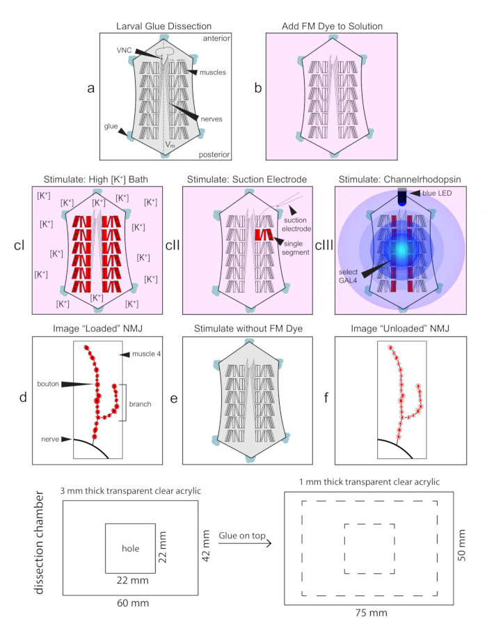
  3. Place a single elastomer-coated glass coverslip into the custom-made plexiglass dissection chamber (Figure 1, bottom) in preparation for the larval dissection.
  4. Prepare the glue pipettes from borosilicate glass capillary using a standard microelectrode puller to obtai....

Access restricted. Please log in or start a trial to view this content.

Results

Larval glue dissection diagram for FM dye stimulation in Drosophila neuromuscular junction (NMJ) studies.
Figure 1: Flowchart of FM1-43 dye loading protocol at the Drosophila NMJ. The larval glue dissection produces a flattened neuromusculature preparation, with the ventral nerve cord (VNC) projecting segmental nerves from the ventral midline (Vm) to t.......

Access restricted. Please log in or start a trial to view this content.

Disclosures

No conflicts of interest declared.

Materials

List of materials used in this article
NameCompanyCatalog NumberComments
SylGard 184 Silicone Elastomer KitFisher ScientificNC9644388To put on cover glass for dissections
Microscope Cover Glass 22x22-1Fisherbrand12-542-BTo put SylGard on for dissections
Aluminum Top Hot Plate Type 2200ThermolyneHPA2235MTo cure the SylGard
Plexi glass dissection chamberN/AN/AHandmade
Borosilicate Glass CapillariesWPI1B100F-4To make suction and glue micropipettes
Laser-Based Micropipette PullerSutter InstrumentP-2000To make suction and glue micropipettes
Tygon E-3603 Laboratory TubingComponent Supply Co.TET-031AFor mouth and suction pipette
P2 pipette tipUSA Scientific1111-3700For mouth pipette
0.6-mL Eppendorf tube capFisher Scientific05-408-120Used to put glue in for dissection
Vetbond 3MWPIvetbondGlue used for dissections
Potassium ChlorideFisher ScientificP-217Forsaline
Sodium ChlorideMillipore SigmaS5886For saline
Magnesium ChlorideFisher ScientificM35-500For saline
Calcium Chloride DihydrateMillipore SigmaC7902For saline
SucroseFisher ScientificS5-3For saline
HRP:Alexa Fluor 647Jackson ImmunoResearch123-605-021To label neuronal membranes
HEPESMillipore SigmaH3375For saline
PaintbrushWinsor & Newton94376864793To manipulate the larvae
Dumont Dumostar Tweezers #5WPI500233Used during dissection
7 cm McPherson-Vannas Microscissors (blades 3 mm)WPI14177Used during dissection
FM1-43Fisher ScientificT35356Fluorescent styryl dye
Digital TimerVWR62344-641For timing FM dye load/unload
LSM 510 META laser-scanning confocal microscopeZeissFor imaging the fluorescent markers
Zen 2009 SP2 version 6.0ZeissSoftware for imaging on confocal
HeNe 633nm laserLasosTo excite HRP:647 during imaging
Argon 488nm laserLasosTo excite the FM dye during imaging
Micro-ForgeWPIMF200To fire polish glass micropipettes
StimulatorGrassS48To control the LED and electrical stimulation
All-trans RetinalMillipore SigmaR2500Essential co-factor for ChR2
Zeiss Stemi Microscope with camera portZeiss2000-CUsed during channelrhodopsin stimulation
LED 470nmThorLabsM470L2Used for ChR activation
T-Cube LED DriverThorLabsLEDD1BTo control the LED
LED Power SupplyCincon Electronics Co.TR15RA150To power the LED
Optical Power and Energy MeterThorLabsPM100DTo measure LED intensity
40X Achroplan Water Immersion ObjectiveZeissUsed during electrical stimulation and confocal imaging

Explore More Articles

Channelrhodopsin StimulationNeuromuscular JunctionFM Dye CyclingBlue Light ActivationCalcium InfluxVesicle ExocytosisEndocytosis