This video demonstrates the detection of Hepatitis C virus replication by analyzing NS3 protein expression in transfected human hepatoma cells. Western blotting reveals viral protein levels as a measure of replication activity.
프로토콜
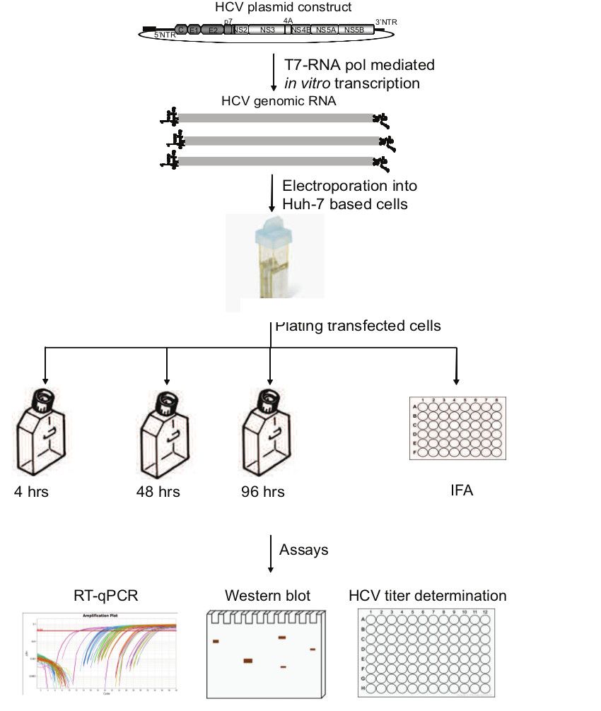
A general outline of the protocol is illustrated in Figure 1.
1. Cells
Prepare complete growth media that contain 10-15% fetal bovine serum (FBS), 10 mM nonessential amino acids, 10 mM HEPES (4-(2-hydroxyethyl)-1-piperazineethanesulfonic acid), penicillin (100 units/ml), streptomycin (100 mg/ml), and 2 mM L-glutamine.
Maintain Huh-7.5.1 cells in complete growth media containing the above supplements for in vitro analysis of the hepatitis C virus (HCV) replication cycle.
Culture viral strains in Huh-7.5.1 cells with the specified supplemented growth media at 37 °C with 5% CO2.
2. In Vitro HCV ribonucleic acid (RNA) Transcription
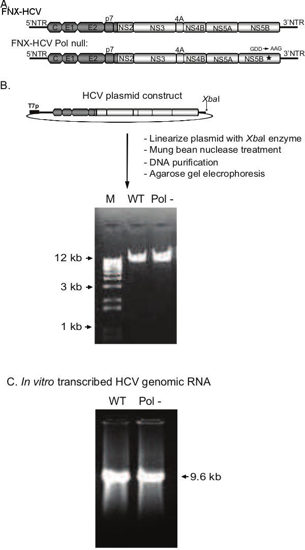
Use an intragenotype chimeric virus FNX-HCV and FNX-HCV Pol null (Pol-) HCV for evaluation of viral replication (Figure 2A).
Linearize the viral plasmids with the XbaI restriction enzyme and then treat with mung bean nuclease to generate blunt ends. Purify the digested plasmids by anion-exchange chromatography. Verify the integrity of the linearized plasmid by subjecting the DNA to agarose gel electrophoresis (Figure 2B).
Transcribe the linearized viral plasmids using the T7 RNA polymerase.
Purify the newly synthesized DNase-treated RNA using an RNA purification kit.
Verify the RNA production by agarose gel electrophoresis (Figure 2C).
Quantify the RNA by spectrophotometry.
Store the generated RNA at -80 °C in 10 μg working aliquots. NOTE: To minimize variation in RNA quality within each experimental design, transcribe all RNA for each viral sample and control at the same time.
3. HCV RNA Transfection and Sample Collection
Harvest the Huh-7.5.1 cells using trypsin.
To rinse cells, centrifuge and resuspend the suspension twice with cold low-serum media. Resuspend the cells in low serum media at 1 x 10⁷ cells per ml.
Mix a total of 10 μg of transcribed viral RNA with 400 μl of resuspended cells (4 x 10⁶ cells) in a 0.4-cm electroporation cuvette. Transfect the cells via electroporation at 270 V, 100 Ω, and 950 μF.
Resuspend the electroporated cells in 10 mL complete growth medium with 15% FBS. NOTE: Increased survivability of electroporated cells is seen when the Huh-7.5.1 cells are cultured in 15% FBS.
Plate the cells in both T-25 flasks (~1.2 x 10⁶ cells per flask) and 48-well plates (1 x 10⁴ cells per well) for each of the following time points: 4, 48 and 96 hr.
Replace the media at 4-8 hr post-transfection with fresh supplemented growth media with 10% FBS to remove dead cell debris from the cultured flasks and plates.
Harvest cell culture supernatants at the 48, 96 hr time points and remove cellular debris from collected samples by centrifugation of cells at 1,500 rpm for 10 min at 4 ºC.
Store the cell-free supernatants at -80 ºC. Lyse the cells for protein and RNA analysis by Western blot and reverse transcription-quantitative PCR at the indicated time points.
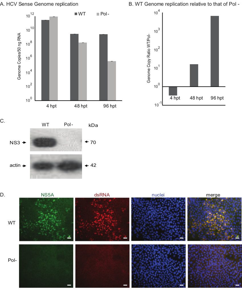
4. Western Blotting Analysis for Detecting HCV Protein Expression (Figure 3C)
Use cell lysates from viral RNA transfected at 96 hr post transfection for protein western blotting analysis.
Resolve the cell lysate using sodium dodecyl sulfate–polyacrylamide gel electrophoresis (SDS-PAGE) and transfer to a polyvinylidene difluoride (PVDF) membrane.
Block the membrane using a blocking solution containing 5% skim milk and 0.2% Tween 20 in phosphate-buffered saline (PBS).
Incubate the membrane with primary mouse monoclonal antibody NS3 (1 in 1,000 dilution) and beta-actin (1 in 5,000 dilution).
Add goat anti-mouse IgG conjugated to horseradish peroxidase (1 in 5,000 dilution) and detect by chemiluminescence.
결과
Figure 1. General outline of HCV replication analysis workflow. Linearized HCV plasmid construct containing T7 RNA polymerase promoter (T7p) subjected to in vitro transcription. The purified HCV RNA genome is electroporated into Huh-7.5.1 cells and plated in flasks and 48-well plate. At 4 hr, 48 hr, and 96 hr post-transfection, the cellular RNA and culture supernatants are harvested from flasks. The cells from 48-well plate are utilized for harvesting protein lysate and are fixed for Immunofluorescence assay. Genome copy number analysis by RT-qPCR, Western blotting and measuring viral titer are done to assess the HCV replication.
Figure 2. Production of HCV genomic RNAs by in vitro transcription of constructed plasmid DNAs. A) Genomic organization of the J6CF/JFH-1 intragenotype 2a chimeric viruses, FNX-HCV and FNX-HCV Pol null. The J6CF strain region (5’NTR to part of NS2) is depicted in dark grey, and the JFH-1 strain region (part of NS2 to 3’NTR) is displayed in light grey. NS5B polymerase catalytic domain mutation (GDD to AAG) is indicated with an asterisk. B) Steps involved in generating linearized HCV plasmid. The gel picture shows the linearized, blunt-ended plasmid DNA produced by XbaI and mung bean nuclease digestion, ready for in vitro transcription. 0.8% agarose gel was used for resolving the DNA. C) Gel picture depicts the HCV genomic RNAs produced by in vitro transcription using the T7 RNA polymerase system. WT: wild-type; Pol-: Polymerase null; M: marker.
Figure 3. Assessing the growth phenotypes of HCV constructs. A) Evaluating the genome replication kinetics of wild-type (WT) and polymerase null (Pol-) viruses. The genome copy numbers of the sense RNA strand assessed by RT-qPCR are presented in the bar graph. The viral genome copies of Pol- virus declined over the period of time indicating replication deficient phenotype. B) Relative genome replication level of wild-type HCV is compared to that of Pol- virus. C) Western blot analysis of HCV protein expression. HCV protein NS3 is detected, and beta-actin is used as a cellular control. D) Immunofluorescence assay for investigating viral replication. At 96 hr post-electroporation, the cells were fixed and subjected to immunostaining for HCV NS5A protein and double-stranded RNA (dsRNA), a marker for HCV RNA replication intermediates. The nuclei were visualized with Hoechst stain (Scale bar: 50 μm). hpt: hours post-transfection.
재료
이 논문에 사용된 재료 목록
이름
회사
카탈로그 번호
댓글
Dulbecco’s modified Eagle’s medium (DMEM)
Fisher Scientific
10-017-CV
Non essential amino acid
Fisher Scientific
MT25025CI
HEPES
Life Technologies
15630080
Glutamax
Life Technologies
35050061
Opti-MEM reduced serum medium,no phenol red
Life Technologies
11058-021
Huh-7.5.1
The Scripps Research Institute
The cell line was kindly provided by Dr. Francis Chisari to Dr. Arumugaswami under executed MTA between The Scripps Research Institute and Cedars-Sinai Medical Center
Plasmids (pFNX-HCV, pFNX-HCV Pol null, pFNX-Rluc, and pFNX-Rluc Pol null)
Cedars-Sinai Medical Center
The HCV plasmids were synthesized by Dr. Arumugaswami using overlapping oligo-nucleotides.
PVDF membrane package
Bio-Rad
162-0263
Blotting grade blocker non fat dry milk
Bio-Rad
170-6404XTU
Tween-20
Bio-Rad
170-6531XTU
Anti-hepatitis C virus NS3 antibody [8 G-2]
Abcam
ab65407
Anti-hepatitis C virus NS3 antibody [H23]
Abcam
ab13830
Goat anti-mouse IgG conjugated with horseradish peroxidase (HRP)
Jackson ImmunoResearch Laboratories Inc.
115-035-003
Amersham ECL prime western blotting detection reagents