복부 시험 II: 타악기
1. 복부의 일반적인 타악기
- 환자에게 절차 설명
- 9개의 복부 지구의 각에 빛 타악기를 능력을 발휘합니다.
- 타악기로 환자의 얼굴을 보면서 불편함의 징후가 나타나세요. 환자에게 부드러움이 발생하는지 물어보십시오. 타악기에 부드러움은 비정상이며 다발성 염증을 나타낼 수 있습니다.
- 타악기 노트의 강도, 피치 및 지속 시간을 들어보십시오. 일반적으로, 창자 루프에서 공기에 의해 생성 된 타일패닉 소리가 들릴 것입니다. 타막 소리는 상대적으로 길고, 높은 피치, 그리고 큰 소리로. 가끔 씩씩한 부분(타프타니보다 낮은 피치, 짧고 조용한 소리)은 유체와 대변에 의해 생성되며 정상입니다. 둔함의 넓은 영역은 확대 기관 또는 심포지션 및 추가 기동에 의해 더 평가되어야 질량을 제안한다.
- 비용 마진 위의 하부 전방 가슴을 타악기로 합니다. 일반적으로 비장과 간은 늑골 케이지로 덮여 있으며 간 가장자리는 때때로 비용 마진보다 1-2cm 낮습니다. 간 을 통해 오른쪽에 둔한 소리가 예상된다. 왼쪽에는 위기포와 결장의 비장 굴곡이 들어야 합니다.
- 음모 심기 위의 타악기. 이 지역에 있는 타악기에 둔한 확대자궁 또는 경화방을 나타냅니다.
2. 간 간 경간 확인
타악기에 증가 간 범위는 확대 간을 나타냅니다, 이는 병리학 적 과정의 다양한 기초할 수 있습니다. 확대 된 간 (증가 된 범위를 가지고) 간 칙칙의 하부와 상부 경계를 모두 식별하는 것이 필수적이다, 간은 만성 폐쇄성 폐 질환의 결과로 아래로 변위 (이 경우 간 간 간이 정상입니다).
- 오른쪽 중간 클로비큘러 라인을 찾는 것으로 시작합니다. 오른쪽 중간 쪽줄에 가볍게 타악기를 위쪽으로 올라가서 배꼽 아래 의 폭타 부위에서 시작됩니다. 타악기 메모를 주의 깊게 듣기 위해 아래로 구부려야 할 수도 있습니다.
- tympany가 피부 연필로 둔함 (낮은 테두리)으로 바뀌는 곳에 표시합니다.
- 오른쪽 중간 쪽줄에 있는 타악기는 간 둔함의 상부 경계를 확인하기 위하여 젖꼭지 선에서 아래로 시작합니다.
- 폐 위에 공명하는 소리가 피부 연필로 간 을 둔하게 변화시키는 복벽의 요점을 표시하십시오.
- cm의 둔함의 상하 경계 사이의 거리를 측정합니다. 간 범위는 나이, 성별 및 신체 유형에 따라 다릅니다. 간 간 은 일반적으로 6-12 cm (평균 간 범위는 여자를 위한 7 cm 및 남자를 위한 10.5 cm)입니다.
- 간 간 범위가 증가 하는 경우, 타 악기 측면 및 내측. 중간선에서 의 정상 간 범위는 4-8cm입니다.
3. 타커스는 비장 마비를 감지하기 위해 전방 가슴을 떠났다.
비장은 왼쪽 미다실리 라인의 약간 후방에 위치하고 9번째와 11번째 갈비뼈 사이의 둔한 타원형 영역을 생산하고 있습니다. 정상 비장의 작은 표면만 감지 할 수있을만큼 피상적이며 비장 칙칙함은 종종 위 또는 대장 용액에 의해 가려져 있습니다. 그러나 확대된 비장은 중간선, 전방 및 아래쪽으로 확장되며, 트라우베의 우주 타악기 및/또는 카스텔의 기동이라는 두 가지 특수 타악기 기동에 의해 감지될 수 있습니다.
- 트라우베의 공간, 전방 축축선, 그리고 비용 마진을 남겼습니다.
- 확대된 비장의 다른 병리학적 조건내 확장과 함께 트라우베의 공간에 대한 타악기에 대한 칙칙함을 생성할 수 있습니다.
- 환자 척추와 왼팔이 약간 납치되면서 내측에서 트라우베 공간의 측면 경계까지 타악기가 있습니다. 타악기에 둔하거나 tympany의 영역의 감소는 비장에서 발생할 수 있습니다.
- Castell의 방법 (비장 타악기 기호 확인).
- 낮은 늑간 공간에서 전방 축산 선에서 타악기.
- 환자에게 심호흡을 하고 다시 타악기를 마시라고 한다. 일반 크기의 비장은 영감 중에 내려오더라도 타악기 점 위에 위치하며 타악기 톤은 만료와 영감 모두에 대해 타악기 톤이 있습니다. 타악기 노트가 둔하거나 영감 (긍정적 인 비장 타악기 기호)에 둔해지면 비장 (그림 3)이 의심됩니다.
4. 확률 복부의 평가.
타악기는 확률복부의 원인을 진단하는 데 도움이됩니다. 확률 복부를 통해 Tympany는 장 방해로 인한 공기 축적을 나타냅니다. 확률 복부의 측면에 타악기가 둔한 메모를 생성할 때, 그것은 유체 축적 또는 선동과 일치합니다.
- 변속의 칙칙함" 기동은 선동이 의심될 때 수행됩니다.
- 환자가 척추 자세를 취하면, 배꼽에서 측면 방향으로 타악기를 하고, 타일플라닉 소리가 피부 연필로 둔하게 변하는 지점의 표시를 합니다.
- 환자에게 측면(측면 탈구 위치)으로 롤을 하고, 상단에서 시작하여 타악기를 반복하고, 타일패닉 소리가 칙칙하게 변할 때 피부에 두 번째 표시를 하도록 요청합니다. 복부 유체가 존재하는 경우, 가스/유체 인터페이스의 테두리는 배꼽쪽으로 위쪽으로 이동됩니다(그림 2).

그림 2. 지루한 테스트를 이동합니다. 선동이 존재하는 경우에, 환자가 측면 탈구 위치에 있을 때 복부 측면에 둔하게 tympany 변경되는 지점은 배꼽쪽으로 위로 이동됩니다.
출처: 알렉산더 골드파브, MD, 의학 조교수, 베스 이스라엘 Deaconess 의료 센터, MA
의료 타악기는 바디 월을 두드려 서 유도 하는 소리 사이의 피치의 차이에 따라. 도청에 대한 청각 반응은 신체 벽이 진동하는 용이성에 따라 달라지며 기본 장기, 뇌졸중의 강도 및 신체 벽의 상태에 의해 영향을받습니다. 공명 (폐에서 들리는), tympany (공기가 채워진 창자 루프에서 들었음), 칙칙함 (유체 또는 고체 장기에서 들었음)의 세 가지 주요 의료 타악기 소리가 있습니다. 둔함 대 tympany 또는 공명의 대비는 장기와 질량의 크기와 마진을 결정할 뿐만 아니라 유체 축적 및 통합 영역을 식별할 수 있습니다. 타악기는 200년 전에 처음 도입된 이래로 신체 진단의 복잡한 부분으로 남아 있으며 폐와 복부 검사에 특히 유용합니다.
복부 검사의 일환으로, 타악기는 육안 검사 및 auscultation을 따릅니다. 심사관은 9개의 복부 지역 (상복부 지역, 오른쪽 hypochondriac 지구, 좌측 hypochondriac 지역, 탯줄 지역, 오른쪽 요추 지역, 좌편요추 지역, 저위 지역, 오른쪽 잉구부위 및 좌측 인기구)의 각각을 통해 먼저 타악기를 해야 합니다. 타악기에 의해 유도 된 부드러움은 비정상적이며 복막 염증을 의심해야합니다. 공기로 채워진 창자 루프가 복벽에 가장 가까운 곳에 위치하기 때문에 복강의 대부분의 부분에 대한 타악기는 주로 타막 소리를 불러 냅니다. 둔함의 넓은 영역의 존재는 유기, 복부 내 질량, 또는 액체에 대한 평가를 즉시 해야합니다.
복부 가스의 양 그리고 분포를 평가하는 것 이외에, 복부 검사는 타악기에 의한 간 및 비장 크기의 추정을 포함해야 합니다. 간과 비장은 흉곽에 의해 덮여 있기 때문에, 심사관은 뿐만 아니라 낮은 전방 가슴에 타악기해야합니다. 일반적으로, 하나는 간 위에 오른쪽 전방 가슴의 타악기에 둔한 소리를 듣고 기대, 위 기포와 결장의 비장 굴곡을 통해 왼쪽 전방 가슴의 타악기에 타막 소리(그림 1).
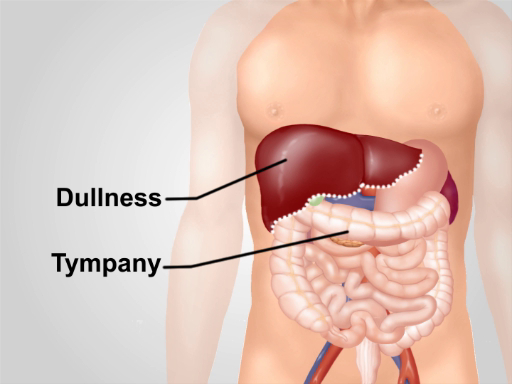
그림 1. 복부 지역에 대한 일반적인 타악기 노트. 오른쪽 아래 전방 가슴에 있는 간 위에 둔함의 영역을 제외하고, tympany는 이 지역에서 들리는 주요 소리입니다.
1. 복부의 일반적인 타악기
- 환자에게 절차 설명
- 9개의 복부 지구의 각에 빛 타악기를 능력을 발휘합니다.
- 타악기로 환자의 얼굴을 보면서 불편함의 징후가 나타나세요. 환자에게 부드러움이 발생하는지 물어보십시오. 타악기에 부드러움은 비정상이며 다발성 염증을 나타낼 수 있습니다.
- 타악기 노트의 강도, 피치 및 지속 시간을 들어보십시오. 일반적으로, 창자 루프에서 공기에 의해 생성 된 타일패닉 소리가 들릴 것입니다. 타막 소리는 상대적으로 길고, 높은 피치, 그리고 큰 소리로. 가끔 씩씩한 부분(타프타니보다 낮은 피치, 짧고 조용한 소리)은 유체와 대변에 의해 생성되며 정상입니다. 둔함의 넓은 영역은 확대 기관 또는 심포지션 및 추가 기동에 의해 더 평가되어야 질량을 제안한다.
- 비용 마진 위의 하부 전방 가슴을 타악기로 합니다. 일반적으로 비장과 간은 늑골 케이지로 덮여 있으며 간 가장자리는 때때로 비용 마진보다 1-2cm 낮습니다. 간 을 통해 오른쪽에 둔한 소리가 예상된다. 왼쪽에는 위기포와 결장의 비장 굴곡이 들어야 합니다.
- 음모 심기 위의 타악기. 이 지역에 있는 타악기에 둔한 확대자궁 또는 경화방을 나타냅니다.
2. 간 간 경간 확인
타악기에 증가 간 범위는 확대 간을 나타냅니다, 이는 병리학 적 과정의 다양한 기초할 수 있습니다. 확대 된 간 (증가 된 범위를 가지고) 간 칙칙의 하부와 상부 경계를 모두 식별하는 것이 필수적이다, 간은 만성 폐쇄성 폐 질환의 결과로 아래로 변위 (이 경우 간 간 간이 정상입니다).
- 오른쪽 중간 클로비큘러 라인을 찾는 것으로 시작합니다. 오른쪽 중간 쪽줄에 가볍게 타악기를 위쪽으로 올라가서 배꼽 아래 의 폭타 부위에서 시작됩니다. 타악기 메모를 주의 깊게 듣기 위해 아래로 구부려야 할 수도 있습니다.
- tympany가 피부 연필로 둔함 (낮은 테두리)으로 바뀌는 곳에 표시합니다.
- 오른쪽 중간 쪽줄에 있는 타악기는 간 둔함의 상부 경계를 확인하기 위하여 젖꼭지 선에서 아래로 시작합니다.
- 폐 위에 공명하는 소리가 피부 연필로 간 을 둔하게 변화시키는 복벽의 요점을 표시하십시오.
- cm의 둔함의 상하 경계 사이의 거리를 측정합니다. 간 범위는 나이, 성별 및 신체 유형에 따라 다릅니다. 간 간 은 일반적으로 6-12 cm (평균 간 범위는 여자를 위한 7 cm 및 남자를 위한 10.5 cm)입니다.
- 간 간 범위가 증가 하는 경우, 타 악기 측면 및 내측. 중간선에서 의 정상 간 범위는 4-8cm입니다.
3. 타커스는 비장 마비를 감지하기 위해 전방 가슴을 떠났다.
비장은 왼쪽 미다실리 라인의 약간 후방에 위치하고 9번째와 11번째 갈비뼈 사이의 둔한 타원형 영역을 생산하고 있습니다. 정상 비장의 작은 표면만 감지 할 수있을만큼 피상적이며 비장 칙칙함은 종종 위 또는 대장 용액에 의해 가려져 있습니다. 그러나 확대된 비장은 중간선, 전방 및 아래쪽으로 확장되며, 트라우베의 우주 타악기 및/또는 카스텔의 기동이라는 두 가지 특수 타악기 기동에 의해 감지될 수 있습니다.
- 트라우베의 공간, 전방 축축선, 그리고 비용 마진을 남겼습니다.
- 확대된 비장의 다른 병리학적 조건내 확장과 함께 트라우베의 공간에 대한 타악기에 대한 칙칙함을 생성할 수 있습니다.
- 환자 척추와 왼팔이 약간 납치되면서 내측에서 트라우베 공간의 측면 경계까지 타악기가 있습니다. 타악기에 둔하거나 tympany의 영역의 감소는 비장에서 발생할 수 있습니다.
- Castell의 방법 (비장 타악기 기호 확인).
- 낮은 늑간 공간에서 전방 축산 선에서 타악기.
- 환자에게 심호흡을 하고 다시 타악기를 마시라고 한다. 일반 크기의 비장은 영감 중에 내려오더라도 타악기 점 위에 위치하며 타악기 톤은 만료와 영감 모두에 대해 타악기 톤이 있습니다. 타악기 노트가 둔하거나 영감 (긍정적 인 비장 타악기 기호)에 둔해지면 비장 (그림 3)이 의심됩니다.
4. 확률 복부의 평가.
타악기는 확률복부의 원인을 진단하는 데 도움이됩니다. 확률 복부를 통해 Tympany는 장 방해로 인한 공기 축적을 나타냅니다. 확률 복부의 측면에 타악기가 둔한 메모를 생성할 때, 그것은 유체 축적 또는 선동과 일치합니다.
- 변속의 칙칙함" 기동은 선동이 의심될 때 수행됩니다.
- 환자가 척추 자세를 취하면, 배꼽에서 측면 방향으로 타악기를 하고, 타일플라닉 소리가 피부 연필로 둔하게 변하는 지점의 표시를 합니다.
- 환자에게 측면(측면 탈구 위치)으로 롤을 하고, 상단에서 시작하여 타악기를 반복하고, 타일패닉 소리가 칙칙하게 변할 때 피부에 두 번째 표시를 하도록 요청합니다. 복부 유체가 존재하는 경우, 가스/유체 인터페이스의 테두리는 배꼽쪽으로 위쪽으로 이동됩니다(그림 2).
그림 2. 지루한 테스트를 이동합니다. 선동이 존재하는 경우에, 환자가 측면 탈구 위치에 있을 때 복부 측면에 둔하게 tympany 변경되는 지점은 배꼽쪽으로 위로 이동됩니다.
타악기는 복부 검사의 중요한 부분입니다. 따라서 올바른 기술을 배우는 것은 위장 병리학을 빠르고 정확하게 진단하는 것을 목표로 하는 모든 의사에게 필수적입니다. 아시다시피 의료용 타악기는 신체 벽을 두드릴 때 발생하는 소리 사이의 피치 차이를 기반으로 합니다. 복부 타악기 중에 생성되는 소리는 장기 비대, 복강 내 종괴 및 체액 축적과 같은 병리학을 감지하는 데 도움이 될 수 있습니다. 이 비디오는 복부 검사 중에 충격을 받아야 하는 주요 해부학적 영역과 이 절차의 단계 및 결과를 보여줍니다.
먼저 예상되는 복부 타악기 소리와 그 해석에 대해 이야기해 보겠습니다. 공기로 채워진 대장 루프가 복벽에 가장 가까운 곳에 위치하기 때문에 복강의 대부분의 부분에 대한 타악기는 주로 고막 소리를 유발합니까? 이 소리는 비교적 길고 높으며 큽니다.
비장이나 간과 같은 빽빽한 장기 조직에 대한 타악기는 둔한 소리를 내나요? 그러므로, 둔함과 고막의 대비는 이러한 기관의 가장자리를 결정할 수 있게 해주며, 따라서 간비대 또는 비장비대와 같은 상태를 감지하는 데 도움이 됩니다. 둔한 소리는 액체와 배설물로 채워진 타악기 영역에서도 생성됩니다. 따라서 퍼커션을 통해 복부 돌출의 원인을 예측할 수 있으며, 이는 복수와 같은 상태를 진단하는 데 도움이 됩니다.
이러한 배경을 염두에 두고 복부 타악기에 대한 자세한 단계별 절차를 검토해 보겠습니다. 검사를 시작하기 전에 환자에게 절차를 설명하고 동의를 구합니다. 환자를 적절하게 드레이프하여 몸통 부위를 노출시키고 9개의 복부 부위 각각에 가벼운 타격을 가합니다.
타악기 음표의 강도, 음높이, 지속 시간을 들어보세요. 일반적으로 대장 루프의 공기에 의해 생성되는 고막 소리가 들립니다. 타악기를 칠 때 환자의 얼굴에 불편한 징후가 없는지 관찰하십시오. 환자에게 압통을 겪고 있는지 물어보십시오. 타악기의 압통은 비정상적이며 복막 염증을 나타낼 수 있습니다.
복부 부위 후에는 늑골 가장자리 위의 아래쪽 앞쪽 가슴을 두드립니다. 오른쪽의 둔한 소리가 나고, 간장 너머로 소리가 날 것으로 예상됩니다 ... 왼쪽에서는 위 기포와 결장의 비장 굴곡 위로 고막이 들리는 소리가 들립니다. 그런 다음 치골 교감 영역으로 이동합니다. 다시 말하지만, 이것은 고막에서 소리가 나는데, 둔감함은 자궁이 커졌거나 방광이 팽창했음을 나타냅니다.
다음 단계는 간 범위를 결정하는 것입니다. 올바른 쇄골 중간선을 찾는 것부터 시작합니다. 배꼽 아래의 고막 영역에서 시작하여 오른쪽 쇄골 중간 선을 가볍게 타악기로 위쪽으로 이동합니까? 스킨 펜슬로 고막이 칙칙하게 변하는 부분을 표시하십시오. 이것은 간의 아래쪽 경계입니다. 그런 다음 젖꼭지 라인에서 시작하여 다시 오른쪽 중간 쇄골 라인을 아래로 이동시킵니다. 공명음이 둔감으로 바뀌는 지점을 확인하고 스킨 펜슬로 표시하십시오. 이것은 간의 위쪽 경계입니다. 위쪽 테두리와 아래쪽 테두리 사이의 거리를 센티미터 단위로 측정합니다. 간 스팬은 일반적으로 6-12cm입니다.
간 스팬 측정 후 비장 비대를 감지하기 위해 퍼커스합니다. 이를 위한 두 가지 동작에는 Traube의 공간 타악기와 Castell의 방법이 포함됩니다. 외상 공간의 타악기를 위해서는 환자의 왼팔이 약간 외전되어 있는지 확인하고 이 부위의 내측에서 외측 경계로 타악기를 합니다. 타악기의 전반적인 둔감함 또는 고막 면적의 감소는 비장 비대를 나타낼 수 있습니다. Castell의 방법의 경우, 하부 늑간 공간의 앞쪽 겨드랑이 선에서 타악기를 사용합니다. 환자에게 심호흡을 하고 다시 타악기를 하도록 요청하십시오. 일반적으로 타악기 음색은 영감과 소기 모두에 고막입니다. 타악기 음이 둔하거나 영감에 둔해지면 비장 비대증을 의심해야 합니다.
마지막으로, 타악기는 돌출된 복부의 원인을 감지하는 데 도움이 됩니다. 환자의 복부가 돌출되어 있는 경우, 배꼽에서 측면 방향으로 타악기를 하고 피부 연필로 고막 소리가 둔감으로 변하는 지점을 표시합니다. 그런 다음 환자에게 옆으로 굴리고 위에서부터 타악기를 반복하고 고막 소리가 둔하게 변할 때 두 번째 표시를 하도록 요청합니다. 복부액이 존재한다면, 옆구리의 둔감함은 누운 자세에서의 둔감한 수준과 비교하여 배꼽 쪽으로 위쪽으로 이동하게 됩니다.
방금 복부 검사 중에 수행되는 타악기에 대한 JoVE의 비디오를 시청했습니다. 이제 중요한 복부 타악기 단계를 알고 타악기 소견을 적절하게 해석하여 수행할 수 있는 감별 진단을 이해해야 합니다. 언제나 그렇듯이 시청해 주셔서 감사합니다!
화상 진찰 기술에 있는 급속한 어드밴스에 있는 에도 불구하고, 복부 타악기는 신체 검사의 필수적인 부분으로 남아 있습니다. 올바른 타악기 기술은 이 방법이 효과적이기 위해 매우 중요합니다. 타악기 스트라이크는 복부 전체에 동일하게 유지되어야 합니다. 환자의 인터뷰 와 전체 신체 검사 중에 얻은 모든 정보와 각 병리학 적 징후의 차동 진단에 대한 좋은 지식은 모든 사실 인정의 적절한 해석에 필수적입니다. 예를 들면, 타악기에 간 범위의 거짓 증가는 오른쪽 폐 통합 및/또는 흉막 삼출에서 유래할 수 있습니다. 하나는 또한 방법의 제한과 각 진단 기동의 감도를 알고 있어야합니다. 타악기는 간장 및 비장을 검출하는 데 적당히 정확하지만 비정상적인 연구 결과는 추가 임상 평가를 촉구해야합니다.
Chapters in this video
0:00
Overview
0:50
Abdominal Percussion Sounds
1:54
Abdominal Percussion Procedure
6:22
Summary
Videos from this collection: