This video demonstrates stable genetic modification of neural stem cells (NSCs) in the ventricular-subventricular zone (V-SVZ) through stereotaxic injection of a virus-carrying therapeutic gene in a mouse model. The virus enters NSCs, undergoes reverse transcription, integrates into the genome, and enables stable and efficient genetic modification for neurotherapeutic applications.
A subscription to JoVE is required to view this content. Sign in or start your free trial.
Method Article
Stereotaxic Injection of Viruses for Stable Genetic Modification in a Mouse Model
852 views
⸱
August 29th, 2025
In This Article
Abstract
Protocol
All procedures involving animal models have been reviewed by the local institutional animal care committee and the JoVE veterinary review board.
1. Stereotaxic Injection of Lateral Ventricle (LV) into the Ventricular-Subventricular Zone (V-SVZ)/Striatum Border or into the LV
- Microinjection of LV
- Select and sterilize tools needed for surgery (scalpel, drill, and small tweezers).
- Anesthetize a 6-8 week-old mouse by intraperitoneally (ip) injecting a veterinary-supervised mixture of ketamine and medetomidine. Weigh each animal and dose each with 50-75 mg ketamine and 0.5-1 mg medetomidine per kg of mouse body weight (around 100-125 µl of the ketamine/medetomidine working solution per mouse).
- Assess the anesthetic plane by pinching the toes, tail, or ear and ensuring that the animal shows no reaction.
- Once the mouse is anesthetized, inject butorphanol subcutaneously at a final dose of 0.4-0.5 mg per kg mouse weight to minimize post-surgical pain.
- Shave the area between the ears and disinfect the skin using an iodophor such as iodopovidone or 70% ethanol. Cleanse using sterile cotton-tipped applicators. Be careful not to excessively wet the animal, as this can exacerbate hypothermia.
- Place the animal in prone position on a stereotaxic frame and carefully fix the head using the ear bars and the palate support of the apparatus. Keep the mouse with a heating pad set at 37 °C and apply ophthalmic lubricant to the eyes.
- Make a 1 cm long incision on the head skin longitudinally using a scalpel, and gently retract the skin to expose the skull using fine tweezers.
- Carefully clean the bone surface with a sterile cotton-tipped applicator. Cleanse the exposed skull bone of any remaining tissue.
- Mount the sterilized syringe on the stereotactic device using the syringe holder.
- Move the syringe holder along x, y, and z axes until the tip of the syringe needle is positioned on the bregma, the conjunction point where the sagittal (longitudinal and medial) suture is perpendicularly intersected by the coronal suture (Figure 1b). Ensure that the "zero" position of the dorso-ventral (DV) axis is at the skull surface at bregma.
- Move the syringe to the x and y destination coordinates (see Table 1 and Figure 1b).
- Annotate the x, y, and z destination coordinates on the Vernier scale in order to be able to come back to the injection site later on. Mark the bone at the x and y coordinates using a surgical marker pen.
- Move the syringe away from the working area.
- Using an electric drill, make a hole in the skull carefully so as not to damage the brain. Do not drill the pial surface, as this may damage the brain surface.
- Load the syringe with 1 µl of the 106 TU/µl (transducing units per microliter) viral solution. Use a 33-gauge sharp beveled needle whose tip has an angle of 10-12°. Position the syringe needle at a 90° angle with respect to the brain surface.
- Move the syringe back to the site of injection and move it down until the tip touches the pial surface.
- Penetrate the brain with the syringe to the z coordinate in the DV axis.
- Slowly release the viral suspension at a rate of 0.2 µl/min in order to minimize damage to the brain tissue due to excessive fluid pressure.
- Wait for 5-10 min to minimize the backflow of viral suspension, and then retract the syringe very slowly.
| Region of injection | Coordinates | ||
| Antero-posterior (AP) | Medio-lateral (ML) | Dorso-ventral (DV) | |
| SEZ/striatum border | +0.6 mm | +1.2 mm | -3.0 mm |
| Lateral ventricle | -0.3 mm | +1.0 mm | -2.6 mm |
Table 1: Stereotaxic coordinates for the injections. For the antero-posterior (AP) and medio-lateral (ML) axes, x and y coordinates are given as a distance (in mm) from the bregma. "-" indicates "towards posterior". For the DV coordinates, "zero" is the surface of the skull at the bregma point, and DV coordinates indicate the distance (in mm) down from this point.
Access restricted. Please log in or start a trial to view this content.
Results
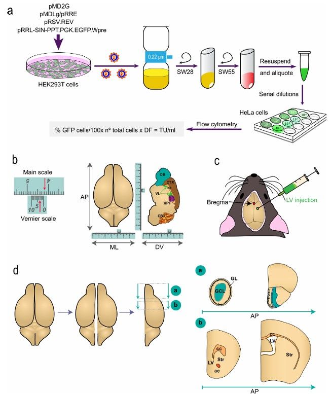
Figure 1: Schematic representation of the different parts of the procedure. (a) Generation of LVs for in vi...
Access restricted. Please log in or start a trial to view this content.
Materials
| Name | Company | Catalog Number | Comments |
|---|---|---|---|
| Equipment: | |||
| Vernier stereotaxic instrument | NeuroLab, Leica | 39463001 | |
| Syringe holder | KD Scientific | KDS-311-CE | |
| 33-gauge syringe | Hamilton | P/N 84851/00 | 85RN |
| Electric drill | Fine Science Tool | 98096 | |
| Thermal blanket | Ufesa | AL5512/01 | 230-240 V, 100-110 W, type C-AL01 |
| Shaver | Jata | MP373N | Model: beauty, 3 V, 300 mA, type HT-03. |
| Reagents: | |||
| Medetomidine | Esteve | Domtor | Comercial solution at 1 mg/ml. |
| Ketamine | Merial | Imalgene 500 | Comercial solution at 50 mg/ml |
| Medetomidina/ketamine mixture | Prepare a working mixture of medetomidine at a final concentration of 0.2 mg/ml dilution and ketamine at a final concentration of 15 mg/ml in saline solution. Use as anesthesia injecting a volume to get a final concentration of 0.5-1 mg medetomidina per kg body weight and 50-75 mg ketamine per kg body weight | ||
| Butorphanol | Pfizer | Torbugesic | Stock solution at 10 mg/ml. Used as analgesia at 1 mg/ml in saline solution. |
| 0.9% saline solution | Braun | 13465412 | |
| Surgical marker pen | Staedler | 313-9 | Permanent lumocolor |
| Ophthalmic lubricant | Siccafluid | 0.5 g/dosis, carbomer 974P | |
| Povidone-iodine | Betadine | 694109.6 | 10% povidone-iodine |
