This video demonstrates virus-mediated gene transfer into the mouse dorsal cochlear nucleus (DCN) to enable light-induced auditory stimulation. A viral vector encoding opsin, a light-sensitive ion channel, is injected into the DCN following a craniotomy and cerebellar aspiration for direct access. The expressed opsins allow neurons to be activated by light, providing a model for studying auditory pathway responses using optogenetics.
A subscription to JoVE is required to view this content. Sign in or start your free trial.
Method Article
Virus-Mediated Gene Transfer Into the Mouse Dorsal Cochlear Nucleus for Light-Induced Auditory Stimulation
651 views
⸱
August 29th, 2025
In This Article
Abstract
Protocol
All procedures involving animal models have been reviewed by the local institutional animal care committee and the JoVE veterinary review board.
1. Cerebellar Aspiration
- Using a 5 French suction, aspirate the lateral-most portion of the left cerebellum overlying the DCN until the DCN is visual (Figure 1, Right Panel). Remove approximately 1/4 to 1/3 of the left cerebellum. The main landmark adjacent to the DCN is the ampulla of the superior semicircular canal.
NOTE: Aspiration of the cerebellum is the key step of the protocol. Directed aspiration of the cerebellum is ....
Access restricted. Please log in or start a trial to view this content.
Results
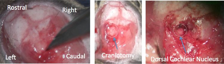
Figure 1: Partial cerebellar aspiration demonstrates access to the dorsal cochlear nucleus. Left Panel: After the .......
Access restricted. Please log in or start a trial to view this content.
Materials
| Name | Company | Catalog Number | Comments |
|---|---|---|---|
| Stereotaxic holder | Stoelting | 51500 | |
| 5 French suction | Symmetry Surgical | 2777914 | |
| Dental Points | Henry Schein | 100-8170 | |
| Bone rongeur | Fine Surgical Tools | 16020-14 | |
| 10 µl Hamilton syringe | Hamilton | 7633-01 | |
| 34 gauge, needle | Hamilton | 207434 | |
| Rongeurs | Fine Surgical Tools | 16021-14 |
