This video demonstrates the stereotaxic injection of a hydrogel into the stroke core of a post-stroke mouse, highlighting its role in scaffold formation that mimics the extracellular matrix, promotes cellular infiltration, and supports angiogenesis, underscoring its neurotherapeutic potential in stroke recovery.
A subscription to JoVE is required to view this content. Sign in or start your free trial.
Method Article
Stereotactic Injection of Hydrogel into a Post-Stroke Mouse Model
495 views
⸱
August 29th, 2025
In This Article
Abstract
Protocol
All procedures involving animal models have been reviewed by the local institutional animal care committee and the JoVE veterinary review board.
Injection of hydrogel or other biomaterials
- Perform the injection of biomaterials at a user-defined time. In this experiment, injections were performed between 5 and 7 days post-stroke. 2 - 6 µL of hydrogel is injected.
- Turn on the heating pad to maintain the mouse's body temperature during anesthesia (37 °C).
- Prepare autoclaved scissors, forceps, cotton, vet eye ointment, surgical glue or suture material, sterile alcohol, and beta iodine wipes. Prepare the bone drill with sterile drill bit heads and the 25 µL glass Hamilton syringes with metal 30 G needles.
- Turn on the bead sterilization to 160 °C to allow for the sterilization of instruments between mice.
- Attach the injection pumps to the stereotaxic device. Set the flow rate to 1 µL/min and set the volume to 4 µL.
- Prepare a clean cage for the mice after surgery.
- Place the mouse into the induction chamber with an isoflurane concentration of 4% to anesthetize it until spontaneous movement stops and its breathing comes to a slow, steady rate.
- Once asleep, transfer and secure the mouse to the stereotaxic device with a maintenance isoflurane concentration of 1.5%.
- Apply the vet eye ointment to both eyes.
- Shave any fur that may have regrown off the mouse’s head, and aseptically prepare the area by using an alcohol wipe followed by a beta iodine wipe. Repeat three times.
NOTE: If necessary, use an extra alcohol wipe at the end to clean off all the beta iodine, as it causes skin irritation and leads the mice to itch their skull after surgery.
- Using the small scissors and/or forceps, reopen the skin above the brain. Use cotton with sterile saline or PBS (Phosphate-buffered saline) to clean any debris from the skull. A white-yellow circle (the stroke, Figure 1B) and the burr hole from the drill will be visible. If the dead tissue is not visible, likely no stroke occurred. If the burr hole is not centered above the stroke, re-drill to center it.
- Orient the injection pump over the mouse and lock it at 90°. Clean the syringe with sterile saline or PBS solution before backloading the material.
- Once clean, disassemble the glass syringe so that there is no needle. Backload the syringe using a 25 µL positive displacement pipette with 10 µL of hydrogel (or other biomaterial here with or without cells).
- Using the syringe plunger, push the gel all the way to the front of the syringe. Then, reassemble the syringe and continue pushing until the gel visibly exudes from the needle.
- Place the syringe onto the pump. The back of the pump may need to be adjusted so the syringe fits. Adjust the flow rate to 30 µL/min, select Withdrawal or Inject depending on the direction it needs to move, and then press Start. Hit Stop when the back of the pump is at the correct location.
- Screw the back of the pump so the syringe is secure. If adjustments were previously made, make sure the flow rate is set back to 1 µL/min and is set to inject.
- Move the pump in the x and y directions to orient it over the hole. Move the pump down in the z direction until the needle of the syringe touches the top of the hole.
- Reset the Z on the digital display console. Then, move the syringe down in the z direction 0.750 mm. If the syringe bends or there is resistance, the skull either healed or was not completely drilled through, and needs to be re-drilled.
- Press Start on the pump console to inject 4 - 6 µL of the hydrogel at a rate of 1 µL/min.
- Set a 5-minute timer when the pump stops injecting to allow the gel to begin cross-linking.
- After 5 minutes, slowly pull up the syringe by turning the z-nob. Watch to see if any material is accidentally pulled or leaking from the hole. Unlock the pump and move it away from the mouse when the needle is far enough away from the skull.
- Using forceps, surgical glue, or sutures, close the skin above the head. Place the mouse in the clean cage and observe its recovery. It should take about 5 – 10 minutes for the mouse to recover from anesthesia.
Access restricted. Please log in or start a trial to view this content.
Results
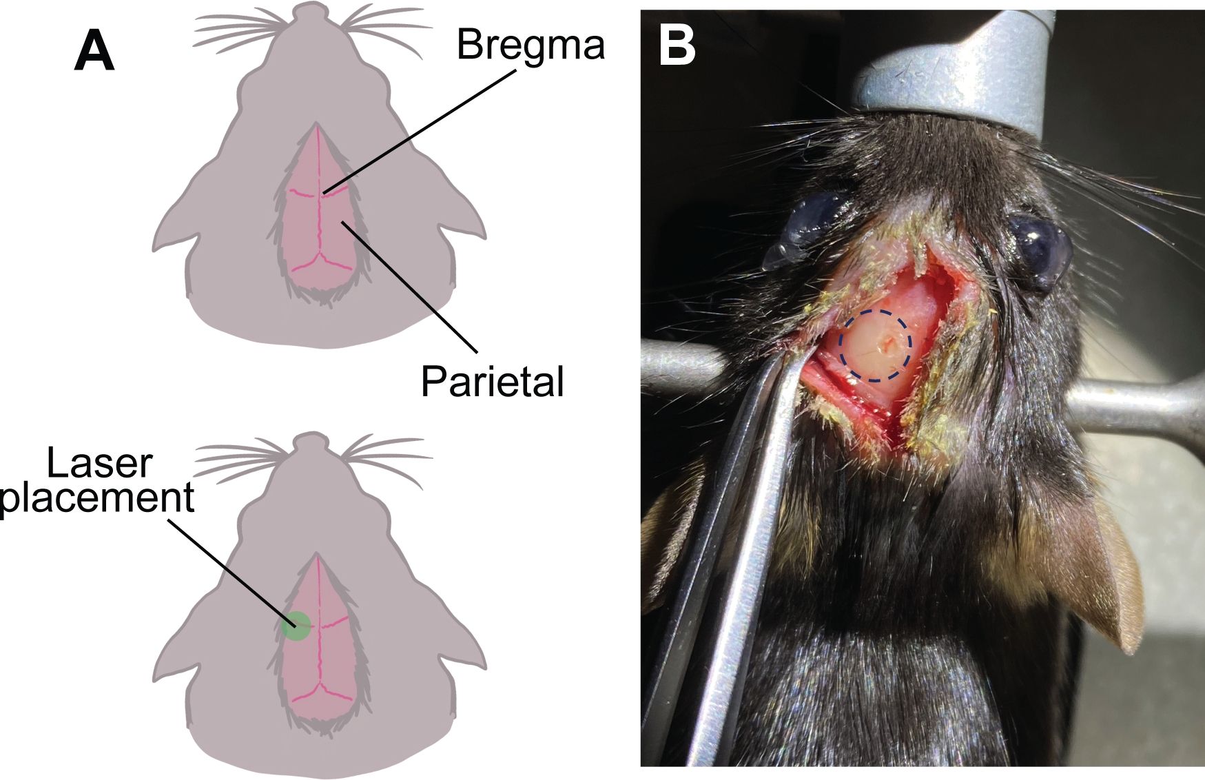
Figure 1: Schematic representation of bregma and localization of stroke. (A) The p...
Access restricted. Please log in or start a trial to view this content.
Materials
| Name | Company | Catalog Number | Comments |
|---|---|---|---|
| 25uL Hamilton Syringe 702RN, no needle | Fishcer Scientific | 14824663 | Syringes used to inject biomaterials |
| 25uL Positive displacement pipette | Gilson | M-25 | |
| Adjustable Stage Platform | Kopf Instruments | 901 | |
| C57BL/6 Mice | Jackson Laboratory | 664 | 8-12 weeks of age |
| Cage Assembly Rob | Thorlabs | ER3-P4 | 3" Long, diameter 6mm, 4 pack - for attaching laser to sterotax |
| Cotton Swabs | VWR | 89031-288 | |
| CP 25 pipette tips | Gilson | F148012 | |
| Heating Plate | Kopf Instruments | HP-4M | |
| IMPAC 6-Integrated Multi Patient Anesthesia Center | VetEquip | 901808 | |
| Jewlers Forceps #5 | GFS chemicals | 46085 | |
| Miltex Adson Dressing Forceps, Disecting-grade | Intefra Miltex | V96-118 | |
| Mini-pump variable flow | Thomas Scientific | 70730-064 | Pump for perfusions |
| Mouse Gas Anethesia Head Holder | Kopf Instruments | 923-B | |
| Nanojet Control Box | Chemyx | 10050 | |
| Nanojet pump header | Chemxy | 10051 | Attach to stereotaxic device for injecting biomaterial |
| Needle RN 30G PT STY 3, 0.5 inch | Fishcer Scientific | NC9459562 | |
| Non-rupture ear bars 60º | Kopf Instruments | 922 | |
| Puralube Vet Ointment | Dr. Foster Smith | 9N-76855 | |
| Rectal Probe Mouse | Kopf Instruments | Ret-3-ISO | |
| Sol-M U-100 Insuline syringe with 1/2 unit markings 0.5 mL | VWR | 10002-726 | To inject rose bengal |
| StainTray Slide Staining System | Simport Scientific | M920-2 | For staining slides |
| Sterotaxic device | Kopf Instruments | 940 | Small Animal Stereotaxic Instrument |
| Student Adson Forceps -1x2 teeth | Fine Science Tools | 91127-12 | |
| Student Fine Forceps - straight/broad Shanks | Fine Science Tools | 91113-10 | |
| Temperature Controller | Kopf Instruments | TCAT-2LV | |
| Tissue-Tek OCT compound | VWR | 25608-930 | |
| Vetbond Tissue Adhessive 3mL | Santa Cruz Biotechnology | sc-361931 |
