This video demonstrates a protocol for performing sonodynamic therapy on a brain tumor in a mouse model using magnetic resonance-guided focused ultrasound (MRgFUS). A sonosensitizer precursor is administered prior to treatment, which accumulates in tumor cells and is activated by focused ultrasound to produce reactive oxygen species (ROS). These ROS induce oxidative stress, resulting in tumor cell death and reduced proliferation.
A subscription to JoVE is required to view this content. Sign in or start your free trial.
Method Article
Treatment of Brain Tumors in a Mouse Model Using Sonodynamic Therapy
507 views
⸱
August 29th, 2025
In This Article
Abstract
Protocol
All procedures involving animal models have been reviewed by the local institutional animal care committee and the JoVE veterinary review board.
1. Magnetic resonance imaging (MRI) procedures
1. Take the MRI bed with the animal Stereotactically fixed and place it in the MRI cradle previously connected to the MRI scanner (see Table of Materials), by slotting the MRI bed hole at its end to the peg on the MRI cradle, as shown in Figure 1. Attach the inlet and outlet anesthesia tubes to the corresponding tubes in the MRI machine.
2. Slide the MRI cradle containing the animal into the MRI bore, making sure to keep the same position as where the phantom was placed.
3. Perform a localizer to see the location of the animal's brain and then a post-contrast T1-weighted MRI scan covering the entire brain.
4. Export the MRI scan as a set of uncompressed DICOM files, one for each slice.
2. Focused ultrasound treatment (Figure 2)
1. Remove the animal from the MRI cradle after completion of the scan and place it on the platform by slotting the front of the bed into its corresponding peg at the back of the platform and slotting the back of the bed into its corresponding peg.
2. Connect the inlet tube of the MRI bed to a source of isoflurane for anesthesia and set the gas flow rate to 1.0 mL/min and the vaporizer to 2.0% for maintenance of anesthesia. Connect the outlet tube to a charcoal filter canister for anesthesia absorption.
3. Transfer the set of DICOM files to the computer and place them in a folder in the main working directory of the current study.
4. In the software, go to the main initialization page and turn on Jog Mode by clicking on Guided Focus Finding and then clicking on Jog Mode On.
5. Place a dab of centrifuged ultrasound gel on the animal's head, with enough gel to cover the entire scalp above the skull.
6. Pour deionized (DI) and degassed water into the water bath up to 80% and then slot the water bath onto its corresponding columns on the platform.
7. Lower the water bath filled with DI water until the bottom membrane touches the ultrasound gel on the animal's head, forming a coupling surface between the water and the gel. Make sure that the ultrasound gel covers the entire head of the animal to the water bath and that there are no air bubbles in the ultrasound gel between the water bath and the mouse's scalp.
8. Submerge the ultrasound transducer into the water bath, ensuring no air bubbles are forming on the transducer surface.
9. Lower the transducer arm using Jog Mode Down toward the partly submerged transducer, and couple it to the transducer by aligning the magnetic slots with each other while the transducer surface remains submerged.
10. Turn off Jog Mode, and then Exit Focused Finding, as done during the initialization step. Make sure to confirm the home position as done before.
11. Go to the treatment tab and then upload the postcontrast T1-weighted MRI files by clicking the folder icon in the top middle of the screen. Select the folder containing the DICOM files that were uploaded to the computer previously and open them. This step should automatically load all DICOM files in the top right panel.
12. Next, in the Sonication Mode tab, select the proper treatment mode by choosing either Burst or Continuous Wave. If in burst mode, in the Sonication Settings tab, enter the burst length, burst period, and number of periods. These parameters will correspond to each sonication location. If in continuous wave mode, only enter the sonication duration.
NOTE: In this study, continuous wave mode was used for a 120 s duration.
13. Click on the target icon in the top middle of the page and select the proper locations on the correct MRI slice where the focused ultrasound (FUS) focal region is to be aimed. A light red square with a red circle will be present when clicked. More than one selection can be chosen.
14. Left of the MRI image is a table, which will populate with the 3D coordinates of the focal regions selected. In the last column of the table, enter the power level to be sonicated at each focal spot, which can be calculated separately for each transducer used to obtain the desired pressure or intensity levels. Click on each focal spot in the table to confirm which will result in the coordinates being highlighted, and the corresponding focal region on the image will turn blue. NOTE: Refer to the transducer datasheet to determine how to translate the power level entered into the table into pressure or intensity, as these values are transducer-specific.
15. Once satisfied with all focal regions, select Motion Test. This will prompt the software to move the transducer to all of the focal spots to ensure the movements are possible. After confirmation, the software will indicate Motion Test Complete. If an error occurs, adjust the focal regions so that they can fit within the 3D axes of the transducer motors.
16. Once ready, select Begin Sonication to begin the sonication protocol, moving the transducer and applying the correct FUS parameters at each focal region selected.
17. Once the sonication protocol is finished, uncouple the transducer from the transducer arm, remove the water bath from the platform, and wipe off the ultrasound gel from the animal's scalp.
18. Unscrew the bite and ear bars, remove the animal from the MRI bed, and then place the animal on a warm pad until the animal wakes up from anesthesia. At this point, return the animal to its cage.
19. For subsequent animals, repeat the same protocol, starting from section 6. 20. Once the desired animals are treated, clean all surfaces touched by animals with 70% ethanol (see Table of Materials).
21. Quit out of the software and then shut down and turn off each piece of equipment.
Access restricted. Please log in or start a trial to view this content.
Results
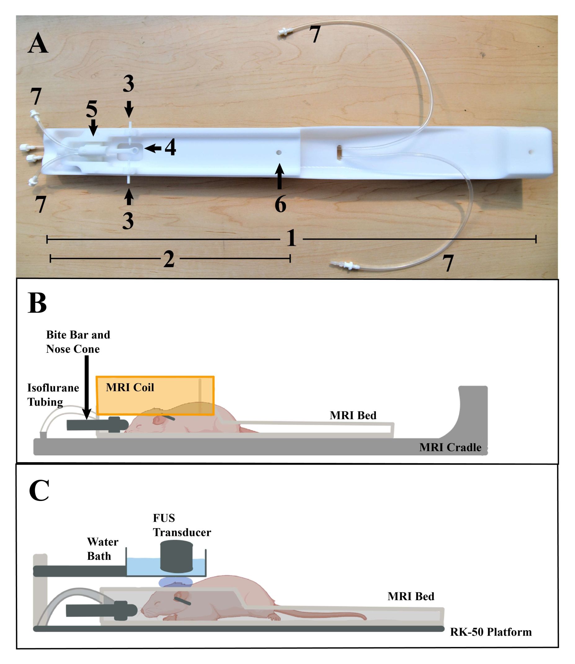
Figure 1: Animal placement. (A) MRI bed and cradle, with various parts labeled: 1...
Access restricted. Please log in or start a trial to view this content.
Materials
| Name | Company | Catalog Number | Comments |
|---|---|---|---|
| 7T PET-MR system | Bruker | Biospec 70/30 | |
| Aluminum foil | Reynolds Brand | ||
| Amplifier | FUS Instruments | 2175 | |
| Athymic nude mice | Charles River Laboratories | Strain Code 490 | |
| Charcoal isoflourane waste container | Patterson scientific | 78909457 | |
| Computer | FUS Instruments | 2269 | |
| Cover glass | Fisherbrand | 12-545J | |
| Desktop monitor | ASUS | VZ239H | |
| Electronic shaver | Wahl | 93235-002 | |
| Fetal bovine serum | Thermo Fisher Scientific | 16000044 | |
| Function generator | Siglent | QS0201X-E01B | |
| Gadolinium contrast agent (Gadavist) | McKesson Corporation | 2068062 | |
| Gauze | Henry Schein | 101-4336 | |
| Heat lamp | |||
| Heat pad | Kent Scientific | RT-0501 | |
| Induction chamber | Patterson scientific | 78933388 | |
| Isofluorane vaporizer | Patterson scientific | 78916954 | |
| Isoflurane | Covetrus | 29405 | |
| Isoflurane system | Patterson Scientific | 78935903 | |
| IVIS spectrum | Perkin Elmer | 124262 | |
| Nair | Church and Dwight Co. | 42010553 | |
| Ophthalmic ointment | Puralube vet ointment | ||
| RK-50 MRgFUS system | FUS Instruments | 2182 | |
| Scale | |||
| Scalpel blade | Covetrus | 7319 | |
| Scalpel handle | Fine Science Tools | 91003-12 | |
| Screwdriver set | Jakemy | JM-8160 | |
| Skin marker | Time Out | D538,851 | |
| Stereotactic frame | Kopf Instruments | 5000 | |
| Stereotactic MRI prototype plastic imaging fixture | FUS Instruments | ||
| Transducer and matching box | FUS Instruments | T515H750-118 | |
| Ultrasonic degasser | FUS Instruments | 2259 | |
| Ultrasound gel | ParkerLabs | 01-08 | |
| Water bath | FUS Instruments | ||
| Xylazine | Covetrus | 1XYL006 |
