This video demonstrates the preparation of a luciferase-expressing enterohemorrhagic Escherichia coli suspension from frozen stock for use as an oral inoculum in animal models. This enables real-time, bioluminescent tracking of intestinal colonization during in vivo infection studies.
A subscription to JoVE is required to view this content. Sign in or start your free trial.
Method Article
Preparation of Bioluminescent Enterohemorrhagic Escherichia coli Inoculum
484 views
⸱
September 26th, 2025
In This Article
Abstract
Protocol
1. Bioluminescent EHEC (enterohemorrhagic Escherichia coli) Bacteria Preparation for Oral Inoculation
NOTE: The timeline flowchart of the experimental procedures for EHEC preparation and mouse oral gavage is presented in Figure 1 to aid in experimental preparation.
- Streak E. coli O157:H7 EDL933 bacteria harboring the luciferase plasmid onto an LB (Luria-Bertani) agar plate with 50 µg/mL kanamycin from -80 °C stock. Grow the bacteria for 16 - 18 h at 37 °C.
- Pick a single colony from the overnight plate and culture in 3 mL LB medium with 50 µg/mL kanamycin for 16 - 18 h in a 37 °C incubator at 220 rpm.
- Subculture the bacteria (1:100 dilution) into kanamycin (50 µg/mL)- containing LB broth for 2.5 - 3 h in a 37 °C incubator at 200 rpm. (For instance, add 2 mL overnight cultured bacteria to 200 mL LB medium with kanamycin (50 µg/mL) supplemented.)
- Incubate the bacteria for 2.5 - 3 h and measure the optical density (OD) value at 600 nm (OD600) until the value is between 0.9 to 1. The bacterial cell number is at a density of about 108 colony-forming units (CFU)/mL.
- Centrifuge the re-cultured bacteria at 8,000 x g for 30 min, 4 °C.
- Discard the supernatant without agitating the bacteria pellet, and wash the pellet with 100 mL 0.9% sterile normal saline by gentle agitation.
- Repeat steps 1.5 and 1.6 once.
- After the wash, centrifuge the bacterial culture at 8,000 x g for 30 min, 4 °C, and discard the supernatant gently.
- Condense the pellet 100-fold with 0.9% sterile normal saline. For instance, if 200 mL of bacteria are centrifuged, add 2 mL of normal saline to suspend the pellet.
- Confirm the bacteria CFU (it should be approximately 109 CFU/100 μL after condensation in normal saline) by plating the concentrated bacteria through a 10-fold serial dilution.
Access restricted. Please log in or start a trial to view this content.
Results
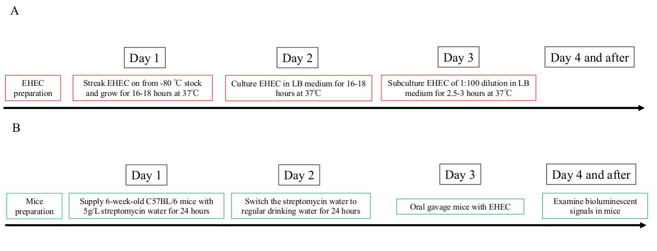
Figure 1: Timeline of the experimental preparation flow chart.
Overview of the timing needed to prepare biolu...
Access restricted. Please log in or start a trial to view this content.
Materials
| Name | Company | Catalog Number | Comments |
|---|---|---|---|
| Shaker incubator | YIH DER | LM-570R | bacteria incubation |
| Orbital shaking incubator | FIRSTEK | S300 | bacteria incubation |
| Glycerol | SIGMA | G5516-1L | use for bacteria stocking solution |
| Sodium chloride | Sigma | 31434-5KG-R | chemical for making LB medium, 10 g/L |
| Tryptone | CONDA pronadisa | Cat 1612.00 | chemical for making LB medium, 10 g/L |
| Yeast Extract powder | Affymetrix | 23547-1 KG | chemical for making LB medium, 5 g/L |
| Agar | CONDA pronadisa | Cat 1802.00 | chemical for making LB agar |
| Kanamycin | Sigma | K4000-5G | antibiotics, use for seleciton |
| Streptomycin | Sigma | S6501-100G | antibiotics, eliminate the microbiota in mice |
| High-speed centrifuge | Beckman Coulter | Avanti, J-26S XP | use for centrifuging bacteria |
| Fixed-Angle Rotor | Beckman Coulter | JA25.5 | use for centrifuging bacteria |
| Fixed-Angle Rotor | Beckman Coulter | JLA10.5 | use for centrifuging bacteria |
| Centrifuge bottles | Beckman Coulter | REF357003 | use for centrifuging bacteria |
| Centrifuge bottles | Thermo Fisher scientific | 3141-0500 | use for centrifuging bacteria |
| Eppendorf biophotometer plus | Eppendorf | AG 22331 hamburg | for measuring the OD600 value of bacteria |
