This video demonstrates a method for culturing and observing individual bacterial cells encapsulated inside giant vesicles anchored to a supported lipid bilayer. The system enables stable, long-term imaging of single-cell growth dynamics in a confined, physiologically relevant microenvironment.
A subscription to JoVE is required to view this content. Sign in or start your free trial.
Method Article
Single-Cell Culture of Bacteria Within Giant Vesicles
428 views
⸱
September 26th, 2025
In This Article
Abstract
Protocol
1. Preparation of a giant vesicle (GV) Observation System (Bacterial Cell Culture System)
- Preparation of small vesicles (SVs) for constructing a supported bilayer membrane
- Pour 20 μL of the POPC (1-palmitoyl-2-oleoyl-sn-glycero-3-phosphocholine) solution and 4 μL of the biotin-PEG-DSPE (1,2-distearoyl-sn-glycero-3-phosphoethanolamine-N-[biotinyl(polyethyleneglycol)-2000) solution into a glass tube.
- Evaporate the organic solvent by air flow to form a lipid film and place this sample in a desiccator for 1 h to completely evaporate the organic solvent.
- Add 200 μL of 200 mM gluc....
Access restricted. Please log in or start a trial to view this content.
Results
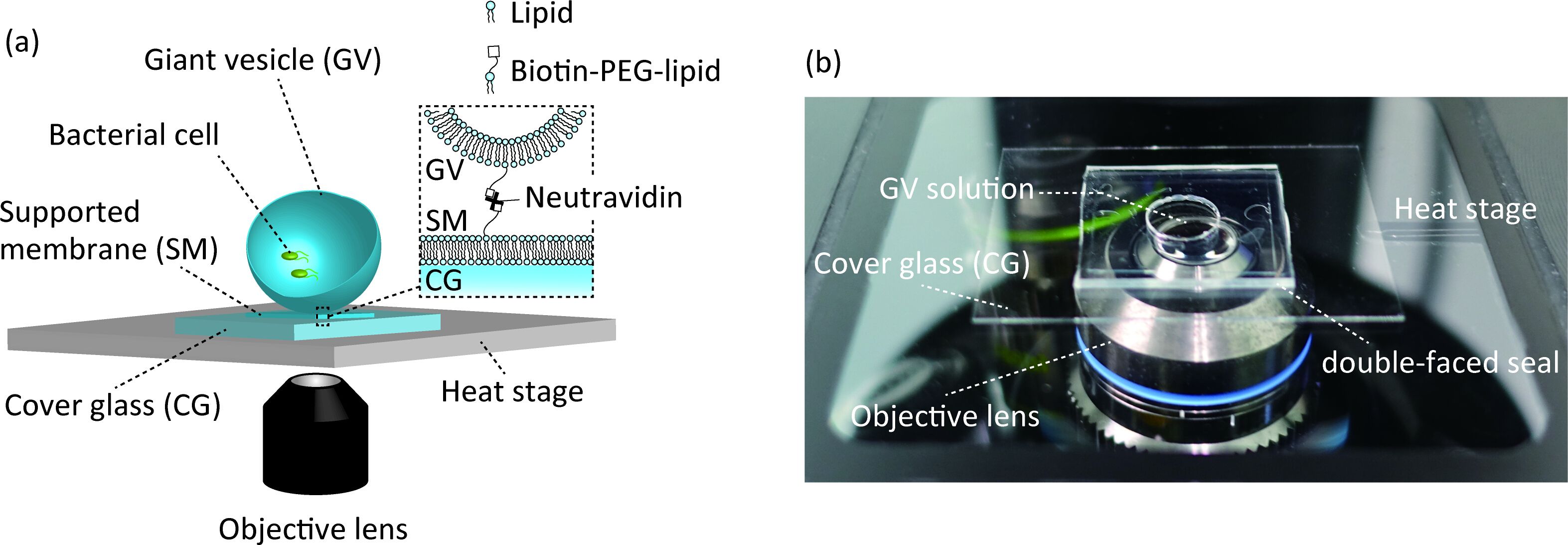
Figure 1: The observation system of bacterial cell culture inside GVs. (a
Access restricted. Please log in or start a trial to view this content.
Materials
| Name | Company | Catalog Number | Comments |
|---|---|---|---|
| Bactotryptone | BD Biosciences | 211705 | |
| Chloroform | Wako Pure Chemicals | 032-21921 | |
| Cover glass (18 × 18 mm) | Matsunami Glass Ind. | C018181 | thickness 0.13–0.17 mm |
| Cover glass (30 × 40 mm) | Matsunami Glass Ind. | custom-order | thickness 0.25–0.35 mm |
| Desktop centrifuge | Hi-Tech Co. | ATT101 | swing rotor type |
| Double-faced seal (10 × 10 × 1 mm) | Nitoms | T4613 | |
| Glucose | Wako Pure Chemicals | 049-31165 | |
| Inverted microscope | Olympus | IX-73 | |
| Methanol | Wako Pure Chemicals | 133-16771 | |
| Microscopic heating stage system | TOKAI HIT | TP-110R-100 | |
| Mineral oil | Nacalai Tesque | 23334-85 | |
| Neutravidin | Thermo Fisher Scientific | 31000 | |
| Objective lens | Olympus | LUCPLFLN 40×/0.6 NA | |
| Polycarbonate membranes | Avanti Polar Lipids | 610005 | |
| sCMOS camera | Andor | Zyla 4.2 plus | pore size 100 nm |
| Sodium chloride | Wako Pure Chemicals | 191-01665 | |
| Sucrose | Wako Pure Chemicals | 196-00015 | |
| Ultrasonic bath | AS ONE | ASU-3D | |
| Yeast extract | BD Biosciences | 212750 | |
| 0.6 mL lidded plastic tube | Watson | 130-806C | |
| 1.5 mL lidded plastic tube | Sumitomo Bakelite Co. | MS4265-M | |
| 1-palmitoyl-2-oleoyl-sn-glycero-3-phosphocoline | Avanti Polar Lipids | 850457P | |
| 1,2-distearoyl-snglycero-3-phosphoethanolamine-N-[biotinyl(polyethyleneglycol)-2000] | Avanti Polar Lipids | 880129P | POPC |
| Biotin-PEG-DSPE |
