This video demonstrates a two-step ultracentrifugation method using sucrose gradients and cushions to purify and concentrate viral particles from cell culture supernatant. The procedure enables separation based on buoyant density, allowing recovery of intact virus suitable for downstream applications.
Method Article
Preparation of Virus-Enriched Suspension Using Stepwise Sucrose Centrifugation
90 views
⸱
September 30th, 2026
In This Article
Abstract
Protocol
1. Concentration of Viral Particles by Ultracentrifugation
NOTE: We utilize a double-sucrose method of purification that involves two steps: a sucrose gradient step and a sucrose cushion step (Figure 1).
- Load the conical ultracentrifugation tubes in the following order to create a sucrose gradient: 0.5 mL 70% sucrose (dissolved 1x phosphate-buffered saline [PBS]), 0.5 mL 60% sucrose (dissolved in DMEM), 1 mL 30% sucrose (dissolved in Dulbecco's Modified Eagle Medium [DMEM]), and 2 mL 20% sucrose (dissolved in 1x PBS).
- Add the virus-containing supernatant carefully to the gradient. As the total volume of supernatant from four 15 cm plates is 100 mL, use six ultracentrifugation tubes per spin to process the full volume of the virus supernatant.
- Each ultracentrifugation tube used for this step has a volume capacity of up to 30 mL (including the volume of sucrose). Distribute viral supernatant equally among tubes, leaving at least 10% headspace to prevent spillage.
- Adjust the culture volume to ~20 - 22 mL per plate when using more than four 15 cm plates for each experiment, so that the final volume of the pooled viral supernatant can easily be accommodated within six ultracentrifugation tubes.
- Fill ultracentrifugation tubes to at least three-fourths their total volume capacity, otherwise breakage of tubes can occur during centrifugation, resulting in possible loss of sample and/or equipment damage.
- Balance the tubes with 1x PBS and centrifuge samples at 70,000 x g for 2 h at 17 °C
NOTE: To prevent disruption of the sucrose layer during acceleration, set the ultracentrifuge to slowly accelerate the rotor to 200 rpm during the first 3 min of the spin. Similarly, set the ultracentrifuge to slowly decelerate the rotor from 200 rpm to 0 rpm over 3 min at the end of the spin. - Carefully collect 30 - 60% sucrose fractions into clean tubes (Figure 1). Add cold 1x PBS to the pooled fractions and bring up the volume to 100 mL; mix by pipetting up and down several times.
- Proceed to the sucrose cushion step by carefully layering the viral preparation on a sucrose cushion. For this, add 4 mL of 20% sucrose (in 1x PBS) to the tube, followed by ~20 - 25 mL of the viral solution per tube. If tubes are less than three-fourths full, top up with sterile 1x PBS.
- Carefully balance and centrifuge the samples at 70,000 x g for 2 h at 17 °C, as before. Pour off the supernatant and allow the remaining liquid to drain by inverting tubes on paper towels.
- Aspirate remaining droplets in order to remove all liquid from the pellet. At this step, the virus-containing pellets should be barely visible as small translucent spots.
- Resuspend pellets by adding 70 µL of 1x PBS to the first tube and thoroughly pipetting the suspension, subsequently transferring the suspension to the next tube and mixing as before, continuing until all the pellets are resuspended.
- Rinse the tubes with an additional 50 µL of cold 1x PBS and mix as before. Ensure that the combined volume of the final suspension is ~120 µL, and appears slightly milky; clear it by centrifugation at 10,000 x g for 30 s on a tabletop microcentrifuge.
- Transfer the supernatant to a fresh microfuge tube, make 10 µL aliquots, and store them at -80 °C.
NOTE: Avoid performing repeated freeze-thaw cycles on the lentiviral samples. Except when centrifugation is required, efforts should be made to carry out the remaining steps in tissue culture hoods or designated tissue-culture rooms using appropriate biosafety measures
Results
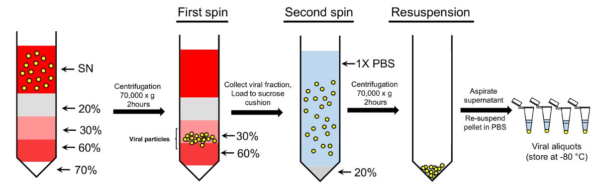
Figure 1: Concentration of the viral particles using double-sucrose gradient protocol. Viral particles collected from the supernatant (SN) are loaded onto gradient sucrose gradient. 70%, 60%, 30%, and 20% sucrose solutions are used to create the gradient. Following centrifugation, the particles collected from the 30-60% sucrose fractions are further loaded onto a 20% sucrose cushion and precipitated. The final pellet containing purified viral particles is resuspended in 1x PBS for further usage.
Materials
| Name | Company | Catalog Number | Comments |
|---|---|---|---|
| Equipment | |||
| Optima XPN-80 Ultracentrifuge | Beckman Coulter | A99839 | |
| Allegra 25R tabletop centrifuge | Beckman Coulter | 369434 | |
| Conical bottom ultracentrifugation tubes | Seton Scientific | 5067 | |
| Conical tube adapters | Seton Scientific | PN 4230 | |
| SW32Ti swinging-bucket rotor | Beckman Coulter | 369650 | |
| 50mL conical centrifuge tubes | Corning | 430291 | |
| Tissue culture pipettes, 25 mL | Corning | 4489 |
