This video demonstrates the generation of recombinant measles viruses using liposomes to deliver plasmids encoding the viral antigenome and essential helper proteins into producer cells. The transcribed and replicated RNA leads to the formation of viral particles expressing immunostimulatory and fluorescent reporter proteins. Fluorescence microscopy is then used to visualize syncytia formation, confirming successful recombinant virus production.
Method Article
Generation of an Immunostimulatory Protein-Encoding Recombinant Measles Virus
76 views
⸱
September 30th, 2026
In This Article
Abstract
Protocol
NOTE: [O], [P], and [M] indicate subsections applicable to: oncolytic viruses (OVs) in general, (most) paramyxoviruses, or measles virus (MV) only, respectively. [B] indicates sections specific for bispecific T cell engager (BTE) transgenes.
1 Cloning of Immunomodulator-encoding Transgenes into Measles Virus Vectors
- [O] Design insert sequence.
- [O] Decide on an immunomodulator of interest based on literature research or on exploratory data such as genetic screens and derive the relevant complementary DNA (cDNA) sequence from appropriate databases such as GenBank, the European Nucleotide Archive, or the international ImMunoGeneTics information system (IMGT).
- [O] Add additional features to the transgene sequence (Figure 1). A preceding Kozak sequence and species-specific codon optimization can enhance expression. Signal sequences are required for secretion. Include sequences encoding N- and/or C-terminal protein tags for detection and purification. Include restriction sites for insertion into the vector.
NOTE: [M] Additional transcription units (ATUs) harboring MV polymerase gene start and stop signals are necessary for transgene expression from recombinant MV genomes. Suitable anti-genomic cDNA vectors, controlled by cytomegalovirus (CMV) promoters and containing ATUs with unique restriction sites for introduction of positive-sense transgenes at defined positions, have been developed previously. [P] Adequate positioning of the transgene is crucial, as it affects viral replication and transgene expression due to the expression gradient typical for paramyxoviruses. Introduction into an ATU close to the 3' end of the anti-genome (i.e., in the leader position or downstream of the P gene) generally results in high transgene expression at the cost of reduced viral replication. Increased replication and lower levels of transgene expression can be expected when using the ATU downstream of the H gene. Packaging of the genome of several paramyxoviruses, including measles virus, requires binding of six nucleotides by each nucleocapsid protein. For insertion of transgenes into such viruses, ensure that the number of nucleotides of the complete genome will be divisible by six. This is also referred to as "rule of six". If necessary, include additional nucleotides in the insert (upstream of the Kozak sequence or downstream of the stop codon) without introducing frame shifts or premature stop codons. [M] Avoid particular sequences in the transgene that are similar to MV gene start (AGGRNCMARGW) and stop (RTTAWANAAAA) signals and RNA editing sequences (AAAAAGGG). - [O] Purchase oligonucleotides of the desired sequence or assemble from available sequences using standard molecular cloning.
NOTE: [O] For polymerase chain reaction (PCR) amplification of the transgene, design a forward primer including the upstream restriction site and the first 15-20 nucleotides of the insert and a reverse primer including the last 15-20 nucleotides of the insert followed by the downstream restriction site.
- [O] Clone insert into DNA encoding the viral (anti-)genome.
- [O] Clone the insert into DNA vectors or DNA anti-genomes of RNA viruses by standard molecular cloning techniques (i.e., enzymatic restriction followed by DNA ligation).
- [O] Prevent re-ligation of the vector by using non-compatible restriction sites or by dephosphorylation prior to ligation.
- [O] Isolate the ligation product by agarose gel electrophoresis and subsequent gel purification using commercially available kits. In general, optimal ligation efficiency is achieved at a 3:1 molar ratio of insert to vector.
- [O] Transform the ligation product into competent bacteria suited for efficient recovery of large plasmids (i.e., perform heat shock of E. coli) and identify bacterial clones harboring the correct DNA by colony PCR.
- [O] Isolate amplified DNA from a single bacterial clone using commercially available DNA preparation kits. Confirm genomic integrity by control digest with appropriate restriction enzymes (e.g., HindIII for MV genomes [M]). Confirm correct insertion and integrity of the transgene by sequencing.
NOTE: [M] For transgenes inserted in the MV H-ATU, perform Sanger sequencing with the following primers: H-9018 [forward primer, binds to MV genome position 9018 in the H open reading frame (ORF)]: 5' GTGTGCTTGCGGACTCAGAATC 3'; L-9249+ (reverse primer, binds to MV genome position 9249 in the L ORF): 5' CAGATAGCGAGTCCATAACGG 3'.
- [O] Clone the insert into DNA vectors or DNA anti-genomes of RNA viruses by standard molecular cloning techniques (i.e., enzymatic restriction followed by DNA ligation).
2. Rescuing Recombinant Measles Virus Particles Encoding Immunomodulators
- [O] Generate recombinant virus particles from (anti-)genomic DNA via transfection of virus producer cells according to the standard protocol for the respective virus. Follow guidelines for working under sterile conditions. Perform cell culture under hoods, in particular all steps involving virus in class II biological safety cabinets.
- [M] For rescue of measles viruses from cDNA, plate MV producer (African green monkey kidney-derived Vero) cells evenly on a 6-well plate 24 h before transfection. Seed 2 x 10⁵ cells in 2 mL Dulbecco's Modified Eagle's Medium (DMEM) containing 10% fetal bovine serum (FBS) per well to achieve 65–75% confluency at the time of transfection.
NOTE: [P] Reduce cell numbers if the cells overgrow prior to virus-induced syncytia formation. - [P] Transfect the cells with DNA encoding the viral anti-genome, required helper plasmids, and, if desired, a plasmid encoding a fluorescent reporter to assess transfection efficiency.
- [M] Mix 5 µg of recombinant DNA encoding the measles virus anti-genome, 500 ng each of mammalian expression plasmids encoding measles virus N and L proteins, and 100 ng each of plasmids encoding P protein in a total volume of 200 µL DMEM. If desired for assessing transfection efficiency (and if not already present within the viral anti-genome construct), include 100 ng of a fluorescent reporter plasmid.
- Add 18.6 µL of liposomal transfection reagent, immediately mix by flicking the tube, and incubate for 25 min at room temperature (RT).
- [P] Replace the medium with 1.8 mL of DMEM, 2% FBS, 50 µg/mL kanamycin (or other antibiotics to prevent contamination) per well, then add the transfection mix dropwise to the well and swirl carefully. Incubate cells overnight at 37 °C, 5% CO2. Replace medium with 2 mL of fresh DMEM, 2% FBS, and 50 µg/mL kanamycin the next day; repeat this when medium becomes acidic.
CAUTION: [O] Handle cells and materials according to biosafety regulations, as virus may be present from transfection through the following steps. Dispose of (potentially) infectious waste appropriately.
- [M] For rescue of measles viruses from cDNA, plate MV producer (African green monkey kidney-derived Vero) cells evenly on a 6-well plate 24 h before transfection. Seed 2 x 10⁵ cells in 2 mL Dulbecco's Modified Eagle's Medium (DMEM) containing 10% fetal bovine serum (FBS) per well to achieve 65–75% confluency at the time of transfection.
Results
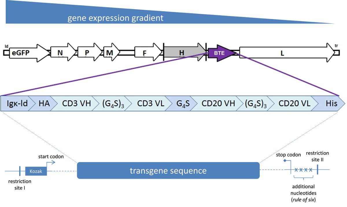
Figure 1: Schematic representation of a measles virus genome. Enhanced green fluorescent protein (eGFP) is encoded in an additional transcription unit downstream of the leader (ld) sequence. N, P, M, F, H, and L designate genes encoding the measles virus structural proteins nucleoprotein, P protein, matrix protein, fusion protein, hemagglutinin protein, and large protein (polymerase), respectively, which are differentially expressed as visualized above. A bispecific T cell engager (BTE) transgene is inserted into an additional transcription unit (ATU) downstream of the H open reading frame. The transgene encodes an immunoglobulin kappa (Igκ) leader peptide for efficient secretion as well as influenza hemagglutinin (HA) and hexa-histidine (His) protein tags for detection and purification. Genes coding for variable heavy-chain (VH) and variable light-chain (VL) domains of antibodies targeting CD3 and CD20, respectively, are connected via peptide linker sequences containing glycine (G) and serine (S) residues. The transgene sequence is preceded by a Kozak sequence. Downstream of the coding region, additional nucleotides have been included as an example to comply with the rule of six. The insert cassette is flanked by two restriction sites enabling insertion into the respective ATU.
Materials
| Name | Company | Catalog Number | Comments |
|---|---|---|---|
| Rapid DNA Dephos & Ligation Kit | Roche Life Science, Mannheim, Germany | 4898117001 | |
| CloneJET PCR Cloning Kit | Thermo Fisher Scientific, St. Leon-Rot | K1231 | |
| Agarose | Sigma-Aldrich, Taufkirchen, Germany | A9539-500G | |
| QIAquick Gel Extraction Kit | QIAGEN, Hilden, Germany | 28704 | |
| NEB 10-beta Competent E. coli | New England Biolabs (NEB), Frankfurt/Main, Germany | C3019I | |
| QIAquick Miniprep Kit | QIAGEN, Hilden, Germany | 27104 | |
| Restriction enzyme HindIII-HF | New England Biolabs (NEB), Frankfurt/Main, Germany | R3104S | |
| Dulbecco's Modified Eagle's Medium (DMEM) | Invitrogen, Darmstadt, Germany | 31966-021 | |
| Fetal bovine serum (FBS) | Biosera, Boussens, France | FB-1280/500 | |
| FugeneHD | Promega, Mannheim, Germany | E2311 | may be replaced by transfection reagent of choice |
| Kanamycin | Sigma-Aldrich, Taufkirchen, Germany | K0129 | |
| Vero cells | ATCC, Manassas, VA, USA | CCL81 | |
| 6-well plates | Neolab, Heidelberg, Germany | 353046 |
