A way to study the integration of newborn dentate granule cells in adult animals is described. This technique uses an engineered retrovirus to label newborn neurons, followed by electrophysiological recordings to determine in vivo functional integration.
Method Article
Studying the Integration of Adult-born Neurons
Opens in a new tab
In This Article
Summary
Loading...
$$\rightleftharpoonup{xx}$$
$$\longleftharp{xx}$$,
$$\longrightharp{xx}$$,
Abstract
Loading...
$$\rightleftharpoonup{xx}$$
$$\longleftharp{xx}$$,
$$\longrightharp{xx}$$,
Neurogenesis occurs in adult mammalian brains in the sub-ventricular zone (SVZ) of the lateral ventricle and in the sub-granular zone (SGZ) of the hippocampal dentate gyrus throughout life. Previous reports have shown that adult hippocampal neurogenesis is associated with diverse brain disorders, including epilepsy, schizophrenia, depression and anxiety (1). Deciphering the process of normal and aberrant adult-born neuron integration may shed light on the etiology of these diseases and inform the development of new therapies.
SGZ adult neurogenesis mirrors embryonic and post-natal neuronal development, including stages of fate specification, migration, synaptic integration, and maturation. However, full integration occurs over a prolonged, 6-week period. Initial synaptic input to adult-born SGZ dentate granule cells (DGCs) is GABAergic, followed by glutamatergic input at 14 days (2). The specific factors which regulate circuit formation of adult-born neurons in the dentate gyrus are currently unknown.
Our laboratory uses a replication-deficient retroviral vector based on the Moloney murine leukemia virus to deliver fluorescent proteins and hypothesized regulatory genes to these proliferating cells. This viral technique provides high specificity and resolution for analysis of cell birth date, lineage, morphology, and synaptogenesis.
A typical experiment often employs two or three viruses containing unique label, transgene, and promoter elements for single-cell analysis of a desired developmental process in vivo. The following protocol describes a method for analyzing functional newborn neuron integration using a single green (GFP) or red (dTomato) fluorescent protein retrovirus and patch-clamp electrophysiology.
Protocol
Loading...
$$\rightleftharpoonup{xx}$$
$$\longleftharp{xx}$$,
$$\longrightharp{xx}$$,
1. Virus Injection
High-titer engineered retrovirus (1×10⁹ unit/ml) is produced by co-transfection of retroviral vectors and VSVG into HEK293T cells, followed by ultracentrifugation of viral supernatant. For sources and production methods, see the excellent JoVE demonstration (3).
Note: Young adult (4-6 weeks old) female C57Bl/6 mice (Charles River) are housed under standard conditions. All procedures follow the National Research Council's Guide for the care and use of laboratory animals under a protocol approved by the Stony Brook University IACUC.
- Thaw 2ul of frozen retrovirus per animal on ice.
- Anaesthetize (100 μg ketamine + 10 μg xylazine per gram body weight) and mount the animal on a stereotaxic frame (Steolting). Remove the hair on the head and wipe the skin with 70% ethanol.
- Expose the skull and drill four shallow holes using a dental drill (0.6mm drill bit) at the following coordinates:
- anterioposterior = -2 mm from bregma; lateral = ±1.6 mm; ventral = 2.5 mm; anterioposterior = -3 mm from bregma; lateral = ±2.6 mm; ventral = 3.2 mm.
- anterioposterior = -2 mm from bregma; lateral = ±1.6 mm; ventral = 2.5 mm; anterioposterior = -3 mm from bregma; lateral = ±2.6 mm; ventral = 3.2 mm.
- Mount a 1μl Hamilton, flat-tip syringe and inject 0.5 μl retrovirus per site at a rate of 0.25 μl/min into the dentate gyrus. (See the above coordinates for ventral depth.) Pause 2 minutes after each injection before slowly withdrawing the syringe to prevent fluid backflow. Between injections, briefly rinse the external surface of the syringe tip with sterile PBS and an applicator to remove traces of blood.
- Close the wound with sterile surgical suture material (Type P-1; Size 6-0) and return the animal to standard housing conditions until the desired time stages after injection.
2. Slice Preparation
To obtain high-quality acute slices from adult mice, we use a modified cutting solution for slice preparation (see below).
- Pre-chill cutting solution to 0-4 °C on ice and bubble with 95% O2-5% CO2 for at least 30 minutes.
- Anaesthetize and transcardially perfuse an animal with ice-cold oxygen-saturated cutting solution.
- Quickly remove the brain into a petri-dish with filter-paper pre-wetted with cold, oxygenated cutting solution. Cut off the cerebellum and slice a mounting surface at least 1mm anterior to the (visible) injection coordinates. Glue the brain -onto a vibrotome stage and mount it into the cutting chamber filled with cold and oxygenated cutting solution.
- Prepare coronal or horizontal slices (300-350μm) and transfer them with a large-bore pipette to an incubating chamber containing room-temperature ACSF bubbled with 95% O2/5% CO2.
- Incubate the slices, bubbling continuously, at 32°C for 30-60 minutes. Return the chamber to room temperature for the duration of the recording.
3. Electrophysiological Experiments
- Slices are transferred into the recording chamber which is continuously perfused with oxygen-saturated ACSF at 32°C - 34°C.
- A bipolar tungsten stimulation electrode is placed in the molecular layer of the dentate gyrus using a low magnification microscope objective (10X).
- Newborn DGCs in the sub-granular zone/ granular cell layer are identified by the expression of GFP or dTomato. Whole-cell patchclamp recording is performed on newborn neurons under high magnification (40X).
- Firing properties of the recorded cells are obtained by injection of a series of currents under current-clamp mode.
- Electric stimuli are delivered by the stimulation electrode via a stimulation isolator controlled by the recording software, and evoked postsynaptic currents in the newborn DGCs are recorded.
4. Data Analysis
Amplitudes of evoked postsynaptic responses are analyzed at different developmental stages of adult-born neurons.
5. Representative Results
Using the above protocol, GFP expression in newborn dentate gyrus neurons similar to Figure 1 is common. Note that newborn neurons are easily visualized and both dendrites and axons are robustly labeled. Expression in thin DGC axons and small spines depends on the quality and titer of the virus, the promoter used and the length of in vivo expression. In our hands, newborn cells and sub-cellular organelles are usually visible within a few days following injection, and spine expression can be traced from the earliest stages of development. Whole-cell recordings from newborn neurons at different time stages allow the study of unique newborn cell properties, e.g. action potentials (Figure 2a), as well as the process of synaptic integration into existing neural circuits during maturation (Figure 2b).
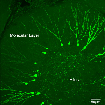
Figure 1 2-photon confocal image of newborn neurons in a horizontal dentate gyrus section of an adult mouse. Green cells are 21dpi (days post injection) GFP-expressing newborn DGCs labeled with pUX-GFP retrovirus.
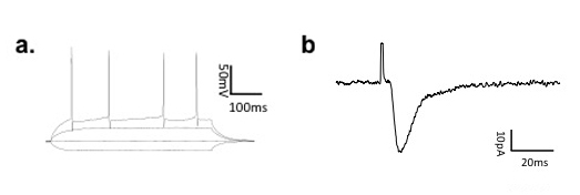
Figure 2 a) Action potential firing properties of a newborn DGC at 21 dpi. b) representative evoked excitatory postsynaptic currents (EPSCs) recorded on a newborn DGC at 21 dpi.
Discussion
Loading...
$$\rightleftharpoonup{xx}$$
$$\longleftharp{xx}$$,
$$\longrightharp{xx}$$,
Continuous neurogenesis in the hippocampus of adult mammalian brains provides a unique experimental model system to study neuronal development and integration in the mature brain. The protocol presented here combines retroviral labeling and electrophysiological methods to study the integration of newborn dentate granule neurons in the brains of adult mice.
To ensure that your experiments are successful, we recommend the following during critical steps of the procedure:
To avoid unwanted infection and inflammation, all tools should be sterilized (by autoclaving, any type of sterilizer, or 70% ethanol) before surgery. Use sterile PBS to wash the tip of the syringe needle before injection.
For virus injection, animals must to be well mounted on the stereotaxic device and sources of unnecessary movement must be minimized during injection. Make sure the head is well fixed and firm, and adjust the nose position of the mouse to level bregma and lambda prior to calculating injection coordinates. Extract virus into the syringe quickly to minimize exposure to room temperatures - retroviruses are particularly temperature sensitive. Inject virus slowly at the recommended rate to minimize pressure damage. Using the recommended flat-tipped syringe (vs. common beveled tip) will ensure an evenly distributed dentate gyrus infection area.
Before preparing slices, cutting solution and incubating ACSF must be bubbled with 95% O2-5% CO2 for at least 30 minutes to allow oxygen to saturate the solutions. Animals should be perfused quickly and tissue maintained in cold, oxygen-saturated solution for best slice quality.
For recording, increase the stimulus intensity gradually until there is a postsynaptic response. Change location of the stimulation electrode within the molecular layer of dentate gyrus as necessary.
In summary, the recommended approach offers a way to explore distinct properties and possible functional roles of adult-born neurons during early-stage integration into active, mature circuitry.
Disclosures
Loading...
$$\rightleftharpoonup{xx}$$
$$\longleftharp{xx}$$,
$$\longrightharp{xx}$$,
No conflicts of interest declared.
Acknowledgements
Loading...
$$\rightleftharpoonup{xx}$$
$$\longleftharp{xx}$$,
$$\longrightharp{xx}$$,
This work was funded by NINDS and American Heart Association to S.Ge. This approach was originally established and developed by the author in Dr. Hongjun Song's laboratory in the Johns Hopkins University. We would like to thank Dr. Hongjun Song for his guidance and support.
Materials
| Name | Company | Catalog Number | Comments |
|---|---|---|---|
| |||
References
Loading...
$$\rightleftharpoonup{xx}$$
$$\longleftharp{xx}$$,
$$\longrightharp{xx}$$,
- Zhao, C., Deng, W., Gage, F. H. Mechanisms and functional implications of adult neurogenesis. Cell. 132, 645-660 (2008).
- Ge, S., Goh, E. L., Sailor, K. A., Kitabatake, Y., Ming, G. L., Song, H. GABA regulates synaptic integration of newly generated neurons in the adult brain. Nature. 439, 589-593 (2006).
- Gavrilescu, L. C., Van Etten, R. A. Production of replication-defective retrovirus by transient transfection of 293T cells. J Vis Exp. , (2007).
Reprints and Permissions
Request permission to reuse the text or figures of this JoVE article
Request Permission