Dense array electroencephalography is being used increasingly to study social cognitive functions in infants and adults. Here we present an established methodology that represents a significant improvement on conventional methodologies for studying EEG in infants and adults.
Method Article
Investigating Social Cognition in Infants and Adults Using Dense Array Electroencephalography (dEEG)
In This Article
Summary
Abstract
Dense array electroencephalography (dEEG), which provides a non-invasive window for measuring brain activity and a temporal resolution unsurpassed by any other current brain imaging technology1,2, is being used increasingly in the study of social cognitive functioning in infants and adults. While dEEG is enabling researchers to examine brain activity patterns with unprecedented levels of sensitivity, conventional EEG recording systems continue to face certain limitations, including 1) poor spatial resolution and source localization3,4,2) the physical discomfort for test subjects of enduring the individual application of numerous electrodes to the surface of the scalp, and 3) the complexity for researchers of learning to use multiple software packages to collect and process data. Here we present an overview of an established methodology that represents a significant improvement on conventional methodologies for studying EEG in infants and adults. Although several analytical software techniques can be used to establish indirect indices of source localization to improve the spatial resolution of dEEG, the HydroCel Geodesic Sensor Net (HCGSN) by Electrical Geodesics, Inc. (EGI), a dense sensory array that maintains equal distances among adjacent recording electrodes on all surfaces of the scalp, further enhances spatial resolution4,5,6 compared to standard dEEG systems. The sponge-based HCGSN can be applied rapidly and without scalp abrasion, making it ideal for use with adults7,8, children9,10,11, and infants12, in both research and clinical4,5,6,13,14,15 settings. This feature allows for considerable cost and time savings by decreasing the average net application time compared to other dEEG systems. Moreover, the HCGSN includes unified, seamless software applications for all phases of data, greatly simplifying the collection, processing, and analysis of dEEG data.
The HCGSN features a low-profile electrode pedestal, which, when filled with electrolyte solution, creates a sealed microenvironment and an electrode-scalp interface. In all Geodesic dEEG systems, EEG sensors detect changes in voltage originating from the participant's scalp, along with a small amount of electrical noise originating from the room environment. Electrical signals from all sensors of the Geodesic sensor net are received simultaneously by the amplifier, where they are automatically processed, packaged, and sent to the data-acquisition computer (DAC). Once received by the DAC, scalp electrical activity can be isolated from artifacts for analysis using the filtering and artifact detection tools included in the EGI software. Typically, the HCGSN can be used continuously for only up to two hours because the electrolyte solution dries out over time, gradually decreasing the quality of the scalp-electrode interface.
In the Parent-Infant Research Lab at the University of Toronto, we are using dEEG to study social cognitive processes including memory, emotion, goals, intentionality, anticipation, and executive functioning in both adult and infant participants.
Protocol
1. Preparing the Experimental Setup
- Prior to the participant's arrival at the lab, ensure that the Geodesic EEG System (GES) components and the experiment control computer and software (Net Station) are operating properly.
- Remove all unneeded and potentially distracting items from the experiment room.
- Set up the experiment room in accordance with the particular experimental paradigm being implemented. Here we use paradigms ranging from computer-based tasks administered via E-Prime presentation software to various parent-infant and experimenter-infant interaction paradigms.
- Ensure that the video cameras are charged and arrange the cameras at angles ideal for the current experimental setup.
2. Exchanging Information
- Upon the participant's arrival at the lab, describe the Geodesic Sensor Net application protocol and explain the experiment.
- After ensuring that the participant has a good understanding of the experimental protocol, make sure that he or she (or his or her legal guardian) signs a consent form before proceeding with the experiment.
- For EEG studies, it is often useful to obtain from the participant certain information that may be expected to have an influence on the recorded electrical brain activity. For example, information about drowsiness, last meal, and last sleep might be of interest. In the Haley lab, such information is obtained by administering standardized questionnaires. To help the experiment proceed in a timely manner, it is advisable to have two experimenters present: one to administer the questionnaires, and one to carry out the HCGSN preparations detailed below.
3. Preparing for HydroCel Geodesic Sensor Net Application
- Prepare the HydroCel Saline electrolyte solution of distilled water, potassium chloride, and baby shampoo, making sure to stir thoroughly until all of the potassium chloride is dissolved in the solution.
- Using a measuring tape, measure the circumference of the participant's head, recording the measurement in centimeters.
- Retrieve the appropriately sized HCGSN-i.e., the one that encompasses the range of head circumferences in which the participant's head circumference falls.
- Submerge the sensor end of the HCGSN in the electrolyte solution and allow it to soak for 5 minutes, being sure to keep the connector end away from all liquids. It is advisable to use a timer or stopwatch to guarantee accurate timing.
- Open the Net Station software on the control computer and begin an experimental control session to allow zero and gain measurements to be taken before connecting the HCGSN connector and collecting data.
4. Applying the HydroCel Geodesic Sensor Net
In order to allow for the valid and consistent analysis of collected EEG data, the net application procedure must be highly standardized. Such standardization is achieved by taking measurements of the participant's head (as described above) to ensure the accurate and consistent placement of electrodes from participant to participant. If optimal net positioning is not initially achieved, remove the net and reapply.
- While waiting for the sensor net to finish soaking in the electrolyte solution, guide the participant into the experiment room and instruct him or her to sit on a chair. The chair should be without wheels to avoid unwanted movement during the sensor net application. In the case of an infant participant, have the infant's guardian sit on the chair with the infant sitting upright on his or her lap, holding the infant by the waist so that the infant's entire head is accessible and the infant remains still.
- Using a measuring tape and a china marker pencil, locate and mark the vertex by taking the following measurements:
- Ear to ear over the top of the head
- Nasion to inion over the top of the head
- Remove the sensor net from the electrolyte bucket and place it on a clean, dry towel. Gently pat the sensor net with the towel to remove excess electrolyte solution.
- Give the connector end of the HCGSN to the participant or to a second experimenter to hold during the net application.
- Pick up the HCGSN, putting both hands in the interior of the net. Hold the net such that your thumbs are pushed firmly (but gently) on either side of the central, most frontal electrode and your pinkie fingers are pushed firmly against the band connecting the most posterior row of electrodes. Be careful not to overstretch the net. Your remaining six fingers should be loose within the net to allow the center portion to fall limp.
- Crouch or kneel down so that you are at eye level with the participant.
- From back to front, pull the sensor net over the participant's head. It is often useful to have a second experimenter present when applying the sensor net on an infant participant, to distract the infant and minimize head movement.
- Using your fingertips, pull gently on the band of the net and adjust it so that the vertex electrode sits on the vertex point previously marked with the china marker pencil. Tighten the ear and chin straps. Inspect anatomical landmark locations and net symmetry to see whether the net is positioned correctly, and make the necessary adjustments.
5. Measuring Electrode Impedances
EEG is measured as the potential difference (voltage) between the referenced site and the measured site. High impedance at the scalp-electrode interface causes a drop in measured voltage, attenuation of signal amplitude, and an increase in the presence of noise. While post-acquisition filtering in certain studies (e.g., conventional ERP studies) can get rid of this noise, high impedance generally compromises the fidelity of acquired EEG data. It is therefore important to make sure impedances are within the tolerable specifications before beginning to record EEG data.
- Plug the connector end of the HCGSN into the interface cable and turn the lever to lock it in place. Turn on the camera.
- In the control room, open up a new session in Net Station, enter the participant's information, and click Begin Session. Because the video and dEEG signals are synched, a live video feed from the experiment room should appear on the screen.
- Select the Panels dropdown menu, open Impedance, and click the Measure button. A montage of the HCGSN sensor array will appear on the screen. Drag the window to the edge of the screen so that it opens up on the monitor in the experiment room.
- Using a disposable pipette, rake aside the participant's hair so that each electrode is seated directly on the participant's scalp.
- Refer to the Impedance Measurement window displaying the HCGSN montage on the monitor. Sensors that are not making good contact with the scalp will appear red. Make note of the electrode sensor numbers. Use the pipette and a small amount of the already prepared electrolyte solution to improve the impedance of these sensors. Electrodes on the montage will turn green as their impedance is improved.
- When impedances are satisfactory (i.e., when all electrodes on the montage are green), click the Save & Close button on the Impedance Measurement window in the control room.
- In Net Station, open the Dense Waveform Display. Scroll through the waveforms and note any channels that are displaying high amplitude noise due to poor scalp contact.
6. Taking Baseline Recordings
Before beginning the experimental paradigm, take some baseline dEEG recordings of the participant's resting electrical brain activity. Resting baseline recordings are important in continuous dEEG studies because there is a high degree of variability in electrical brain activity from participant to participant. As a result, most continuous dEEG studies must implement a within-subjects experimental design, analyzing the differences between each experimental condition and a pre-experiment baseline phase. For event-related EEG studies, a baseline phase may not be necessary.
- Leave the participant alone in the experiment room and instruct him or her to sit quietly and minimize movement. For infant participants, the parent/guardian should remain in the room with the infant sitting quietly in his or her lap. In the Haley lab, a movie titled Baby Mozart, an audio-visual combination of classical music and colourful moving designs, is played to ensure that infants remain calm and still. While an interactive baseline phase may not always be ideal, it can be used as a pre-baseline phase to calm the infant before taking baseline recordings.
- In Net Station, click the Record button to begin recording video and waveform data. On the Dense Waveform Display, event markers can be inserted to link behavioural events to electrical activity throughout the experiment. Insert a 'baseline' event marker on the waveform display.
- Record baseline for a standardized time period. In the Haley lab, the standard baseline phase lasts 2 minutes.
7. Running the Experiment
In dEEG studies, it is essential to have at least two experimenters present throughout the study. One experimenter will be responsible for interacting with the participant and implementing the behavioural paradigm, while the second experimenter will monitor the Dense Waveform Display.
- Implement the experimental paradigm.
- Monitor the Dense Waveform Display throughout the experiment to determine whether any channels are displaying increasing noise levels. Increasing noise may be reflective of increasing impedances. Acceptable impedance levels can be regained by using more electrolyte solution. This should be done at a break in the experimental protocol.
8. Debriefing the Participant
- When the experiment is complete, carefully loosen and remove the HCGSN from the participant's head. Rinse, disinfect, and dry the net.
- Give the participant a towel with which to wipe off any excess electrolyte solution.
- Give the participant any remaining questionnaires that need to be completed.
9. Analysis
The same EGI software used to acquire dEEG data is also used to analyze the data, allowing for a smooth and easy transition from data collection to data analysis. Because the HCGSN also picks up electrical noise originating from the environment, the data must first be filtered and cleaned before it can be analyzed. All of the necessary tools are included in Net Station.
- In Net Station, click on and open the Waveform Tools panel. Run the acquired data file through the filtering and artifact detection tools after setting the desired parameters. The artifact detection tool identifies spikes resulting from eye blinks or eye movements and detects bad channels. In many cases, additional hand-editing and artifact removal may be necessary (especially when working with infant populations, in which you cannot control eye blinks and movements).
- Segment the data so as to separate the different experimental conditions on the basis of the inserted event markers.
- Apply any desired analysis. In the Haley lab, frequency analysis is used to analyze how characteristic frequencies of brain activity varies between experimental conditions and between different brain regions. A decrease in wave power is potentially reflective of the increased activity of neurons in that region.
10. Representative Data

Figure 1. Raw EEG waveforms showing the fluctuations in recorded voltage (μv) across time (s), at a single electrode (electrode 30). The waveforms represent data collected during the first 1000ms of each of 3 experimental phases: baseline, demonstration, and immediate recall.
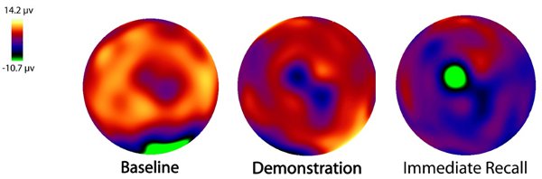
Figure 2. Raw data collected from the entire scalp (128-electrode montage) during three experimental phases (baseline, demonstration, immediate recall) of a participating infant. Data is presented as a topographical map and illustrates differences in surface-level brain electrical activity (μv) across brain regions and experimental phase of the puppet task.
Discussion
The Hydrocel Geodesic Sensor Net from EGI constitutes a non-invasive and easy-to-apply method of obtaining dEEG data from both adult and infant participants. This technology combines high temporal and spatial resolution with an increased opportunity for mobility, making it ideal for use in complex behavioural paradigms to investigate cognitive activities that may be reflected only in subtle changes in electrical activity. Given increasing interest in investigating the development of infant cognition and the relative scarcity of brain imaging techniques suitable for use in the infant population, the use of the HCGSN is likely to increase, leading us to a deeper understanding of infant cognition.
Disclosures
No conflicts of interest declared.
Acknowledgements
The manufacturer of the Geodesic Sensor Net is Electrical Geodesics, Inc (EGI). For contact information, please visit http://www.egi.com/company.
References
- Willis, W. G., Weiler, M. D. Neural substrates of childhood attention-deficit / hyperactivity disorder: Electroencephalographic and Magnetic Resonance Imaging evidence. Developmental Neuropsychology. 27, 135-182 (2005).
- Yang, L., Liu, Z., He, B. EEG-fMRI reciprocal functional neuroimaging. Clinical Neurophysiology. 121, 1240-1250 (2010).
- Dale, M. P., Halgren, E. Spatiotemperal mapping of brain activity by integration of multiple imaging modalities. Current Opinion in Neurobiology. 11, 202-208 (2001).
- Sperli, F., Spinelli, L., Seeck, M., Kurian, M., Michel, C. M., Lantz, G. EEG source imaging in pediatric epilepsy surgery: A new perspective in presurgical workup. Epilepsia. 47, 1-10 (2006).
- Holmes, M. D., Quiring, J., Tucker, D. M. Evidence that juvenile myoclonic epilepsy is a disorder of frontotemporal corticothalamic networks. NeuroImage. 49, 80-93 (2010).
- Holmes, M. D., Tucker, D. M., Quiring, J. M., Hakimian, S., Miller, J. W., Ojemann, J. G. Comparing Noninvasive dense array and intracranial electroencephalography for the localization of seizures. Neurosurgery. 66, 1-10 (2010).
- Muthukumaraswamy, S. D., Johnson, B. W. Changes in rolandic mu rhythm during observation of a precision grip. Psychophysiology. 41, 152-156 (2004).
- Muthukumaraswamy, S. D., Johnson, B. W., McNair, N. A. Mu rhythm modulation during observation of an object-directed grasp. Cognitive Brain Research. 19, 195-201 (2004).
- Arbel, Y. Error processing by individuals with specific language impairment: An ERP study. Dissertation Abstracts International: Section B: The Sciences and Engineering. 67, 3095-3095 (2006).
- Lepage, J., Théoret, H. EEG evidence for the presence of an action observation-execution matching system in children. European Journal of Neuroscience. 23, 2505-2510 (2006).
- Sokhadze, E. Event-related potential study of novelty processing abnormalities in autism. Applied Psychphysiology and Biofeedback. 34, 37-51 (2009).
- Nyström, P. The infant mirror neuron system studied with high density EEG. Social Neuroscience. Special Issue: The Mirror Neuron System. , 3-3 (2008).
- Bernier, R., Dawson, G., Webb, S., Murias, M. EEG mu rhythm and imitation impairments in individuals with autism spectrum disorder. Brain and Cognition. 64, 228-237 (2007).
- Brodbeck, V., Lascano, A. M., Spinelli, L., Seeck, M., Michel, C. M. Accuracy of EEG source imaging of epileptic spikes in patients with large brain lesions. Clinical Neurophysiology. 120, 679-685 (2009).
- Ramon, C. Power spectral density changes and language lateralization during covert object naming tasks measured with high-density EEG recordings. Epilepsy and Behaviour. 14, 54-59 (2009).
Reprints and Permissions
