Method Article

hFTAA Staining of Amyloid Deposits: A Technique to Visualize Amyloid Fibrils Using Oligothiophene-Based Dye for Fluorescence Imaging in Tissue Sections

July 8th, 2025

In This Article

Abstract

Loading...
$$\rightleftharpoonup{xx}$$ $$\longleftharp{xx}$$, $$\longrightharp{xx}$$,

Source: Nyström, S. et al., Imaging Amyloid Tissues Stained with Luminescent Conjugated Oligothiophenes by Hyperspectral Confocal Microscopy and Fluorescence Lifetime Imaging. J. Vis. Exp. (2017).

This video describes a technique to stain amyloids in tissue sections using a luminescent-conjugated oligothiophene dye - heptamer-formyl thiophene acetic acid, or hFTAA - for fluorescence imaging. Amyloids are β-sheet-rich abnormal protein aggregates. Their detection in tissue samples is helpful in the diagnosis and treatment of amyloid-associated diseases or proteinopathies.
Protocol

Loading...
$$\rightleftharpoonup{xx}$$ $$\longleftharp{xx}$$, $$\longrightharp{xx}$$,

1. LCO Staining Solution

NOTE: If hFTAA is purchased from a commercial vendor, please follow the vendor's instructions instead of section 1.

  1. Resuspend the lyophilized hFTAA in 2 mM NaOH to prepare a stock solution of 1 mg/mL. Keep the stock in a glass vial at 4 °C. The stock can be stored for one year.
  2. On the day of staining, prepare a working solution by diluting the stock 1:10,000 in Phosphate-buffered saline (PBS).

2. Preparation of Tissue Samples

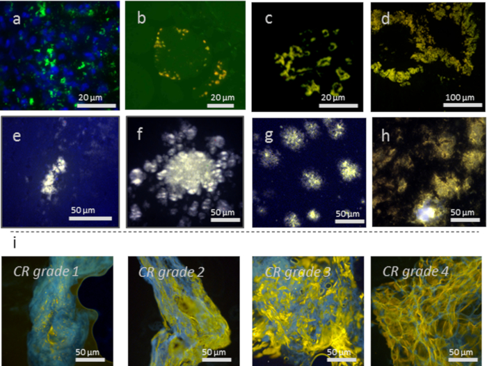
NOTE: Many tissue types can be imaged using hFTAA as an amyloid marker. See Figure 1 for examples. hFTAA is sensitive to aggregate conformation. The staining should hence preferably be performed on undisrupted aggregates with no epitope exposure. Optimal spectral quality is achieved if the tissue fixation is kept to a minimum. Hence fresh frozen material, gently fixed in ethanol at the time of staining, is preferred. However, it is possible to detect amyloid deposits also in tissue that has been fixed with e.g., formalin. hFTAA generally penetrates the tissue well. Select a specimen thickness that is compatible with the intended imaging technique.

  1. If formalin-fixed, paraffin-embedded sections are used, deparaffinized in xylene overnight. Dip the sections in consecutive baths of 99% ethanol, 70% ethanol, dH2O, and PBS, 10 min in each. Allow the tissue sections to dry under ambient conditions. Caution: Xylene is always handled in a chemical fume hood. Xylene and other organic solvents are harmful.
    1. Thaw cryosections in room temperature. Fix the tissue sections in 10% formalin overnight and rehydrate by dipping them in consecutive baths of 99% ethanol, 70% ethanol, dH2O, and PBS, 10 min in each. Allow the tissue sections to dry under ambient conditions.
  2. Add droplets of the hFTAA working solution (approximately 200 μL) to the tissue sections to cover it. The droplet should stay in place by surface tension. Incubate for 30 min at room temperature for staining.
  3. Rinse off the staining solution with 500 μL PBS using a pipette and then immerse the slide in the PBS bath for 10 min. Allow the section to dry under ambient conditions.
  4. Mount using the fluorescence mounting medium. Allow mounting medium to settle overnight.
    NOTE: hFTAA staining can be performed in combination with other staining methods such as immunofluorescence, cell- or organelle-specific markers, etc. To perform co-staining, run your complete staining protocol of choice and add hFTAA staining at the end, starting from step 2.2. See Figure 2 for examples. For immunofluorescence, preferably select a secondary antibody that is excited at 640 nm or higher. In this wavelength range, hFTAA does not absorb and hence cannot be excited and will not fluoresce. This ensures that no bleed-through is seen between hFTAA and antibody.

Access restricted. Please log in or start a trial to view this content.

Results

Loading...
$$\rightleftharpoonup{xx}$$ $$\longleftharp{xx}$$, $$\longrightharp{xx}$$,

Fluorescence microscopy of CR grades in extracellular matrix, diagram illustrating protein aggregation.
Figure 1: Various tissue types and proteins aggregates showing h-FTAA staining of: (a) Mallory-Denk bodies consisting of keratin aggregates in a liver (counterstained with DAPI), (b) p62-positive r inclusions in sporadic inclusion-body myositis (s-I...

Access restricted. Please log in or start a trial to view this content.

Disclosures

Loading...
$$\rightleftharpoonup{xx}$$ $$\longleftharp{xx}$$, $$\longrightharp{xx}$$,
No conflicts of interest declared.

Materials

List of materials used in this article
NameCompanyCatalog NumberComments
hFTAA/Amytracker545 Ebba Biotech
Dako fluorescene mounting mediumAgilent technologies GM304
LeicaDM6000 Leica
Lumen 200 Prior
Spectraview system ASI spectral imaging

Reprints and Permissions

Request permission to reuse the text or figures of this JoVE article

Request Permission

Tags

Oligothiophene DyeDeparaffinizationPBS WashesFluorescence MicroscopyMounting MediumSpectral Analysis

Related Articles