1. Synchronized Culture of C. elegans
- Before seeding, culture the Escherichia coli (E. coli) strain OP50 overnight at 37 °C in 300 mL of Luria-Bertani (LB) broth liquid medium. Store the cultured OP50 at -4 °C.
- To make LB broth liquid medium, use 10 g of tryptone, 5 g of yeast extract, 10 g of NaCl, and 1.5 mL of 1 N NaOH, and add to 1 L with deionized water. Autoclave.
NOTE: OP50 and C. elegans strains are available from the Caenorhabditis Genetics Center (University of Minnesota, St. Paul, MN, USA).
- To make nematode growth medium (NGM) agar, use 3 g of NaCl, 2.5 g of peptone, 17 g of agar, and 975 mL of deionized water. Autoclave. Cool to 55 °C, then sterilely add, in order, 1 mL of 1 M MgSO4, 1 mL of 1 M CaCl2, 1 mL of 5 mg/mL cholesterol in EtOH, and 25 mL of 1 M potassium phosphate pH 6.0 in 90-mm Petri dishes.
- For 1 M potassium phosphate pH 6.0, use 108.3 g of KH2PO4 and 46.6 g of K2HPO4, and add deionized water to 1 L. Autoclave.
- Spread 1-2 mL of the cultured OP50 on the NGM agar plates. To make a thin layer of OP50, incubate the plates overnight at room temperature before adding any nematodes.
NOTE: The NGM agar plates inoculated OP50 can be stored at room temperature for 2-3 weeks.
- Add at least 100 worms onto an NGM agar plate with OP50, and culture at 20 °C until the adult stage. At least three plates are required.
- To collect eggs in utero, transfer gravid hermaphrodites from the three NGM agar plates each with 5 mL of S buffer in a 15-mL conical tube, and wash the worms 3 times with 15 mL of S buffer using centrifugation at 300 x g for 30 s at room temperature.
- To make S buffer, use 5.9 g of NaCl and 50 mL of 1 M potassium phosphate pH 6.0, and add to 1 L with deionized water. Autoclave.
- Dissolve the worms in an alkaline hypochlorite solution for axenization and bulk egg isolation (0.5 mL of fresh bleach or equivalent: 5-6% sodium hypochlorite, 0.1 mL of 10 M NaOH, approximately 4.5 mL of S buffer), and stand the solution for 10-15 min at room temperature with mixing by inverting.
NOTE: Within 15 min, adult worms should dissolve, leaving a hazy solution of eggs liberated from their carcasses (the liberated eggs should be confirmed using a stereoscopic microscope). Instead of 0.1 mL of 10 N NaOH, 0.2 mL of 5 N NaOH can also be used.
- After hypochlorite treatment, wash the egg pellet 3 times with 15 mL of S buffer, and resuspend in 5-6 mL of S buffer. Hatch the released eggs during an overnight incubation at 20 °C in S buffer without E. coli for an age-synchronous culture of L1 stage larvae.
- To determine the approximate number of L1 stage larvae, count the worms using a stereoscopic microscope in 10 µL of S buffer after resuspending the larvae at least 3 times, and calculate the average. Then, transfer the L1 stage larvae to five NGM agar plates with OP50 (1,500-3,000 worms per plate using a 90-mm Petri dish), and culture at 20 °C until they have grown to the young adult stage when the self-fertilization begins and a few eggs are laid (ordinarily after 3 days).
2. Extraction of Cellular Fraction from C. elegans
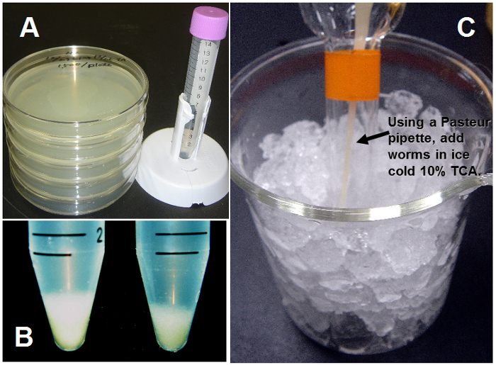
- Collect the young adult stage (5-day-old animals) worms from the five NGM agar plates with S buffer (Figure 1A).
- To select only living worms using the sucrose method 6 for flotation on 30% (w/v) sucrose, mix the worms suspended in 3-4 mL of S buffer with an equal volume of ice-cold 60% (w/v) sucrose in a 15-mL conical tube. Spin the tube at 1,500 x g for 15 s at 4 °C and remove the floating worms into a fresh tube by moving them off the wall of the tube with a Pasteur pipette.
- Wash the worms 3 times with S buffer by centrifugation at 1,500 x g for 30 s at 4°C. Check the wet volume of washed worms after centrifugation using a 1,000 µL micropipette tip (Figure 1B).
- Add the washed worms to an equal volume of ice-cold 10% (w/v) trichloroacetic acid (TCA; final concentration of 5%) for protein precipitation (Figure 1C). Instead of TCA, perchloric acid (PCA) or metaphosphoric acid can be used.
- Homogenize the worms with the precipitant using 40 strokes of a pestle in a Teflon homogenizer (Potter-Elvehjem tissue grinder) with rotation at up to 1,300 rpm on ice.
- Transfer the homogenate into a fresh 1.5-mL microtube with a Pasteur pipette, and sonicate using an ultrasonic homogenizer for 3 min (3 times of 1 min) with a 20% duty cycle on ice.
- Clarify the homogenate by centrifugation at 8,000 x g for 10 min at 4 °C. Neutralize the supernatants with 4 M KOH (0.25 volume to 10% TCA) for 20 min on ice, and centrifuge at 8,000 x g for 10 min at 4 °C. The supernatant (as a test sample) can be stored at -80 °C until the following assays.
3. Lactate Assay Using a Colorimetric Assay Kit
- Measure the concentration of lactate in the test samples using a colorimetric assay kit (Table of Materials). Carry out duplex examinations for the test samples. Add 5 or 10 µL of the test samples to a 96-well plate and adjust the volume to 50 µL per well with the Lactate Assay Buffer provided with the kit.
- For the lactate standard curve, dilute 100 mM L(+)-Lactate Standard to 1 mM with Lactate Assay Buffer. Add 0, 2, 4, 6, 8, and 10 µL of the 1 mM L(+)-Lactate Standard, which is provided with the kit, into a series of wells.
- Add 50 µL of Reaction Mix (containing 46:2:2 of Lactate Assay Buffer, Lactate Enzyme Mix, and Lactate Probe in DMSO, anhydrous; all reagents are provided with the kit) or Background Control Mix (containing 48:2 of Lactate Assay Buffer and Lactate Probe) into each well and incubate at room temperature for 30-60 min while the samples are protected from light.
- Measure the absorbance of each well at 570 nm using a microplate reader and subtract the absorbance of the Background Control Mix from the absorbance of the Reaction Mix.
- Plot the lactate standard curve. Calculate the lactate concentrations of the test samples from the lactate standard curve.