All procedures involving sample collection have been performed in accordance with the institute's IRB guidelines.
1. Formalin fixation of tissues
- Place 3-5 mm brain tissues collected after necropsy into 10% phosphate-buffered formalin solution (1:20 to 1:50 tissue/formalin ratio) for 24-72 h.
CAUTION: Formalin is a toxic fixative.
- Record the approximate tissue weight (size), tissue type (brain areas), and volume of formalin. It is important for laboratories to document and maintain records on fixation times.
- For longer tissue storage after formalin fixation and prior to processing, place the tissue in 70% ethanol.
2. Tissue processing
- Following the specimen fixation, dissect the tissue to include the important brain areas e.g., cross sections of the brainstem, cerebellum (3 lobes), or the hippocampus (both hippocampi) each cut 3 to 5 mm thick and placed into processing cassettes.
- Process the tissue cassettes for paraffin wax infiltration, embedded into paraffin blocks and sectioned (3 to 6 µm) on a microtome.
3. Preparation of Materials / Staining dishes
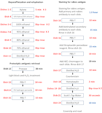
- Set up the staining dish (Table of Materials) as shown in Figure 1. Fill each dish with 250 mL of the solution.
- Preparation of the 3-Amino-9-ethylcarbizole (AEC) substrate stock solution
- Dissolve one 20 mg tablet of 3-amino 9-ethylcarbazole (AEC) into 5 mL of N,N, dimethylformamide using a glass pipette.
CAUTION: AEC is a carcinogen.
- Preparation of the protease (e.g., Pronase) stock solution for the antigen retrieval
- Dissolve 7 mg of the protease in 200 mL of phosphate-buffered saline (PBS).
- Preparation of the rinse buffer PBS-T
- Add 10 mL of Tween 80 to 990 mL of PBS. Mix it well to form a homogenous solution.
4. Deparaffinization and tissue rehydration
- Make a 5 µm paraffin section using a microtome, float it on a water bath at 38 ˚C, and collect it onto glass slides. Label the slides with a reagent-resistant pen/marker.
- Place the slides onto a tray and melt in a 55-60 ˚C oven for 1 hour. Do not raise the temperature above 60 ˚C as it can destroy the viral antigen.
- Remove slides from the oven and immediately deparaffinize in 3 consecutive xylene rinses of 5 min each in dishes 1, 2, and 3.
- Rehydrate the sections on the slide by sequential immersions in decreasing dilution of ethanol to deionized water: (4 through 11 is a dip rinse) dish 4: xylene/100% ethanol (1:1); dish 5: 100% ethanol; dish 6: 100% ethanol; dish 7: 95% ethanol; dish 8: 95% ethanol; dish 9: 80% ethanol; dish 10: 70% ethanol; dish 11: deionized water (Figure 1. Dish set-up).
CAUTION: Xylene is a hazardous chemical and work should be conducted in a fume hood.
5. Proteolytic antigen retrieval
- Treat slides with the protease (2.5 µg/mL of PBS) for 30 min for proteolytic antigen retrieval in dish 12.
- Then rinse in PBS-T for 10 min (dish 13).
- Treat with 3% hydrogen peroxide for 10 min (dish 14).
- Again, wash with PBS-T for 10 min (dish 15).
6. Staining procedure
- Handle slides one at a time keeping the remaining slides submerged in the buffer (do not remove the whole slide holder - keep slides wet). Remove one slide and blot off excess buffer (using a paper towel) from around the tissue section being careful not to disturb the tissue section. Incubate slides in a humidity chamber, made by placing moistened paper towels, on the lab bench top at room temperature with normal goat serum (blocking) for 15 min.
- Incubate with the optimal pre-determined dilution primary anti-rabies antibody (1:250 dilution of mouse anti-rabies serum, unpublished) (positive control) and negative control antibodies at room temperature same as above (step 6.1.) for 60 min with no washes in between.
- After 60 min, wash with PBS-T for 10 min (dish 16).
- Incubate with the biotinylated antibody (species-specific) in a humidity chamber at room temperature for 15 min (handling same as step 6.1.).
- Wash with PBS-T for 10 min (dish 16).
- Incubate with Streptavidin- horseradish peroxidase (HRP) complex in a humidity chamber at room temperature for 15 min (handling same as 6.1.).
- Wash with PBS-T for 10 min (dish 16).
- Incubate with peroxidase substrate, amino-ethylcarbazole (AEC), in a humidity chamber at room temperature for 10 min. Make AEC just prior to use. To do so, add 1 mL of AEC stock solution to 14 mL of 0.1M acetate buffer, pH 5.2. Add 0.15 mL of 3% peroxide, H2O2. Filter the mixture just before use (0.45 µm filter).
NOTE: The working solution of AEC is only stable for 2-3 hours. The AEC stock solution can be stored in the refrigerator for longer periods.
- Wash in deionized water for 10 min (dish 17)
- Counterstained with Gill's Hematoxylin diluted 1:2 with deionized water for 2 min (dish 18).
- Rinse off excess Hematoxylin with deionized water dip rinse (dishes 19 and 20).
- Rinse in Scott's Tap water for 30 s (bluing solution) dish 21.
- Wash in deionized water for 10 min (dish 22)
- Remove slides one at a time - mount with a water-soluble mounting medium.
- Read slides on a light microscope.