A subscription to JoVE is required to view this content. Sign in or start your free trial.

Method Article

An Antibody Uptake Assay for Imaging Notch/DeltaD Signaling in Zebrafish Radial Glia Progenitors

597 views

⸱

July 8th, 2025

In This Article

Abstract

Source: Zhao, X. et al., Antibody Uptake Assay for Tracking Notch/Delta Endocytosis During the Asymmetric Division of Zebrafish Radial Glia Progenitors. J. Vis. Exp. (2023)

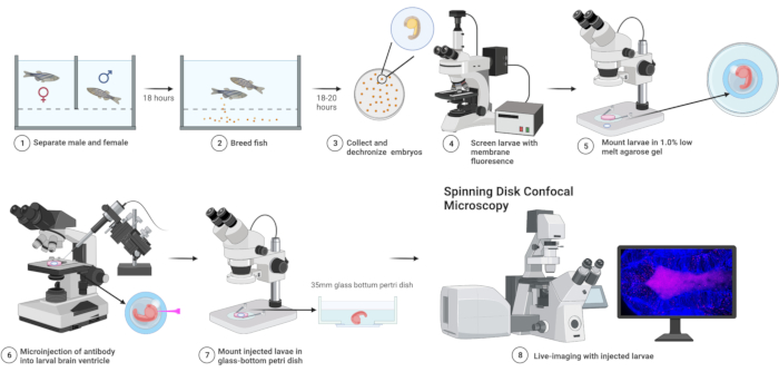
This video demonstrates an antibody uptake assay to image Notch/DeltaD signaling in zebrafish radial glia progenitors. It involves mounting agarose-embedded dechorionated zebrafish embryos, injecting a dye solution targeting DeltaD ligands on RGPs, and imaging under a fluorescence microscope for asymmetric cell division confirming Notch/DeltaD signaling activation.
Protocol

1. Preparation of zebrafish embryos

  1. Set up fish crossing tanks in the afternoon before 5:00 p.m., with one female wild-type fish and one male Tg [ef1a:Myr-Tdtomato] fish by using dividers to separate them in each tank.
  2. Remove the dividers before 11:00 a.m. from all crossing tanks the next morning. Keep quiet and do not disturb the fish while they are mating. Spawning typically occurs within 30 min after removing the dividers. The fertilized eggs remain on the bottom of the tank.
    1. Collect fertilized eggs from the tanks with a mesh filter. Transfer the eggs by washing them off into a Petri dish full of egg water....

Access restricted. Please log in or start a trial to view this content.

Results

Fish breeding and imaging workflow; confocal microscopy diagram with larval injection and imaging.
Figure 1: Flowchart of experimental steps. Transgenic zebrafish expressing the MyR-Tdtomato reporter are outcrossed with AB wild-type on day 1. On day 2, red fluorescent embryos are selected for microinjection, followed by time-lapsing imaging. The whole .......

Access restricted. Please log in or start a trial to view this content.

Disclosures

No conflicts of interest declared.

Materials

List of materials used in this article
NameCompanyCatalog NumberComments
35mm glass bottom culture dish MatTek corporationP35GC-1.5-10-C
Air pressure injector NarishigeIM300
Anti-Mouse-IgG-Atto647N Sigma-Aldrich50185
CaCl2.2H2O Sigma-AldrichC3306
Capillaries, 1.2 mm OD, 0.9 mm ID, with filamentWorld Precision Instruments1B120F-6
CSU-W1 Spinning Disk/High Speed WidefieldNikinN/ANikon Ti inverted fluorescence microscope with CSU-W1 large field of view confocal.
Dumont Medical Tweezers Style 5Thomas Scientific72877-D
Flaming-Brown P897 pullerSutter InstrumentsN/Ahttps://www.sutter.com/manuals/P-97-INT_OpMan.pdf
KClMillipore529552
MgSO4.7H2OSigma-AldrichM2773
MicromanipulatorsWorld Precision InstrumentsWPI M3301R
Mouse anti-Dld AbcamAB_1268496
Mouse IgG blocking buffer from ZenonThermofisher ScientificZ25008
NaClSigma-AldrichS3014
Phenol redSigma-AldrichP0290
Stemi 2000 Zeiss N/A
TricaineSigma-AldrichE10521
UltraPureTM low melting point agarose Invitrogen16520050

Explore More Articles

Fluorescence MicroscopyAsymmetric Cell DivisionEndocytosis ImagingMicroinjection TechniqueEmbryo MountingTime Lapse ImagingDeltaD Ligand