Method Article

Stimulating Neuronal Cell Differentiation Using Gold Nanorods

July 8th, 2025

In This Article

Abstract

Loading...
$$\rightleftharpoonup{xx}$$ $$\longleftharp{xx}$$, $$\longrightharp{xx}$$,

Source: Paviolo., et al. Gold nanorod-assisted optical stimulation of neuronal cells. J. Vis. Exp. (2015).

This video demonstrates a technique to promote neuronal differentiation by exposing nerve cells to gold nanorods and laser light, which forms cell projections supported by the βIII-tubulin. Staining and observing tubulin and the cell nucleus with epifluorescence microscopy reveals the cell outgrowths, indicating the nanorods' efficacy in neuronal differentiation.
Protocol

Loading...
$$\rightleftharpoonup{xx}$$ $$\longleftharp{xx}$$, $$\longrightharp{xx}$$,

1. NG108-15 Neuronal Cell Line Culture and Differentiation

  1. For the cell culture medium, prepare 500 ml of sterile Dulbecco's modified Eagle medium (DMEM) containing 10% (w/v) fetal calf serum (FCS), 1% (w/v) L-glutamine, 1% (w/v) penicillin/streptomycin and 0.5% (w/v) amphotericin B.
    Note: Supplements can be aliquoted, stored at -20 °C, and added to the media on the day required. Cell culture medium can be refrigerated in a sterile condition for a maximum of 1 month.
  2. For the cell differentiation medium, prepare 50 ml of sterile DMEM containing 1% (w/v) L-glutamine, 1% (w/v) penicillin/streptomycin, and 0.5% (w/v) amph....

Access restricted. Please log in or start a trial to view this content.

Results

Loading...
$$\rightleftharpoonup{xx}$$ $$\longleftharp{xx}$$, $$\longrightharp{xx}$$,

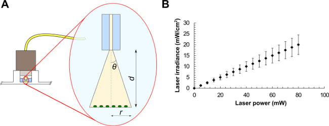
Optical excitation diagram; laser power vs. irradiance chart; photonic study; experimental setup.
Figure 1. Optical fiber experimental setup (A) and average laser irradiances as a function of the laser power for a laser beam of area equals to 0.4 mm2 (B). Beam parameters are (A): the half-angle of the maximum cone of light exiting the fiber (θ), t.......

Access restricted. Please log in or start a trial to view this content.

Disclosures

Loading...
$$\rightleftharpoonup{xx}$$ $$\longleftharp{xx}$$, $$\longrightharp{xx}$$,
No conflicts of interest declared.

Materials

List of materials used in this article
NameCompanyCatalog NumberComments
Au NRSigma Aldrich716812
NG108-15Sigma Aldrich8811230
DMEMSigma AldrichD6546
FCSLife Technologies10100147
L-glutamineSigma AldrichG7513
Penicillin/streptomycinLife Technologies15140122
Amphotericin BLife Technologies15290018
FormaldehydeSigma AldrichF8775
Triton X-100BDHT8532
BSASigma AldrichA2058
Anti-βIII-tubulinPromegaG7121
TRITC-conjugated anti-mouse IgG antibodySigma AldrichT5393
DAPIInvitrogenD1306
UV-Vis spectrometerVarian Medical Systems Inc.Cary 50 Bio
Mini centrifugeEppendorfMini Spin
Sonic bathUnisonics AustraliaFPX 10D
Cell culture incubatorKendroHera Cell 150
Cell culture centrifugeHettichRotofix 32A
Laser diodeOptotech780 nm single mode fibre - coupled LD
Optical fiberThorlabs780 HP
Power meterCoherentLaser Check
ImageJhttp://rsb.info.nih.gov/ij/index.html
Epifluorescent microscopeAxon InstrumentsImageX-press 5000A
PBS
Polystyrene 96 well plate
CO2
t75 flasks

Reprints and Permissions

Request permission to reuse the text or figures of this JoVE article

Request Permission

Tags

Neuronal DifferentiationLaser StimulationBeta III TubulinEpifluorescence MicroscopyCell CultureEndocytosisDAPI StainingAntibody LabelingConfocal Microscopy

Related Articles