All procedures involving sample collection have been performed in accordance with the institute's IRB guidelines.
1. Isolation of the stromal vascular fraction (SVF)
Collect the periaortic arch adipose tissue (PAAT) of 5-6 mice into one 2 mL microcentrifuge tube containing 1 mL freshly prepared digestion medium and mince the tissue using surgical scissors in an Eppendorf tube at room temperature (RT).
- Transfer the mix into 50 mL tubes containing 9 mL of the digestion medium. Homogenize the tissues by pipetting up and down with a 1 mL pipette 10x.
- Incubate the tubes at 37 °C with constant shaking at 100 rpm for 30–45 min and check every 5–10 min to prevent overdigestion. This is critical to improving cell viability and yield.
NOTE: Good tissue digestion will result in a homogenous, light yellow adipose tissue that is visible to the naked eye upon gently swirling the tube.
- Stop the digestion by adding 5 mL of HDMEM containing 10% FBS and 1% v/v PS at RT and mix well by pipetting.
- Centrifuge the cell suspension at 500 x g for 5 min at RT. The SVF will be visible as a brownish pellet. Carefully aspirate the floating adipocytes and decant the remaining supernatant without disturbing the SVF. Dissolve the SVF pellet in 10 mL of culture medium and filter through a 70 µm cell strainer.
- Centrifuge the cell suspension at 500 x g for 5 min, remove the supernatant, and gently resuspend the pellet in 5 mL of erythrocyte lysis buffer in a 15 mL conical tube for 10 min at RT.
- Stop the reaction by adding 10 mL of 1x PBS containing 1% FBS. Centrifuge the cell suspension at 500 x g for 5 min at 4 °C, remove the supernatant, and resuspend the pellet in 10 mL of 1x PBS containing 1% FBS.
- Centrifuge the cells again at 500 x g for 5 min at 4 °C. Remove the supernatant and resuspend the pellet in 5 mL of culture medium in a 15 mL conical tube at 4 °C.
- After a final round of centrifugation (500 x g for 5 min at 4 °C), resuspend the pelleted cells in 5 mL of FACS buffer (PBS containing 10% FBS, 100 units/mL DNA I, and 1% v/v PS) on ice, and count the cells with a hemocytometer.
2. Isolation of NCADSCs by FACS
- Set up and optimize the cell sorter following the instruction manual. Select the 100 µm nozzle, sterilize the collection tubes, install the required collection device, and set up the side streams.
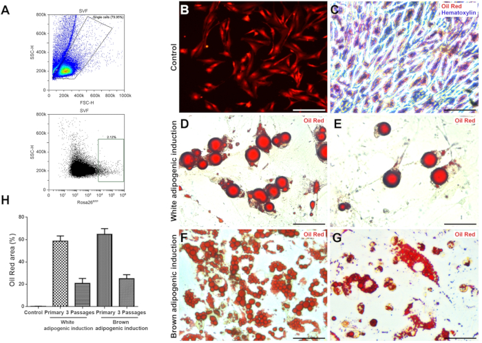
- For sorting RFP+ cells, a 561 nm yellow/green laser and optical filter 579/16 are recommended. Perform the compensation using the negative control and the single-stained positive controls. See Figure 1A for the gating scheme.
- Filter the cells through a 40 µm strainer, centrifuge at 500 x g for 5 min, and resuspend them in 2 mL of FACS buffer at a density of 0.5–1 x 10⁷/mL. Transfer the cells to clearly labeled 5 mL round-bottom polystyrene tubes and load them into the sorter.
- Run the experimental sample tube at 4 °C, turn on the deflection plates, and sort into a 15 mL conical tube precoated with RPMI containing 1% FBS and 1% v/v PS.
NOTE: Protect the samples from strong light to minimize RFP quenching.
3. Culture of NCADSCs
- Plate the sorted cells at a density of 5,000 cells/cm2 in a 12 well culture plate in complete culture medium and incubate at 37 °C in a humid atmosphere with 5% CO2 for 20–24 h.
- Remove the culture medium, wash the cells with prewarmed (37 °C) PBS to remove cell debris, and add fresh culture medium.