Method Article

Differentiation of Induced Pluripotent Stem Cells into Neural Progenitor Cells

July 8th, 2025

In This Article

Abstract

Loading...
$$\rightleftharpoonup{xx}$$ $$\longleftharp{xx}$$, $$\longrightharp{xx}$$,

Source: Phillips, A. W., et al. Developing HiPSC Derived Serum Free Embryoid Bodies for the Interrogation of 3-D Stem Cell Cultures Using Physiologically Relevant Assays. J. Vis. Exp. (2017).

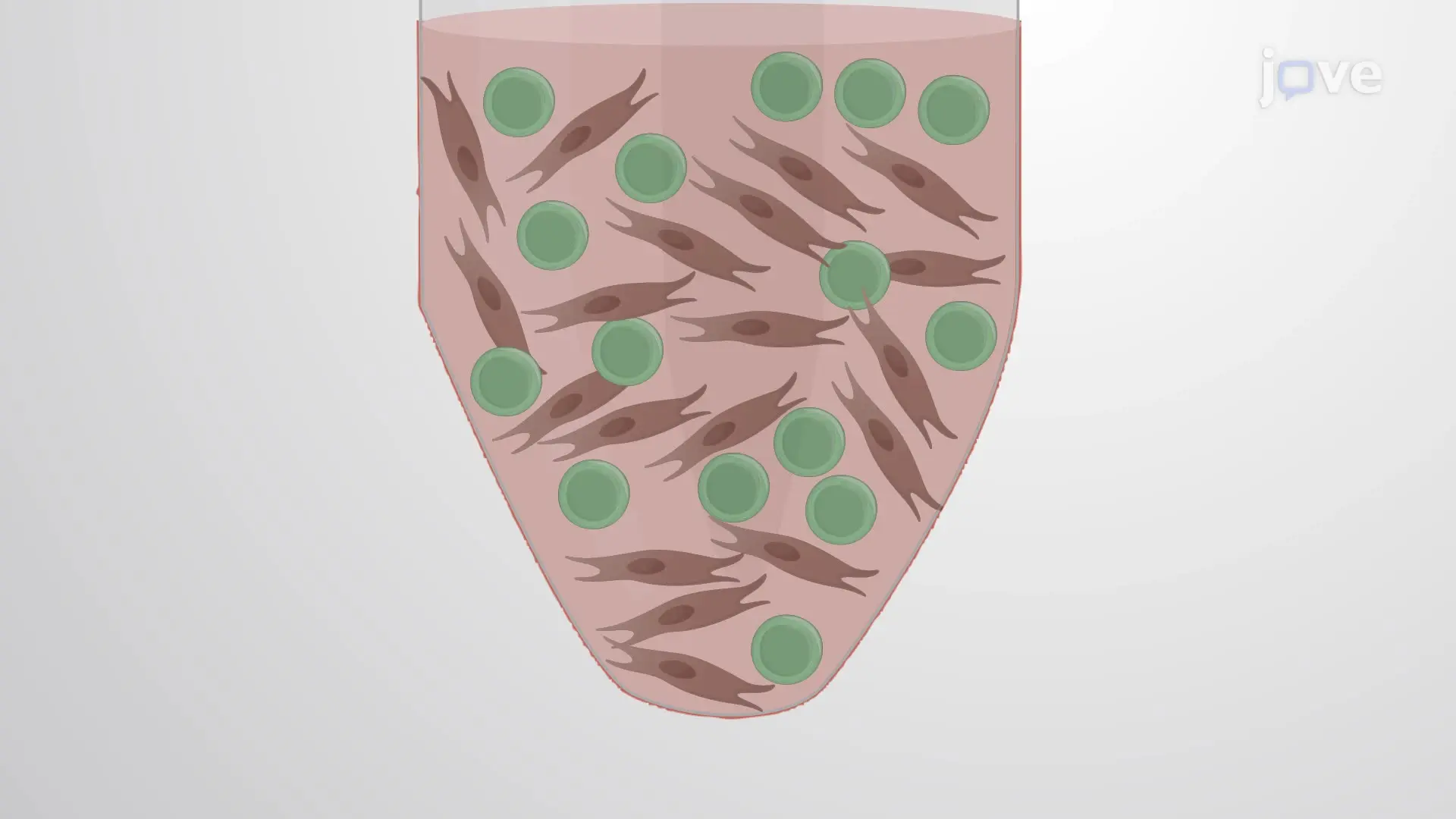
The video demonstrates a technique for differentiating human induced pluripotent stem cells (hiPSCs) into neural progenitor cells (NPCs). Initially, the hiPSCs are cultured on a feeder layer of mouse embryonic fibroblasts (MEFs). Subsequently, the cells are detached and transferred to a biopolymer-coated plate to allow the MEFs to adhere to and separate from the hiPSCs. The suspended hiPSCs are then transferred to a V-bottom plate and centrifuged to form aggregates. Finally, the addition of a differentiation medium induces the differentiation of hiPSCs within the aggregate into NPCs.

Protocol

Loading...
$$\rightleftharpoonup{xx}$$ $$\longleftharp{xx}$$, $$\longrightharp{xx}$$,

1. Generation of Neural Progenitor Cells

  1. Maintain hiPSCs derived from fibroblasts and peripheral blood mononuclear cells (PBMCs) in 6-well plates on a γ-irradiated mouse embryonic feeder (MEF) cell layer in human iPSC medium supplemented with small-molecules.
    NOTE:
    Daily maintenance is a modified version of previously reported procedures.
    1. Plate 6 x 10⁵ MEFs onto each well of a 6-well tissue culture grade plate in 300 μL/well recommended medium (following manufacturer's protocol).
    2. After 48 h, replace MEFs medium with 300 μL/well hiPSC medium for 1 h before adding hiPSCs.
    3. Quickly thaw hiPSCs in a 37 °C water bath and slowly add 1 mL hiPSC medium dropwise. Transfer the hiPSCs and medium to a 15 mL conical tube. Add medium to bring the total volume to 5 mL. Centrifuge for 4 min at 129 x g, aspirate the supernatant, gently resuspend the pellet in 1 mL hiPSC medium with Rho-associated protein kinase (ROCK) inhibitor at 1:1,000 concentration.
    4. Plate the cell suspension (typically seeded at 300,000 cells/well) onto the MEF cell layer and incubate at 37 °C, 5% CO2, 95% humidity.
    5. Monitor iPSC culture closely using a light microscope. After 48 h, remove cultures that have uneven edges, have grown to become cystic-like or appear yellowish-brown under the light microscope to minimize spontaneous differentiation.
      NOTE: After 7 - 10 days, hiPSC colonies should form. Colonies should be round, with clearly defined borders and uniform cell densities, and devoid of loosely packed non-uniform cells.
    6. Manually select round hiPSC colonies to clear the culture of spontaneously differentiated cells. Expand the colonies by placing them on fresh MEF layers. Expand 1:3 every 7 days when cultures near confluency and freeze.
  2. After expansion and freezing cells (for backup), grow hiPSC colonies on plates until they reach 50 - 75% confluence.
  3. Seven-days post plating, use mild enzymatic treatment (5-10 min with 300 μL of enzyme solution) and gentle trituration (2-4 times with a 1,000 µL pipet) to harvest and prepare hiPSCs for neural precursor cell differentiation.
  4. Centrifuge the cell suspension at 129 x g for 4 min, aspirate the supernatant, and resuspend the pellet in 5 mL human iPSC medium. Transfer the cell suspension to a 0.1% gelatin coated 5 cm cell culture dish for 1 h at 37 °C to eliminate MEFs and to maximize the hiPSC yield. Transfer the medium (with non-adherent cells) to another 5 cm gelatin coated dish.
  5. Transfer the non-adherent cells to a 15 mL centrifuge tube using a transfer pipet. Gently rinse the plates with 3 mL medium and add it to the 15 mL tube.
  6. Centrifuge the cell suspension at 129 x g for 4 min, aspirate the supernatant, and gently resuspend the pellet in medium. Determine the cell count using an automated cell counter. Plate 9,000 cells/well in a 96-well low-adhesion V-bottom plate.
  7. Centrifuge the plate at 163 x g for 3 min. Incubate the plate at 37 °C, 95% humidity and 5% CO2. Using a pipette, change 50% medium every other day by removing half of the medium and replacing it with fresh chemically-defined differentiating medium 1 (DM1; Figure 1; Table of Materials) for 14 days.

Access restricted. Please log in or start a trial to view this content.

Results

Loading...
$$\rightleftharpoonup{xx}$$ $$\longleftharp{xx}$$, $$\longrightharp{xx}$$,

Centrifugation process diagram, pellet formation and transfer in experimental setup.
Figure 1: Schematic Outlining the Early Steps in Preparing SFEBs. After iPSCs have been harvested using an enzymatic dissociation, they are plated in 96-well plates and centrifuged at 163 x g for 3 min to induce quick aggregation. After 14 days in vitro (DIV) cell aggr...

Access restricted. Please log in or start a trial to view this content.

Disclosures

Loading...
$$\rightleftharpoonup{xx}$$ $$\longleftharp{xx}$$, $$\longrightharp{xx}$$,
No conflicts of interest declared.

Materials

List of materials used in this article
NameCompanyCatalog NumberComments
SFEB Neuronal Differentiation Cell culture Media. Reagents. Components
STEMdiff Neural Induction Medium (hiPSC Media)STEMCELL Technologies0-5835250ml
PluriQ ES-DMEM Medium (MEF Media)GlobalStemGSM-2001
DM1 Media Components
D-MEM/F-12 (1X), Glutamax liquid, 1:1Invitrogen10565018385ml
Knockout Serum ReplacementInvitrogen1082802820% 100ml
Pen/StrepInvitrogen151401225ml
Glutamax 200mMInvitrogen350500615ml
MEM Non-Essential Amino Acids Solution 10 mM (100X), liquidInvitrogen111400505ml
2-Mercaptoethanol (1,000X), liquidInvitrogen21985023900ul
Small Molecules
Y27632 (ROCKi)Stemgent04-0012-1010uM
Components/Materials
Mouse Embryonic FibroblastsGlobalStemGSC-6301G
96 well V bottom w/LidsEvergreen222-8031-01V
StemPro Accutase Cell Dissociation ReagentThermoFisherA1110501
6 well flat bottomFalcon353046

Reprints and Permissions

Request permission to reuse the text or figures of this JoVE article

Request Permission

Tags

Mouse Embryonic FibroblastsFeeder Layer CultureBiopolymer Coated PlateV Bottom PlateCell Aggregate FormationDifferentiation MediumROCK InhibitorLow Adhesion Plate

Related Articles