$$\rightleftharpoonup{xx}$$
$$\longleftharp{xx}$$,
$$\longrightharp{xx}$$,
All procedures involving animal samples have been reviewed and approved by the appropriate animal ethical review committee.
1. Electrophysiology
NOTE: The goal of this protocol is to obtain whole-cell current-clamp recordings from layer II/III pyramidal cell neurons identified visually by GFP expression in GFP-electroporated mouse brains (or any other fluorescent protein previously electroporated). Using this protocol, it is possible to study the effect of a genetic modification introduced by IUE on the electric properties of the neuron. The acquisition of specific firing modes is a gradual process of differentiation that involves the dynamic expression of a wide repertoire of ion channels and that results in the expression of transient firing modes before late postnatal stages. For example, mature electrical responses are not observed in layers II/III of the somatosensory mouse cortex before P16.
- Prerequisites for the acute slices
- Prepare 1 L of ACSF using high-purity (double-distilled water) containing 119 mM NaCl, 26 mM NaHCO3, 11 mM glucose, 2.5 mM KCl, 1.2 mM MgCl2, 2.5 mM CaCl2, and 1 mM NaH2PO4. Titrate the pH to 7.3 - 7.4 with HCl or NaOH. Adjust the osmolarity to 290 mOsm.
- Bubble ACSF with carbogen (95% O2/5% CO2) for 15 - 20 min using Teflon tubes (~ 1 mm), to stabilize the pH to 7.3 - 7.4.
- Prerequisites for the whole-cell recording
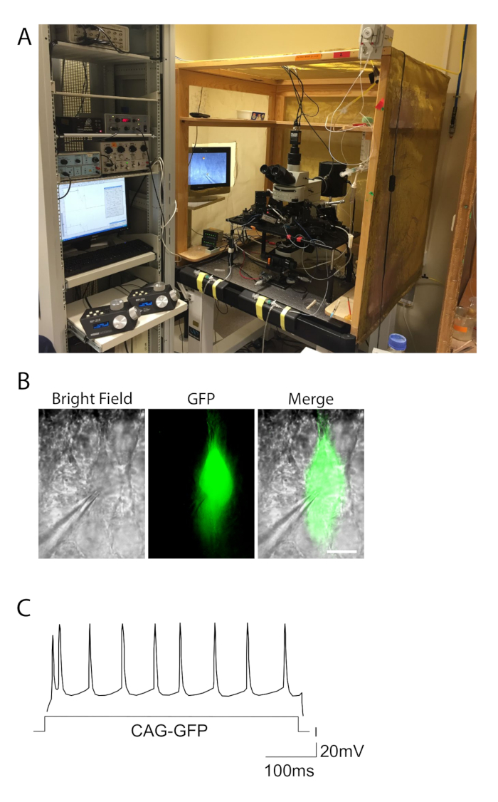
- Make sure that the electrophysiology station is equipped with a recording chamber, a perfusion system, a microscope, electrodes (recording, stimulating, and ground), macro- and micromanipulators, a rigid vibration-resistant table-top and Faraday cage, a stimulator, an amplifier and analog-to-digital (A/D) converter, a computer with acquisition software, and a GFP (or any other fluorochrome) filter for analyzing genetically modified neurons (Figure 1A).
- Prepare the intracellular solution, containing 115 mM potassium gluconate, 2 mM MgCl2, 10 mM HEPES, 20 mM KCl, 4 mM Na2ATP, and 0.3 mM Na3GTP, adjusted to pH 7.2 by KOH and to 290 mOsm by KCl.
- Make patch pipettes by pulling borosilicate glass capillaries. Prepare patch electrodes using a micropipette puller. Use borosilicate capillaries (1.5-mm outer diameter, 0.86-mm inner diameter, 10-cm length). Make patch electrodes showing resistances of 3-10 MΩ when filled with intracellular solution.
- Perfuse the recording chamber with ACSF at a rate of 2 mL/min. Maintain the chamber's temperature at approximately 33 °C.
- Whole-cell recording
- Transfer the slices into the recording chamber using a Pasteur pipette (cut off the long tip) or a small brush. Hold down the slice with a harp. Perfuse the slices constantly with ACSF at a rate of 2 mL/min.
- Patch a GFP-positive neuron.
- Put the slice into the recording chamber and find the area of interest through the microscope at low magnification (10X). Then, find a GFP-positive cell to patch using the 60x objective.
- Fill the recording electrode with intracellular solution. Use the syringe linked to the filter (4-mm filter) and micro-loader tip to fill the recording electrode with intracellular solution.
- Place the glass pipette in the pipette holder. Place the pipette tip in the bath and focus on the tip. Once the pipette is in the bath, apply positive pressure through the back pressure control system.
- Patch a neuron that is fluorescent (Figure 1B).
- Approach the cell of interest under visual guidance while maintaining back pressure in the pipette. Upon the appearance of a small dimple on the cell surface, release the pressure. At this point, a tight seal (resistance larger than 1 GΩ) may be formed. Otherwise, apply a light negative pressure (suction) to facilitate it.
- While the seal is being formed, bring the holding voltage clamp to -60 mV. Once the GΩ seal is formed, apply a pulse of suction to rupture the cell membrane beneath the pipette and go into whole-cell mode.
- Record the activity using current-clamp conditions. Once in whole-cell mode, switch from voltage-clamp to current-clamp mode and start recording. For example, to record cell excitability, apply 500 ms-long depolarizing current injections (100 - 400 pA).
- Calculate the firing rates by plotting the number of action potentials along the train for increasing input currents.
NOTE: Resting membrane potential, input resistance, and membrane capacitance may also be calculated from the recordings.