A subscription to JoVE is required to view this content. Sign in or start your free trial.

Method Article

Inducing Axonal Varicosities with a Micromechanical Stimulus on Neurons

679 views

⸱

July 8th, 2025

In This Article

Abstract

Source: Servello, D., et al. A Microbiomechanical System for Studying Varicosity Formation and Recovery in Central Neuron Axons. J. Vis. Exp. (2018).

This video demonstrates the induction of axonal varicosities by applying micromechanical stimulus on neurons. The system comprises a micropipette attached to a syringe containing buffer via tubing. Using a micromanipulator, the micropipette is positioned above the neurons. Fluid pressure is then applied to induce varicosities in axons.
Protocol

1. Setting Up the Puffing Pipette Apparatus

NOTE: There are five basic components to this set-up: the glass pipette, the tubing, the syringe, the micromanipulator, and the buffer (Hank's) Table 1. Any buffer that has physiologically relevant salt concentrations could be used in this set-up.

  1. Pull a borosilicate pipette to have around 45-µm-diameter opening tip using a micropipette puller and the specifications in Table 2.
    NOTE:
    It was found that 45 µm is a suitable size for the diameter of the pipette opening under experimental conditions. Slight....

Access restricted. Please log in or start a trial to view this content.

Results

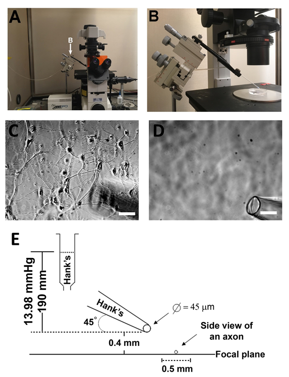
Microscope setup for cell observation; includes micromanipulator, neuron cultures, and focal plane diagram.
Figure 1: Schematic and photograph of puffing apparatus. (A) Photographs showing the overall microscope set-up. (B) The puffing apparatus showing the glass pipette held by the micromanipulator and connected to the rubber tubing. (C) Phase contras.......

Access restricted. Please log in or start a trial to view this content.

Disclosures

No conflicts of interest declared.

Materials

List of materials used in this article
NameCompanyCatalog NumberComments
ubber tubingFisher Scientific14-169-1A
10cc plastic syringe and plungerBecton Dickinson
micromanipulatorSutter Instruments
Cell culture dish (35 mm x 10 mm)Corning3294
Model P-1000 Flaming/Brown Micropipette pullerSutter Instruments
Eclipse TE2000-U McroscopeNikon
Plan Fluor ELWD 20x lensNikon62933objective
Apo TIRF 100x/1.49 oil lensNikonMRD01991objective
HEPESFisher ScientificBP410-500
D-glucoseFisher ScientificD16-500
MgCl2Fisher ScientificBP214-500
NaClFisher ScientificS640-3
KClFisher ScientificBP366-500
CaCl2Fisher ScientificC70-500

Explore More Articles

Neuron CultureMicropipette PositioningFluid Pressure ApplicationFluorescent ImagingTransmitted Light MicroscopyCoverslip PreparationHank's BufferGFP-transfected Neurons