All procedures involving sample collection have been performed in accordance with the institute's IRB guidelines.
1. Dissect and Prepare the Larval Drosophila CNS
NOTE: Methods for larval CNS dissection are clearly illustrated in Hafer and Schedl, but these previously published methods reduce the length of the descending neurons that are important for measuring spike frequency. Here, an additional method is outlined to excise the larval CNS that maintains long, intact descending neurons.
- Saline preparation.
- Prepare the dissection and recording saline to the following in mM: 157 NaCl, 3 KCl, 2 CaCl2, 4 2-[4-(2-hydroxyethyl)piperazin-1-yl]ethanesulfonic acid (HEPES); titrate pH to 7.25.
- Dissect the larval Drosophila CNS.
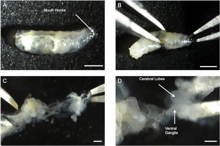
- Identify and extract a wandering third-instar Drosophila melanogaster from the culture vial and place into 200 µL of saline (Figure 1A).
- Grasp the mouth hooks with a pair of fine forceps and grasp the abdomen of the maggot with a second pair of forceps (Figure 1B).
NOTE: Do not apply too much pressure to the abdomen as this can result in tearing of the thin cuticular layer of the maggot.
- Gently pull the mouth hooks and abdomen in different directions to separate the caudal end of the maggot from the head region and expose the viscera of the maggot (Figure 1C).
NOTE: The CNS will be intertwined with the trachea and digestive tract. Do not cut the CNS away from any tissue to prevent cutting the descending peripheral nerves.
- Tease the CNS out of the digestive tract and trachea with forceps (Figure 1D).
- If necessary, disrupt the blood brain barrier by manually transecting the CNS posterior to the cerebral lobes with Vannas spring scissors.
NOTE: This should be done based on the physiochemical properties of the chemical being used. The red line in Figure 2A is the suggested transection point and the transected CNS with intact descending peripheral nerve trunks shown in Figure 2B. The transected CNS is ready for experimentation after completion of step 1.2.5.
2. Extracellular Recording of Drosophila CNS.
- Prepare the CNS for recording.
- Pull a glass pipette electrode from borosilicate glass capillaries to a resistance of 5-15 mΩ.
- Insert the transected CNS into the wax chamber that contains 200 µL of saline. Clamp an uncoated insect pin with the alligator clip soldered to the ground wire and insert the pin into the saline to complete the circuit.
- Using the micromanipulators, orient the electrode to the caudal end of the transected CNS. Eliminate recording of background noise by adjusting the threshold level in the acquisition/analysis software prior to contacting the peripheral nerve trunks.
- Draw any convenient peripheral nerves into the suction electrode by applying slight negative pressure on the syringe.
NOTE: The baseline firing frequency is correlated to the number of neurons drawn into the electrode where more neurons oftentimes result in increased resistance of the electrode.
- Begin extracellular recording of CNS descending nerve activity.
- Start the recording on the data acquisition software and monitor the baseline firing frequency. Allow the firing rate to equilibrate for 5 min prior to collecting baseline firing rate data.
- Discard the preparation and recording if the pattern of firing of control treatment is not a bursting pattern similar to that shown in Figure 3A.
NOTE: Altered pattern of firing suggests abnormal or unstable activity of the central pattern generator, which can alter neural function.
- After 5 min, add 200 µL of saline + vehicle to bring the total volume of the chamber to 400 µL to begin recording control firing rates.
- After baseline has been established (3-5 min), withdraw 200 µL of saline and add 200 µL of the experimental agent solubilized in saline.
NOTE: Dimethyl sulfoxide (DMSO) is the recommended solvent for lipophilic compounds and should not exceed 0.1% v/v.
- Apply drug concentrations in a serial manner (low to high concentrations) and record each concentration for a select period of time (determined by the investigator based on the chemical properties of the drug). Label this time point of drug application in the acquisition/analysis software by including a comment that includes the drug and the final concentration.
NOTE: Firing activity of the CNS will decline by approximately 10-20% after 30-50 min after the dissection and therefore, appropriate untreated controls should be performed, and drug treatments should not exceed this time period.