1. Seed and Treat Cerebral Vascular Endothelial (CVE) cells on Cell Culture Plates
- Seed CVE cells (bEnd.3 cells) on a 96-well extracellular flux cell culture plate: 80 µL/well. Place the cell culture plate at 37 °C in a 5% CO2 incubator. Note: 16 × 103 cells per well will reach 60 - 80% confluency within 24 h.
- If the cells are being exposed to a chemical, add the treatments once the cells are attached to the plate bottom (8 - 24 hours after seeding).
NOTE: For transfection experiments, do not add antibiotics in a complete growth medium.
2. Assessment of Mitochondrial Function Using the Bioanalyzer
- Prior to the start of the assay, hydrate an extracellular flux sensor cartridge with calibrant by adding 150 µL of extracellular flux calibrant solution to the plate and incubating overnight at 37 °C without CO2.
- Using the plate washer station, change the cell culture medium into pH 7.0 extracellular flux assay medium. Incubate the cells at 37 °C without CO2 for 30 - 60 min.
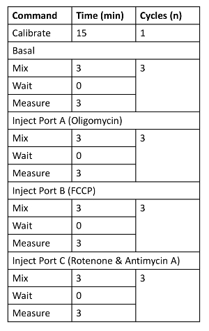
- Prepare working solutions of 8 µM oligomycin, 4.5 µM Carbonilcyanide 4-(trifluoromethoxy) phenylhydrazone (FCCP), 10 µM rotenone, and 10 µM antimycin A in Dulbecco's Modified Eagle's Medium (DMEM). Load 25 µL of the stock solutions into cartridge injection ports: Port A, oligomycin; Port B, FCCP; Port C, rotenone, and antimycin A with extracellular flux assay medium. Implement the assay protocol as described in Table 1.
- Load the hydrated cartridge into the bioanalyzer and perform the calibration by clicking the "Start" button. After the calibration is complete, remove the cartridge bottom plate and load the cell plate by clicking the "Unload Cartridge" prompt. Continue the assay until all measurements are completed.
- Open the data file and obtain the rate values from the bioanalyzer. Export the data to a spreadsheet file.
Table 1. The Instrument Run Protocol.