Method Article

Studying Alpha-Synuclein Accumulation in Primary Embryonic Mouse Dopamine Neurons

July 8th, 2025

In This Article

Abstract

Loading...
$$\rightleftharpoonup{xx}$$ $$\longleftharp{xx}$$, $$\longrightharp{xx}$$,

Source: Er, S. et al., Studying Pre-formed Fibril Induced α-Synuclein Accumulation in Primary Embryonic Mouse Midbrain Dopamine Neurons. J. Vis. Exp. (2020).

This video demonstrates the process of quantifying alpha-synuclein accumulation in embryonic mouse dopamine neurons pre-treated with alpha-synuclein pre-formed fibrils. It outlines the steps for fixing, staining, and quantifying the dopamine neurons containing alpha-synuclein aggregates using immunofluorescence.

Protocol

Loading...
$$\rightleftharpoonup{xx}$$ $$\longleftharp{xx}$$, $$\longrightharp{xx}$$,

All procedures involving animal samples have been reviewed and approved by the appropriate animal ethical review committee.

1. Preparation

  1. Prepare dopamine neuron medium (DPM) with 0.46% D-glucose, 1% L-glutamine, 1% N2, 0.2% primocin, completed with DMEM/F12. Filter the DPM after mixing the ingredients. Store DPM at 4 °C and warm each aliquot only once.
    NOTE: DPM should not contain glia-derived neurotrophic factor (GDNF), as it will reduce α-synuclein accumulation in dopamine neurons.
  2. Prepare siliconized glass pipettes that are extremely hydrophobic, thereby minimizing the attachment to the surface and loss of cells during the initial handling of embryonic neurons.
    1. Add 10 mL of siliconizing fluid to 1 L of distilled water and mix by stirring in a 2 L vessel. Leave the glass pipettes immersed in the siliconizing solution for 15 min.
    2. Rinse the pipettes 3–5x with distilled water. Dry the pipettes overnight at room temperature (RT) or for 1–2 h at 100-120 °C heated sterile space to speed up the drying.
    3. Sterilize the pipettes by standard autoclaving in a sealed autoclave bag.
  3. Prepare poly-L-ornithine (PO) coated 96 well plates with transparent bottoms by adding 60 μL of PO solution into the middle wells of the 96 well plate to be used for seeding of the neurons, leaving at least one row/column of wells at the edges of the plate to avoid edge effects. Keep the coated plate overnight at 4 °C or 4 h at RT.
  4. Prior to plating the cells, aspirate PO completely and wash the cells thrice with 100 μL of 1x Phosphate Buffer Saline (PBS). Aspirate 1x PBS from the wells and keep the lid of the plate open for complete drying.
    NOTE: It is possible to collect used PO and filter it for reuse. This can be repeated twice for the same PO solution.
  5. Add 50 μL of DPM to previously coated wells. Aspirate DPM from the wells with a 100 μL plastic tip and simultaneously scratch the bottom of the well with circular movements to remove the coating at the perimeter of each well. A PO-coated island will remain in the middle of the well.
  6. Under a laminar hood, add 10 μL of DPM to the middle of each coated island to create micro islands.
    NOTE: A plate with DPM-covered micro islands can be kept under the laminar flow hood for 1–2 h during the isolation of cells.

2. Isolation of the ventral midbrain floor from E13.5 mouse embryos

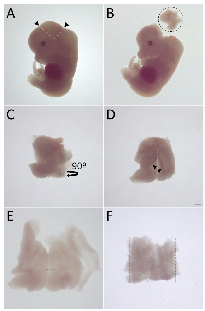
NOTE: Refer to Figure 1 for midbrain floor dissection steps.

  1. Prior to dissection, fill a 10 cm Petri dish with Dulbecco's buffer and keep it on ice.
  2. Euthanize an E13.5 pregnant female mouse according to the institution's guidelines. Place the mouse flat on its back and spray the anterior body with 70% ethanol. Lift the skin above the womb with forceps and make an incision with surgical scissors to expose the uterus.
  3. Carefully remove the uterus and place it into the previously prepared Petri dish on ice.
  4. Using surgical scissors under the laminar hood at RT, carefully remove the embryos from the uterus. Remove all placental residue from the embryos with forceps and place them into a new 10 cm Petri dish filled with Dulbecco's buffer.
  5. Using dissection forceps or needles, cut off the hindquarter of the head from the places marked with black arrows in Figure 1A. Take the cut piece away from the rest of the embryo (Figure 1B).
  6. Place the posterior of the cut piece towards the observer (Figure 1C) and gently cut it open from caudal to cranial (Figure 1D). From 0.5 mm below the cranial opening, cut a 2 mm2–3 mm2 region, shown in Figure 1E.
  7. Collect the ventral midbrain floor (see Figure 1F) in an empty 1.5 mL microcentrifuge tube. Keep the microcentrifuge tube on ice until all midbrain floors are collected in it.
    NOTE: Alternatively, the midbrain floors can be collected with a 1 mL micropipette after dissection of all embryo brains.

3. Establishing primary embryonic midbrain cultures from E13.5 mouse embryos in 96 well plate format

  1. After the collection of midbrain floors from all embryos in the same 1.5 mL tube, remove the residual Dulbecco's buffer and wash the tissue pieces thrice with 500 μL of Ca2+, Mg2+-free Hank's Balanced Salt Solution (HBSS).
  2. Remove HBSS and add 500 μL of 0.5% trypsin to the tube. Incubate it at 37 °C for 30 min.
  3. During incubation, warm 1.5 mL of fetal bovine serum (FBS) at 37 °C, add 30 μL of DNase I to the FBS and mix. Also, fire-polish the tip of a siliconized glass pipette. Make sure that the hole has no sharp edges and is around the same size as a 1 mL micropipette tip.
    NOTE: As an alternative, a low adhesion 1 mL micropipette tip can be used for trituration. However, siliconized glass pipettes seem to give the best results.
  4. As soon as the incubation in step 3.2 ends, add 500 μL of the FBS/DNase mix to the partially digested tissue. Use the glass pipette to triturate the tissue in the FBS/trypsin mix. Triturate until tissues dissociate into tiny, barely visible particles. Avoid bubbles during trituration.
  5. Let the leftover particles precipitate at the bottom of the microcentrifuge tube by gravity. Without pipetting the precipitate at the bottom, collect the supernatant into an empty 15 mL conical polypropylene tube.
  6. Dilute FBS/DNase I from step 3.3 (98:2) with 1,000 μL of HBSS to obtain FBS/DNase-I/HBSS (49:1:50). Mix by pipetting up and down. Add 1,000 μL of the new mix to the leftover particles in the microcentrifuge tube. Triturate again and repeat step 3.5.
  7. Repeat the previous step once more to use up all FBS/DNase-I/HBSS (49:1:50).
  8. Once all the supernatant is collected inside the 15 mL tube (from steps 3.5, 3.7, and 3.8), use a tabletop centrifuge to spin down the supernatant (~3 mL) at 100 x g, for 5 min. Remove the supernatant without touching the pelleted cells at the bottom.
  9. Wash the cell pellet by adding 2 mL of DPM to the tube and spin it down at 100 x g for 5 min. Remove the supernatant and repeat the washing 2x to minimize the debris in the pelleted cells.
    NOTE: Always use fresh, warmed DPM for the cultured neurons. For the washing steps, DPM does not have to be fresh but should be prewarmed to 37 °C.
  10. Dilute the cells with fresh, warm DPM and transfer them to a microcentrifuge tube. The amount of DPM for dilution depends on the number of embryos used for tissue dissection. For example, use 150 μL of DPM to dilute the cells obtained from ten embryos.
  11. Transfer 10 μL of cells in DPM to a microcentrifuge tube. Mix them with 10 μL of 0.4% Trypan blue stain. Count live (i.e., Trypan blue negative) cells using a hemocytometer or an automated cell counter.
    NOTE: Use 30,000 cells for plating per well to obtain ~1,000 dopamine neurons per well. If the cell density is higher than ~30,000 cells per 6 μL, further dilute the cells with DPM before plating so that the seeding volume is no less than 6 μL.
  12. Without touching the bottom of the wells, remove the DPM from the micro islands created at step 1.6.
  13. In order to obtain reproducible cell density at each well, mix the cells by gentle pipetting prior to plating in the well. With a 1–10 μL micropipette, add 6 μL of cells to the middle of the well at the location of each former micro island.
  14. Fill the empty wells at the edges of the plate with 150 μL of water or 1x PBS to minimize evaporation from the wells containing neuronal cultures. Incubate the plate in an incubator at 37 °C, 5% CO2 for 1 h.
  15. After 1 h, remove the plate from the incubator, add 100 μL of DPM into each well with cells, and place it back in the incubator.
  16. Two days after plating (day in vitro 2, or DIV2), remove 25 μL and add 75 μL of fresh DPM to bring the final media volume to 150 μL and avoid evaporation as much as possible.
  17. Exchange half of the medium with fresh DPM (i.e., remove 75 μL and add 75 μL fresh DPM) at DIV5. Do not perform any media changes after DIV5.

4. Induction of α-synuclein aggregates in primary embryonic dopamine neurons by seeding with preformed fibrils

Following any work with α-synuclein pre-formed fibrils (PFFs), clean the laminar hood or any equipment that might have contacted the PFFs with 1% Sodium dodecyl sulfate (SDS), then with 70% ethanol.

  1. Prior to the experiment, dilute the PFFs with 1x PBS to a final concentration of 100 μg/mL. Sonicate the diluted PFFs in microcentrifuge tubes with a bath sonicator at high power with water bath cooling at 4 °C for 10 cycles, 30 s ON/30 s OFF.
    NOTE: It is critical that the fibrils be properly sonicated to generate fragments ~50 nm long. The size of sonicated PFFs can be measured directly from transmission electron microscope images of stained PFFs. Sonication can be achieved as described above in a high-power bath sonicator. Alternatively, a tip sonicator can be used. Sonicated PFFs can be stored at -80 °C in small aliquots to avoid multiple freezing/thawing cycles.
  2. On DIV8, add 3.75 μL of 100 μg/mL of PFFs per well to the 150 μL of medium in the well to a final concentration of 2.5 μg/mL. Use the same amount of 1x PBS for the control group.
  3. Prepare 4% paraformaldehyde (PFA) in 1x PBS and store the aliquots at -20 °C. To do so, follow the steps below.
    NOTE: PFA is toxic; wear a mask and gloves during preparation, work always under a laminar hood, and dispose of all solid and liquid PFA waste according to the institution's directions.
    1. Warm 500 mL of 1x PBS in a 1 L vessel. Put a stir bar in the vessel and put the vessel on a magnetic stirrer with a heating function. Adjust the temperature between 40–60 °C to prevent boiling while keeping the solution warm.
    2. Measure 20 g of PFA powder under the hood in a disposable plastic measuring container. Carefully add the PFA powder into the vessel filled with 1x PBS. Start stirring the solution.
    3. Add 200 μL of 5 M sodium hydroxide into the solution and continue stirring for ~15 min until the PFA dissolves completely.
    4. After the solution appears homogenous, add 168 μL of 5 M hydrogen chloride to balance the pH to ~7. Check the pH with disposable color-fixed pH indicator strips.
    5. Remove the vessel from the heater and allow it to cool down to RT. Filter the solution and aliquot for storage at -20 °C. Thaw the aliquots at RT before the use, and do not refreeze afterwards.
  4. On DIV15, remove all media from the wells by pipetting. Add 50 μL of 4% PFA to each well to fix the cells and incubate for 20 min at RT. After incubation, remove the PFA from the wells and add 100 μL of 1x PBS to each well to wash the cells. Remove 1x PBS and wash 2x more.
  5. Leave 100 μL of 1x PBS in each well to avoid drying. Store the plate at 4 °C until immunochemistry is performed.

5. Immunofluorescent staining and automated imaging of primary embryonic dopamine neurons in 96 well plates

  1. Remove 1x PBS and permeabilize the cells by adding 100 μL of 0.2% Triton X-100 in PBS (PBST) per well and incubating at RT for 15 min.
  2. Remove PBST and add 50 μL of 5% normal horse serum (NHS) per well to the PBST. To block the unspecific antigen activity, incubate at RT for 1 h.
  3. Dilute the primary antibodies against tyrosine hydroxylase (TH) and pS129-αsyn (1:2,000) in 5% NHS in PBST. Add 50 μL of diluted antibodies to each well and incubate overnight at 4 °C.
  4. Remove antibodies and add 100 μL of 1x PBS to each well to wash the cells. Remove 1x PBS and repeat washing 2x.
  5. To prevent the bleaching of fluorescent molecules, start working under minimum light conditions. Dilute the secondary fluorescently labeled antibodies (1:400) in PBST. Add 50 μL of diluted antibodies to each well and incubate at RT for 1 h.
  6. Remove the antibody solution and add 100 μL of 1x PBS to each well to wash the cells. Remove 1x PBS and repeat washing 2x.
  7. Remove 1x PBS, add 50 μL of 200 ng/mL 4',6-diamidino-2-phenylindole (DAPI) per well to stain the nuclei of the cultured cells, and incubate at RT for 10 min.
  8. Wash cells 3x with 100 μL of 1x PBS for 5 min each. Keep 100 μL of 1x PBS in each well after the last wash. Cover the plate with aluminium foil and store it at 4 °C until imaging.
  9. Image primary embryonic dopamine neurons in a 96 well view plate with a high-content plate scanner (see Table of Materials) fitted with a 10x objective.
  10. Adjust the settings based on the specifications of the 96 well plate, such as plate type, manufacturer, size, distance between wells, as well as type and amount of medium.
  11. Select the imaging area of the well to cover all the cells in a micro island. Pick an example well to adjust the autofocus. Base the initial focus on DAPI.
  12. Calibrate the acquisition time for each fluorescent channel based on the intensity of the staining in control wells. Adjust the parameters so that in PFF-treated control wells, one can clearly distinguish dopamine cells harboring pS129-αsyn aggregates in cell soma, allowing for unambiguous quantification of pS129-αsyn positive and pS129-αsyn negative cells.
    NOTE: Wells that do not contain PFFs should not have any staining for pS129-αsyn; therefore, these wells can be used as negative control for adjusting pS129-αsyn intensity.
  13. Image all the selected wells with a 10x objective simultaneously for all channels with immunofluorescence staining with exactly the same parameters.
  14. Optionally, label α-synuclein inclusions in a subset of the wells with antibodies specific for filamentous α-synuclein to confirm that changes in the number of pS129-αsyn-positive inclusions reflect the reduction in protein accumulation rather than inhibition of phosphorylation or dephosphorylation of pS129-αsyn.
  15. Repeat step 5.3, substituting pS129-αsyn antibody with α-synuclein filament antibody (1:2,000). Image the stained aggregated α-synuclein as in step 5.13.

Access restricted. Please log in or start a trial to view this content.

Results

Loading...
$$\rightleftharpoonup{xx}$$ $$\longleftharp{xx}$$, $$\longrightharp{xx}$$,

Embryo dissection sequence, microscope images; experimental setup for developmental study.
Figure 1: Dissection of midbrain floor from E13.5 mouse embryo. (A) Cutting locations at the hindquarter of the head is marked with black arrows and white dashed lines. (B) The piece was removed from the rest of the embryo. The r...

Access restricted. Please log in or start a trial to view this content.

Disclosures

Loading...
$$\rightleftharpoonup{xx}$$ $$\longleftharp{xx}$$, $$\longrightharp{xx}$$,
No conflicts of interest declared.

Materials

List of materials used in this article
NameCompanyCatalog NumberComments
0.4% Trypan Blue stainGibco, Thermo Scientific, Waltham, Massachusetts, USA15250061
15 ml CELLSTAR Polypropylene tubeGreiner Bio-One GmbH, Frickenhausen, Germany188261
4',6-diamidino-2-phenylindole (DAPI)Sigma-Aldrich, St. Louis, Missouri, USA10236276001
5 M HClN/AN/AMedia kitchen, Institute of Biotechnology, University of Helsinki
5 M NaOHN/AN/AMedia kitchen, Institute of Biotechnology, University of Helsinki
96-well ViewPlate (Black)PerkinElmer, Waltham, Massachusetts, USA6005182
99.5% Ethanol (EtOH)Altia Oyj, Rajamäki, FinlandN/A
AquaSil Siliconizing FluidThermo Scientific, Waltham, Massachusetts, USATS-42799
Autoclaved 1.5 ml microcentrifuge tubeN/AN/AMedia kitchen, Institute of Biotechnology, University of Helsinki
Bioruptor sonication deviceDiagenode, Liege, BelgiumB01020001
Ca2+, Mg2+ free Hank’s Balanced Salt Solution (HBSS)Gibco, Thermo Scientific, Waltham, Massachusetts, USA14175-053
CellProfiler and CellAnalyst software packagesN/AN/Afree open-source software
Centrifuge 5702Eppendorf AG, Hamburg, Germany5702000019
CO2 IncubatorHeraCell, Thermo Scientific, Waltham, Massachusetts, USAN/A
Counting chamberBioRad Inc., Hercules, California, USA1450015
DeltaVision Ultra High Resolution Microscope with air table and cabinetGE Healthcare Life Sciences, Boston, Massachusetts, USA29254706
Deoxyribonuclease-I (Dnase I)Roche, Basel, Switzerland22098700
D-glucoseSigma-Aldrich, St. Louis, Missouri, USAG8769
DMEM/F12Gibco, Thermo Scientific, Waltham, Massachusetts, USA21331–020
donkey anti-mouse AlexaFluor 488Thermo Scientific, Waltham, Massachusetts, USAA21202
donkey anti-rabbit AlexaFluor 647Thermo Scientific, Waltham, Massachusetts, USAA31573
donkey anti-sheep AlexaFluor 488Thermo Scientific, Waltham, Massachusetts, USAA11015
Dulbecco's bufferN/AN/AMedia kitchen, Institute of Biotechnology, University of Helsinki
Fetal Bovine Serum (FBS)Gibco, Thermo Scientific, Waltham, Massachusetts, USA10500056
Fisherbrand Plain Economy PTFE Stir Barfisher scientific, Thermo Scientific, Waltham, Massachusetts, USA11507582
ImageXpress Nano Automated Imaging SystemMolecular Devices, San Jose, California, USAN/A
L-glutamineGibco, Thermo Scientific, Waltham, Massachusetts, USA25030–032
mouse monoclonal anti-tyrosine hydroxylaseMillipore, Merck KGaA, Darmstadt, GermanMAB318
N2 supplementGibco, Thermo Scientific, Waltham, Massachusetts, USA17502–048
Normal Horse SerumVector Laboratories Inc., Burlingame, California, United StatesS-2000
paraformaldehyde (PFA)Sigma-Aldrich, St. Louis, Missouri, USA158127
paraformaldehyde powder, 95%Sigma-Aldrich, St. Louis, Missouri, USA158127
pH-Fix, color-fixed indicator stripsMacherey-Nagel, Düren, Germany92110
Phospohate Buffer Saline (PBS)N/AN/AMedia kitchen, Institute of Biotechnology, University of Helsinki
poly-L-ornithineSigma-Aldrich, St. Louis, Missouri, USAP4957
PrimocinInvivoGen, San Diego, California, USAant-pm-1, ant-pm-2
rabbit monoclonal anti-alpha-synuclein filamentAbcam, Cambridge, United Kingdomab209538
rabbit monoclonal anti-phospha-serine129-alpha-synucleinAbcam, Cambridge, United Kingdomab51253
RCT Basic, IKA Magnetic StirrerIKA®-Werke GmbH, Staufen, Germany3810000
recombinant human glia-derived neurotrophic factor (hGDNF)PeproTech, Rocky Hill, NJ, USA or Prospec, Ness-Ziona, Israel450-10 (PeproTech), CYT-305 (Prospec)
recombinant mouse alpha synuclein Pre-Formed Fibrils (PFFs)N/AN/AGift from collaborator, Prof. Kelvin C Luk
Research Stereomicroscope SystemOlympus Corporation, Tokyo, JapanSZX10
sheep polyclonal anti-tyrosine hydroxylaseMillipore, Merck KGaA, Darmstadt, Germanab1542
TC 20 Automated Cell CounterBioRad Inc., Hercules, California, USA1450102
Triton X-100Sigma-Aldrich, St. Louis, Missouri, USA11332481001
trypsinMP Biomedicals, Valiant Co., Yantai, Shandong, China2199700

Reprints and Permissions

Request permission to reuse the text or figures of this JoVE article

Request Permission

Tags

Alpha Synuclein AccumulationPre formed FibrilsDopamine NeuronsImmunofluorescence StainingPhosphorylated Alpha SynucleinFluorescence Plate ScannerPrimary Mouse Embryonic NeuronsCell PermeabilizationAntibody BindingNuclear Staining

Related Articles