This video demonstrates a subpial injection technique for gene delivery to the spinal cord in mice. The procedure involves exposing the dura mater, puncturing the pia mater, and injecting a recombinant adeno-associated virus (rAAV) carrying a fluorescent protein-expressing gene into the subpial space. Following injection and recovery, the virus facilitates gene expression in spinal cord cells, enabling the study of target gene expression.
A subscription to JoVE is required to view this content. Sign in or start your free trial.
Method Article
Subpial Gene Delivery in the Mouse Spinal Cord to Evaluate Gene Expression
1K views
⸱
August 7th, 2025
In This Article
Abstract
Protocol
All procedures involving animal models have been reviewed by the local institutional animal care committee and the JoVE veterinary review board.
1. General Animal and Surgical Preparation
- Before starting the surgical procedure, thaw the virus (AAV9-UBI-GFP [UBI: Ubiquitin Promoter, GFP: Green Fluorescent Protein]; 5 µL aliquots). Prepare a 5% dextran (10,000 MW) solution by mixing dextran powder in distilled water. Mix the virus solution with 5% dextran solution 1:1 to a final dextran concentration of 2.5%.
- Store the virus solution on ice (4 °C).
- Use adult C57BL/6J mice (male and female, 20-30 g). Anesthetize the mice using 5% isoflurane (in O2, 1 L/min) and maintain them at 2-3% inhaled isoflurane (in O2, 1 L/min) by nose cone during surgery, depending on the breathing rate and paw pinch response.
- Shave the back of the animals with shaving clippers and clean the skin with 2% chlorhexidine.
- In chronic recovery studies, follow a strict sterile technique.
- If lumbar subpial injections are to be performed, cut the skin overlaying the Th8-L1 vertebrae with a scalpel and detach the paravertebral muscle from Th10-12 spinal vertebrae using scissors.
- Mount the animal into a standard stereotaxic frame using mouse spinal clamps.
- Shave both sides of the lamina of the Th10-12 vertebrae using a dental drill (drill bit: 0.9 mm, speed: 20,000 rpm) until cracks appear.
- Remove cracked bone fragments with forceps and expose the dorsal surface of the lumbar spinal cord.
- Cut open the dura about 1 cm using a 30 G stainless steel needle and forceps.
2. Opening the Pial Membrane and Inserting the Subpial Needle for AAV9 Delivery
- Mount the 34 G pia-penetrating needle into the Z-arm of an XYZ manipulator using a glass capillary holder (Figure 1A and B).
NOTE: To manufacture the pia-penetrating needle, the original beveled tip of the 34G needle is sharpened using a glass capillary beveller with diamond abrasive plate - coarse (5.0 µm to 50 µm tip sizes) and grinding angle of 15 - 20°. The tip of the needle (1 mm length, measured from the tip) is then gently bent to about 90° (Figure 1B, left insert). - Using a surgical dissecting scope, set to 8-10X magnification, penetrate the pia with the pia-penetrating needle by about 1 mm (Figure 1C) using the X-arm.
- Keep the angle of the penetrating needle to the tissue surface at 5-10°.
- After the pia opening, remove the pia-penetrating needle horizontally from the subpial space (Figure 1D) using the X-arm.
NOTE: Remember the penetrated site by a landmark, such as a blood vessel. - Load a blunt 36G injection needle with AAV9-UBI-GFP virus using a 50-µL microsyringe connected to the injection needle with PE-10 or PE-20 tubing.
- Mount the needle into the Z-arm of a second XYZ manipulator (Figure 1A and B) using a glass capillary holder (Figure 1B, right insert).
NOTE: To manufacture the subpial AAV9 injection needle, the blunt tip of a 36 G needle is polished using a glass capillary beveler with diamond abrasive plate - coarse (5.0 µm to 50 µm tip sizes) to remove the sharp edges. The tip of the needle (2-3 mm length, measured from the tip) is then gently bent to about 90°. The pia-penetrating and subpial injection needles are inserted into 1-2 cm-long 20G slave stainless steel tubing (10 mm from the end of the needle) and glued with epoxy. The use of 20 G (0.91 mm-diameter) tubing is required for a secure attachment to the glass capillary holder. - By manipulating the X, Y, and Z arms of the second manipulator, position the tip of the AAV9 injection needle into the pia-penetrated site and then advance it about 2-3 mm into the subpial space through the previous pial membrane opening using the X-arm ( Figure 1E and F).
NOTE: 1) The AAV9-UBI-GFP is prepared according to previously reported protocols, and the final titers are adjusted to 1.2 x 10¹³ genome copies per mL (gc/mL). 2) There is no need to mark the site of the pial opening because it is readily identifiable (Figure 1D). - Inject the AAV9-UBI-GFP (1.5, 3, or 5 µL) into the subpial space using a 50 µL microsyringe (see Table 1 for experimental groups).
- Remove the injection needle from the subpial space after the AAV9-UBI-GFP injection is complete.
- Close the muscle and skin using 4.0 monofilament suture and surgical clips.
NOTE: There is no need to seal the open vertebra. - Allow the animals to recover on a heating pad.
- For pain control, inject Buprenorphine 0.05 mg/kg/sc every 12 h for 2-3 days post-surgery.
Table 1: Experimental Groups. All experiments were performed in adult C57BL/6J mice.
| Experimental Groups | Site/level of AAV9 delivery (*) | Volume of AAV9 inj, infusion rate | Survival time | Tissue analysis |
| Group A (n = 7) | C2 (bilateral) | 5 µL/5 min | 14 days | Brain + spinal cord |
| Group B (n = 12) | L1-L2 (bilateral) | 1.5 µL or 3µL, 60 sec/µL | 14 days | Brain + spinal cord |
| Group C (n = 6) | C2 + L1-L2 (bilateral at each level) | 5 µL/5 min | 14 days | Spinal cord |
| * - bilateral = two subpial injections, one delivered into the right and one to the left subpial space of the injected segment(s) are performed. | ||||
Access restricted. Please log in or start a trial to view this content.
Results
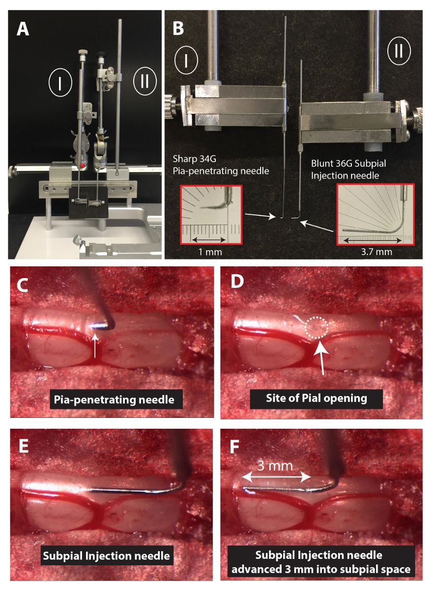
Figure 1: Experimental Setup to Perform Spinal Subpial Injections in an Adult Mouse
Access restricted. Please log in or start a trial to view this content.
Materials
| Name | Company | Catalog Number | Comments |
|---|---|---|---|
| C57BL/6J Mice | Jackson Labs | 664 | |
| Lab Standard Stereotaxic for Mice | Harvard Apparatus | 72-9568 | |
| Mouse Spinal Adaptor | Harvard Apparatus | 72-4811 | |
| XYZ Manipulator | Stoelting | 51604 | |
| Manual Infusion Pump | Stoelting | 51218 | |
| 34G Beveled Nanofill Needle | World Precision Instruments | NF34BV-2 | |
| 36G Blunt Nanofill needle | World Precision Instruments | NF-36BL-2 | |
| Fluriso, Isoflurane | MWI Veterinary Supply | 502017 | |
| Chlorhexidine Solution | MWI Veterinary Supply | 501027 | |
| 20G Stainless Steel Needle | Becton-Dickinson | 305175 | |
| 23G Stainless Steel Needle | Becton-Dickinson | 305145 | |
| 30G Stainless Steel Needle | Becton-Dickinson | 305128 | |
| Cotton Tipped Applicator | MWI Veterinary Supply | 27426 | |
| Glass Capillary Beveller | Narishige International | SM-25B | |
| Slide Microscope Superfrost | Leica Microsystems | M80 | |
| 50μl Microsyringe | Hamilton | 81242 | |
| BD Intramedic PE-20 Tubing | Becton, Dickinson | 427406 | |
| BD Intramedic PE-10 Tubing | Becton, Dickinson | 427401 | |
| 4-0 monofilament suture | VetOne | V1D397 | |
| Glass Capillary Beveller | Narishige | Pipet Micro Grinder EG-40 | |
| 5 min Epoxy (Epoxy Clear) | Devcon | 14310 | |
| Dextran | Polysciences, Inc | 19411 | |
| AAV9-UBC-GFP | UCSD Viral Vector Core Laboratory | ||
