This video demonstrates the intranasal administration of radiolabeled drugs in an anesthetized mouse to achieve nose-to-brain drug delivery. It explains how drugs cross the nasal epithelium and travel via neuronal pathways to reach the brain, as well as how ciliary motion influences drug retention and clearance. Additionally, the video describes an alternative reverse cannulation method, which involves inserting a cannula through the esophagus to deliver drugs against the ciliary motion, enhancing retention and improving transport efficiency.
A subscription to JoVE is required to view this content. Sign in or start your free trial.
Method Article
Intranasal Drug Administration in Mice via Pipette and Reverse-Cannulation for Nose-to-Brain Drug Delivery
3.2K views
⸱
August 7th, 2025
In This Article
Abstract
Protocol
All procedures involving animal models have been reviewed by the local institutional animal care committee and the JoVE veterinary review board.
1. Animals Used for Intranasal Administration Under Inhalation Anesthesia
- House the experimental mice in stainless-steel cages under a 12-h light/dark cycle (light on 8:00 AM–8:00 PM), with a controlled temperature maintained at 23 ± 1°C, humidity of 50% ± 10%, and ad libitum access to food and water.
- Prior to experimentation, anesthetize the mice via inhalation of 2% isoflurane, following initiation at a concentration of 4%. Confirm the requisite level of anesthetization by ch....
Access restricted. Please log in or start a trial to view this content.
Results
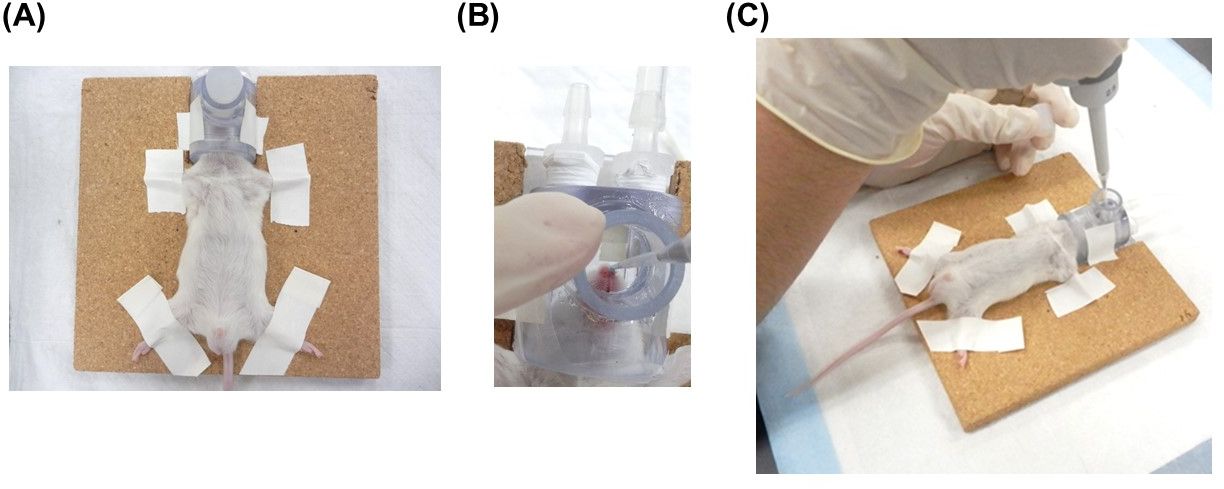
Figure 1: Intranasal administration using a micropipette in conjunction with a temporarily openable inhalation mask.......
Access restricted. Please log in or start a trial to view this content.
Materials
| Name | Company | Catalog Number | Comments |
|---|---|---|---|
| ddY mouse | Japan SLC, Inc. | Male, 4-6 weeks, 20-30 g | |
| Isoflurane | Pfizer | v002139 | |
| Isoflurane setup | SHINANO manufacturing CO. LTD. | SN-487-OTAir, SN-489-4 | |
| Isoflurane mask | SHINANO manufacturing CO. LTD. | For small rodents | |
| Isoflurane mask (Opneable type) | SHINANO manufacturing CO. LTD. | Special orders | |
| Anesthesia Box | SHINANO manufacturing CO. LTD. | SN-487-85-02 | |
| Animal experiments scissors-1 | NATSUME SEISAKUSHO CO., LTD. | B-27H | |
| Animal experiments scissors-2 | NATSUME SEISAKUSHO CO., LTD. | B-13H | |
| Tweezers-1 | FINE SCIENCE TOOLS Inc. | 11272-30 | Dumont #7 Dumoxel |
| Tweezers-2 | NATSUME SEISAKUSHO CO., LTD. | A-12-1 | |
| Cannula tube (PE-50) | Becton, Dickinson and Company. | 5069773 | I.D.: 0.58 mm, O.D.: 0.965 mm |
| Cannula tube (SP-10) | NATSUME SEISAKUSHO CO., LTD. | KN-392 | I.D.: 0.28 mm, O.D.: 0.61 mm |
| Shaver | MARUKAN, LTD. | DC-381 | |
| Stereoscopic microscope | Olympus Corporation | SZ61 | |
| Needle 27G 1/2 in 13 mm | TERUMO CORPORATION | NN-2738R | |
| 1 mL syringe | TERUMO CORPORATION | SS-01T | |
| Syringe pump | Neuro science | NE-1000 | |
| Micro spatula | Shimizu Akira Inc. | 91-0088 | |
| Micropipette (0.5-10 uL) | Eppendorf AG | Z368083 | |
| Pipette chip | Eppendorf AG | 0030 000.811 | |
| Tape | TimeMed Labeling System, Inc. | T-534-R | For fixing mouse |
| [¹⁴C]-Inulin | American Radiolabeled Chemicals Inc. | ARC0124A | 0.1 mCi/mL |
| EtOH | Wako Pure Chemical Industries, Ltd. | 054-00461 |
