Twin-to-twin transfusion syndrome and twin anemia polycythemia sequence are two potentially devastating problems in perinatal medicine. Both disorders occur only in monochorionic twins and result from unbalanced blood flow through placental vascular anastomoses. We provide a simple protocol to accurately evaluate the presence of vascular anastomoses using colored dye injection of placental vessels after birth.
A subscription to JoVE is required to view this content. Sign in or start your free trial.
Method Article
Accurate and Simple Evaluation of Vascular Anastomoses in Monochorionic Placenta using Colored Dye
In This Article
Summary
Abstract
The presence of placental vascular anastomoses is a conditio sine qua non for the development of twin-to-twin transfusion syndrome (TTTS) and twin anemia polycythemia sequence (TAPS)1,2. Injection studies of twin placentas have shown that such anastomoses are almost invariably present in monochorionic twins and extremely rare in dichorionic twins1. Three types of anastomoses have been documented: from artery to artery, from vein to vein and from artery to vein. Arterio-venous (AV) anastomoses are unidirectional and are referred to as "deep" anastomoses since they proceed through a shared placental cotyledon, whereas arterio-arterial (AA) and veno-venous (VV) anastomoses are bi-directional and are referred to as "superficial" since they lie on the chorionic plate. Both TTTS and TAPS are caused by net imbalance of blood flow between the twins due to AV anastomoses. Blood from one twin (the donor) is pumped through an artery into the shared placental cotyledon and then drained through a vein into the circulation of the other twin (the recipient). Unless blood is pumped back from the recipient to the donor through oppositely directed deep AV anastomoses or through superficial anastomoses, an imbalance of blood volumes occurs, gradually leading to the development of TTTS or TAPS. The presence of an AA anastomosis has been shown to protect against the development of TTTS and TAPS by compensating for the circulatory imbalance caused by the uni-directional AV anastomoses1,2. Injection of monochorionic placentas soon after birth is a useful mean to understand the etiology of various (hematological) complications in monochorionic twins and is a required test to reach the diagnosis of TAPS2. In addition, injection of TTTS placentas treated with fetoscopic laser surgery allows identification of possible residual anastomoses3-5. This additional information is of paramount importance for all perinatologists involved in the management and care of monochorionic twins with TTTS or TAPS. Several placental injection techniques are currently being used. We provide a simple protocol to accurately evaluate the presence of (residual) vascular anastomoses using colored dye injection.
Protocol
1. Preparation of the placenta at delivery
- Label the umbilical cords of the twins with one (for the first-born) or two (for the second-born) clamps.
- Inspect the maternal and fetal surface of the placenta for completeness or disruption.
- Record the following data: type of cord insertion (central, eccentric, marginal or velamentous), number of blood vessels in the umbilical cord (usually one vein and two arteries, sometimes only one artery) and color difference between both placental shares. A section of the dividing membranes can be sent to Pathology to confirm the type of chorionicity.
- The placenta can then be placed in a plastic bowl and refrigerated until the final examination (best within one week) and color dye injection.
- The placenta must not be frozen or fixed (do not use formalin).
2. Catheterization of the umbilical vessels
- Wash the placenta with warm water or saline.
- Trim the peripheral membranes, remove the inter-twin dividing membrane and peel off the amnions (for better visualization of the vascular anastomoses and better quality of the placental pictures).
- Transect each umbilical cord at approximately 5 cm distance from the cord insertion.
- Gently squeeze out blood clots from the umbilical vessels and placental vessels.
- The umbilical vein is usually easy to identify due to its larger diameter, compared to the smaller diameter of the two umbilical arteries.
- Cannulate the umbilical vein with an appropriately sized catheter. Avoid false passages.
- Cannulate one umbilical artery with a smaller catheter. Use tweezers to widen the lumen of the umbilical artery. Avoid false passages. Only one of the 2 umbilical arteries needs to be catheterized since an anastomosis (of Hyrtl) connects the 2 arteries near the cord insertion.
- Repeat both steps for the other umbilical cord.
- Placement of the catheter can be facilitated by gentle back and forth massaging of the umbilical vessels. Any type of catheter can be used for this procedure. We choose to use (and recycle) the catheters used at our neonatology ward for umbilical catheterization in neonates.
- Tie a piece of tape around both cords to avoid back flow of the colored dye during dye injection.
3. Injection with colored dye
- Connect a 20 ml syringe filled with colored dye to each catheter.
- Any viscous colored dye can be used to visualize the placental angio-architecture. Use contrasting colors to allow good visualization of the anastomoses (dark colors for the arteries, bright colors for the veins).
- Gently inject (with low pressure) the colored dye in the vein while an assistant gently pushes the dye to allow the colored dye to fill all placental vessels, also the smallest ones.
- Pay particular attention to the small vessels near the vascular equator (the vascular equator is the place where the anastomoses from either twin connect with each other).
- Repeat the previous steps to inject colored dye into the artery. Of note: arteries may be more difficult to inject and require more patience.
- Repeat both steps for the other umbilical cord.
4. Evaluation and documentation of the placenta after colored dye injection
- Carefully examine the vascular equator and record the number and types of anastomoses.
- Place a measuring tape on the placenta to measure the diameters and placental shares on the digital picture.
- Use a high-resolution digital camera and take pictures of the injected placenta. Make sure that the pictures are taken perpendicular to the placenta.
5. Representative Results:
The placental angio-architecture in monochorionic twins varies according to the type of monochorionic twin pregnancy. Injection studies have demonstrated that AA, AV and VV anastomoses are present in respectively 80%, 95% and 20% of uncomplicated monochorionic twin pregnancies1 (Figure 1). AA anastomoses are considered to protect against the development of TTTS and TAPS6. Injections studies have shown that AA anastomoses occur in only 20% of TTTS placentas and 10% of TAPS placentas1,2,7 (Figure 2). In TTTS placentas treated with fetoscopic laser surgery, the scars caused by laser coagulation of the vascular anastomoses can be seen on the placental surface (Figure 3 and 4). TAPS placentas are characterized by the presence of only a few minuscule AV anastomoses2 (Figure 5). The placental share of the TAPS recipient is often plethoric, whereas the placental share of the donor is pale (Figure 6). In monochorionic twin pregnancies with birth weight discordance, the growth restricted fetus often has a velamentous cord insertion and a much smaller placental share (Figure 7). AA anastomoses are often present in monochorionic placentas from twins with birth weight discordance8. Monochorionic-monoamniotic placentas have a characteristic angio-architecture with a short distance between both cord insertions (Figure 8). The incidence of AA anastomoses in monoamniotic-monochorionic placentas is virtually 100% and prevents the development of TTTS in monoamniotic twins9. The figures in this article show the characteristic findings of monochorionic placentas injected at our center with color dye. We routinely use darker colors (blue or green) for arteries and lighter colors (yellow, pink or orange) for veins.

Figure 1. Monochorionic placenta from a normal, uncomplicated monochorionic twin pregnancy showing several AV anastomoses from green arteries to pink veins (white stars), several VA anastomoses from blue arteries to yellow veins (green stars) and 1 large AA anastomosis (identified by the mixing of the blue and green dye; blue star).

Figure 2. Monochorionic placenta in a TTTS pregnancy treated with serial amnioreduction showing the presence of only AV (white stars) and VA (green stars) anastomoses without an AA anastomosis.

Figure 3. TTTS placenta after fetoscopic laser coagulation of the vascular anastomoses using the selective laser technique in which a small residual anastomosis was inadvertently left patent (white star). With the selective laser technique, the vascular anastomoses are first identified and subsequently coagulated one by one.
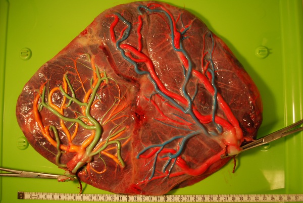
Figure 4. TTTS placenta after fetoscopic laser coagulation using the Solomon technique in which, after identification and coagulation of each individual anastomosis, the complete vascular equator is coagulated from one placental margin to the other.

Figure 5. TAPS placenta in which only a few, minuscule VA anastomoses (white stars) are visible along the vascular equator.

Figure 6. The maternal side of the TAPS placenta (shown in Figure 5) demonstrates the characteristic color difference between both placental shares.

Figure 7. Monochorionic placenta of a twin pregnancy with selective intrauterine growth restriction of one twin. The growth restricted fetus has a velamentous cord insertion and a much smaller placental share. The white star indicates an AA anastomosis.

Figure 8. Monoamniotic placenta: Note the various AV (green star), VA (blue star) and the 2 AA anastomoses (white stars), and the short distance between both cord insertions.
Access restricted. Please log in or start a trial to view this content.
Discussion
TTTS and TAPS are two severe disorders that can occur in monochorionic twins. Both disorders are due to AV anastomoses causing imbalanced intertwin blood flow. In the last 2 decades, fetoscopic laser surgery became available and was shown to be the best method for treatment of TTTS10. The aim of fetoscopic laser treatment is to interrupt the inter-twin circulation through coagulation of the vascular anastomoses on the placental surface. However, the laser treatment for TTTS is far from perfect. Treatment with ...
Access restricted. Please log in or start a trial to view this content.
Disclosures
No conflicts of interest declared.
Acknowledgements
No funding sources.
Access restricted. Please log in or start a trial to view this content.
Materials
| Name | Company | Catalog Number | Comments |
|---|---|---|---|
| Umbilical catheters(various dimensions, Length 40 cm) | Vygon | 1270.08 (8 F) 1270.08 (5 F)1270.04 (4 F)1270.03 (3.5 F)1270.02 (2.5 F) | Any type of catheter can be used |
| 20 ml syringes | BD Biosciences | 300613 | |
| Color dye(different colors) | Royal Talens Schoolverf | 36716010 (blue)36715010 (green)36712350 (yellow)36713570 (pink) | Any viscous color dye can be used |
References
- Lopriore, E., Middeldorp, J. M., Sueters, M., Vandenbussche, F. P., Walther, F. J. Twin-to-twin transfusion syndrome: from placental anastomoses to long-term neurodevelopmental outcome. Curr. Pediatr. Rev. 1, 191-203 (2005).
- Slaghekke, F., Kist, W. J., Oepkes, D. Twin anemia-polycythemia sequence: diagnostic criteria, classification, perinatal management and outcome. Fetal Diagn. Ther. 27, 181-190 (2010).
- Lopriore, E., Middeldorp, J. M., Oepkes, D., Klumper, F. J., Walther, F. J., Vandenbussche, F. P. Residual anastomoses after fetoscopic laser surgery in twin-to-twin transfusion syndrome: frequency, associated risks and outcome. Placenta. 28, 204-208 (2007).
- Lopriore, E., Slaghekke, F., Middeldorp, J. M., Klumper, F. J., Oepkes, D., Vandenbussche, F. P. Residual anastomoses in twin-to-twin transfusion syndrome treated with selective fetoscopic laser surgery: localization, size, and consequences. Am. J. Obstet. Gynecol. , (2009).
- Lewi, L., Jani, J., Cannie, M. Intertwin anastomoses in monochorionic placentas after fetoscopic laser coagulation for twin-to-twin transfusion syndrome: is there more than meets the eye. Am. J. Obstet. Gynecol. 194, 790-795 (2006).
- Denbow, M. L., Cox, P., Taylor, M., Hammal, D. M., Fisk, N. M. Placental angioarchitecture in monochorionic twin pregnancies: relationship to fetal growth, fetofetal transfusion syndrome, and pregnancy outcome. Am. J. Obstet. Gynecol. 182, 417-426 (2000).
- van Meir, H., Slaghekke, F., Lopriore, E., van Wijngaarden, W. J. Arterio-arterial anastomoses do not prevent the development of twin anemia-polycythemia sequence. Placenta. 31, 163-165 (2010).
- De Paepe, M. E., Shapiro, S., Young, L., Luks, F. I. Placental characteristics of selective birth weight discordance in diamniotic-monochorionic twin gestations. Placenta. 31, 380-386 (2010).
- Umur, A., van Gemert, M. J., Nikkels, P. G. Monoamniotic-versus diamniotic-monochorionic twin placentas: anastomoses and twin-twin transfusion syndrome. Am. J. Obstet. Gynecol. 189, 1325-1329 (2003).
- Senat, M. V., Deprest, J., Boulvain, M., Paupe, A., Winer, N., Ville, Y. Endoscopic laser surgery versus serial amnioreduction for severe twin-to-twin transfusion syndrome. N. Engl. J. Med. 351, 136-144 (2004).
- Lopriore, E., Sueters, M., Middeldorp, J. M., Vandenbussche, F. P., Walther, F. J. Haemoglobin differences at birth in monochorionic twins without chronic twin-to-twin transfusion syndrome. Prenat. Diagn. 25, 844-850 (2005).
- Papathanasiou, D., Witlox, R., Oepkes, D., Walther, F. J., Bloemenkamp, K. W., Lopriore, E. Monochorionic twins with ruptured vasa previa: double trouble! Fetal Diagn. Ther. 28, 48-50 (2010).
- Weingertner, A. S., Kohler, A., Kohler, M. Clinical and placental characteristics in four new cases of twin anemia-polycythemia sequence. Ultrasound Obstet. Gynecol. 35, 490-494 (2010).
- Lewi, L., Cannie, M., Blickstein, I. Placental sharing, birthweight discordance, and vascular anastomoses in monochorionic diamniotic twin placentas. Am. J. Obstet. Gynecol. 197, 587-588 (2007).
- Lewi, L., Gucciardo, L., Huber, A. Clinical outcome and placental characteristics of monochorionic diamniotic twin pairs with early- and late-onset discordant growth. Am. J. Obstet. Gynecol. , (2008).
- De Paepe, M. E., Friedman, R. M., Poch, M., Hansen, K., Carr, S. R., Luks, F. I. Placental findings after laser ablation of communicating vessels in twin-to-twin transfusion syndrome. Pediatr. Dev. Pathol. 7, 159-165 (2004).
- De Paepe, M. E., DeKoninck, P., Friedman, R. M. Vascular distribution patterns in monochorionic twin placentas. Placenta. 26, 471-475 (2005).
- Nikkels, P. G., Hack, K. E., vanGemert, M. J. Pathology of twin placentas with special attention to monochorionic twin placentas. J. Clin. Pathol. 61, 1247-1253 (2008).
- Quintero, R. A., Martinez, J. M., Lopez, J. Individual placental territories after selective laser photocoagulation of communicating vessels in twin-twin transfusion syndrome. Am. J. Obstet. Gynecol. 192, 1112-1118 (2005).
- Quintero, R. A., Comas, C., Bornick, P. W., Allen, M. H., Kruger, M. Selective versus non-selective laser photocoagulation of placental vessels in twin-to-twin transfusion syndrome. Ultrasound Obstet. Gynecol. 16, 230-236 (2000).
Access restricted. Please log in or start a trial to view this content.
Reprints and Permissions
