We describe the use of styryl FM dyes to image synaptic vesicle recycling in functional nerve terminals. This protocol can be applied not only to evoked, but also spontaneous and miniature synaptic activities. The protocol expands the variety of synaptic events that can be effectively evaluated.
Method Article
Examination of Synaptic Vesicle Recycling Using FM Dyes During Evoked, Spontaneous, and Miniature Synaptic Activities
In This Article
Summary
Abstract
Synaptic vesicles in functional nerve terminals undergo exocytosis and endocytosis. This synaptic vesicle recycling can be effectively analyzed using styryl FM dyes, which reveal membrane turnover. Conventional protocols for the use of FM dyes were designed for analyzing neurons following stimulated (evoked) synaptic activity. Recently, protocols have become available for analyzing the FM signals that accompany weaker synaptic activities, such as spontaneous or miniature synaptic events. Analysis of these small changes in FM signals requires that the imaging system is sufficiently sensitive to detect small changes in intensity, yet that artifactual changes of large amplitude are suppressed. Here we describe a protocol that can be applied to evoked, spontaneous, and miniature synaptic activities, and use cultured hippocampal neurons as an example. This protocol also incorporates a means of assessing the rate of photobleaching of FM dyes, as this is a significant source of artifacts when imaging small changes in intensity.
Introduction
The functionality of synaptic vesicles is an important determinant of synaptic transmission. These vesicles release neurotransmitters when they fuse with the presynaptic plasma membrane (exocytosis), and they become ready for another cycle of release after being regenerated from the plasma membrane (endocytosis) and reloaded with neurotransmitter. Research into the dynamics of and mechanisms underlying synaptic vesicle recycling has been greatly accelerated by the introduction of styryl FM dyes1. These amphipathic molecules, which have positively charged hydrophilic head groups and hydrophobic tails (multiple dyes in Figure 1A, stereoview of FM1-43 in Figure 1B), can reversibly enter and exit lipid membranes without permeating them. Groups of FM dyes share similar features that influence the range of light that they emit. For example, FM2-10, FM1-43, and FM1-84 have one double bond between two cyclic compounds and show green emission. The difference between them is the length of the hydrophobic tail, which determines its hydrophobicity and therefore the rate of exit from the membrane (departitioning). In the cases of FM5-95 and FM4-64, three double bonds link the cyclic compounds, and they show red emission. These dyes differ with respect to their hydrophilic parts. In all FM dyes, the fluorescence intensity increases when they are inserted into biological membranes, due to an increase in quantum yield in the hydrophobic environment relative to the hydrophilic environment. Thus the changes in FM intensity represent the changes in membrane turnover. The different colors (emission spectra) and hydrophobicities make the FM dyes a versatile research tool in synaptic vesicle recycling.
Based on these features, the FM dyes are mostly used according to the following scheme when analyzing synaptic vesicle recycling (Figure 2). Neurons are bathed in an extracellular solution containing the FM dye, enabling it to be taken up into synaptic vesicles (SVs) as they form via endocytosis (staining). The dye is then washed out by applying a dye-free extracellular solution; this reveals the functional nerve terminals, i.e. only those actively undergoing endocytosis will contain a cluster of synaptic vesicles that are loaded with the dye (Figure 2 bottom). Subsequent exocytosis leads to loss of the FM dye to the extracellular space and a concomitant loss of fluorescence (destaining; due to both the departitioning to a hydrophilic environment and diffusion away from the site of exocytosis). Therefore the changes in FM fluorescence intensity are indicators of synaptic vesicle exo- and endocytosis.
FM dyes have been used to stain and destain the synaptic vesicles in various organisms and preparations2,3. Examples include mammalian neuronal cultures4-9, mammalian brain slices10,11, neuromuscular junctions12,13, retinal bipolar neurons14,15, and hair cells of cochlea16.
Typically in such experiments, both staining and destaining are triggered by extensively stimulating the neurons (evoked activity). Recently, however, synaptic vesicle recycling in response to weak stimulation has also been analyzed, as has recycling in the absence of an external stimulus (spontaneous and miniature synaptic activity)9,17-19. Spontaneous and miniature synaptic activities are defined as those that occur in the absence of external stimuli, with the former involving the spontaneous firing of action potentials (Figure 3). These weak synaptic activities are associated with smaller changes in FM signals than those triggered by extensive stimulation. The measurement requires that the changes in FM fluorescence intensity accurately reflect synaptic vesicle exocytosis or endocytosis but not artifactual changes in intensity. One cause of the artifact is the presence of nonspecific staining of the plasma membrane by FM dyes. Gradual washout of this component will lead to a gradual change in the measured fluorescence intensity, which will be erroneously ascribed to synaptic activities. This factor can be reduced by appropriate methods (see Protocol). The most notable cause of the artifact is the photobleaching of FM dye retained within synaptic vesicles. The photobleaching-related changes in FM intensity must be small in comparison to the biological (synaptic) changes that are measured. The recent development of sensitive cameras, e.g. the electron-multiplying charge-coupled device (EMCCD) camera, makes it possible to minimize photobleaching by shortening exposure time and weakening the intensity of the light used to excite the fluorophore. Another cause of the artifact is a drift in the focusing level of light microscope. The focus drift during an imaging session can be caused by mechanical or thermal effects, and will erroneously lead to a change in the measured fluorescence intensity.
Here we describe protocols and equipment that make it possible to use FM dyes to analyze synaptic vesicle recycling even in the context of weak or no stimulation, in particular, the miniature synaptic activity. We show examples of the staining and destaining of vesicles during evoked and spontaneous synaptic events, using cultured rodent hippocampal neurons, and imaging the destaining phase. We also demonstrate how to evaluate the degree of FM dye photobleaching, in the absence of any FM dye loss due to synaptic activities.
Protocol
1. Primary Culture of Neurons from the Mammalian Brain
All animal procedures performed in this study are approved by the Institutional Animal Care and Use Committee of the University of Iowa.
- Prepare the dissociated cell culture of the CA3-CA1 regions of hippocampus from mice or rats on postnatal days 0-119,20. Plate the hippocampal cells on 12-mm coverslips (thickness number 0) preseeded with the rat glial feeder layer, in 24-well dishes, and at a density of 12,000 cells/well.
- Culture the hippocampal neurons for at least 8 days for functional nerve terminals to develop21. We use neurons on 11-14 days in vitro when individual nerve terminals are relatively isolated without much clustering. Neurons can be used for experiments up to about 21-28 days in vitro e.g. Willig et al.22 and Nosyreva and Kavalali23.
- For functional studies, assess coverslips (before staining) for indicators of healthy neurons using transmitted light microscopy (e.g. phase contrast optics at a magnification of 10-20X). The indicators of good health include: a uniform glial cell layer, a clear cellular margin around the neurons, extended dendrites without beaded structures, a lack of clustered somata, and a lack of bundled neurites.
2. Loading the Synaptic Vesicles with the FM Dye (Staining)
- Staining due to synaptic activity evoked by electrical field stimulation (involves action potentials)
- Prepare staining solution 1 by adding the FM dye to a HEPES-based dye-free solution, e.g. Tyrode's solution (see SOLUTIONS for details). The concentration of FM dye is 10 μM FM1-43 and 2.5 μM FM4-64. Typical concentration ranges are: 2-15 μM FM1-43, or 2.5-20 μM FM4-643,6. For experiments requiring suppression of recurrent glutamatergic synaptic activity and the induction of synaptic plasticity, further add antagonists of ionotropic glutamate receptors: AMPA receptors (e.g. 10 μM CNQX) and NMDA receptors (e.g. 50 μM AP5).
- Transfer a coverslip with cells from the culture medium to the imaging chamber prefilled with plain Tyrode's solution. We use an imaging chamber with the stimulation electrodes attached to the side walls (platinum wires separated by 6.3 mm, RC-21BRFS). This chamber is designed as a closed imaging chamber, but we use it as an open chamber (i.e. without a top coverslip), with a bath volume of ~200 μl. During transfer, do not expose the cultured cells to air, and avoid pinching the cultured cells with the tweezers (pick the coverslip up from the perimeter rather than near the center).
- Perfuse the imaging chamber with plain Tyrode's solution at room temperature (23-25 °C).
- Apply staining solution 1 by constant perfusion.
- Evoke synaptic activity by applying electrical pulses. For maximal staining of the total recycling pool of synaptic vesicles in hippocampal neurons, stimulate the cultures at 10 Hz for 60-120 sec 24. When trying to attain maximal efficiency of field stimulation, keep several features in mind. 1) The height of the bath solution should be as low as possible, while keeping the neurons and stimulating electrodes entirely immersed in a thin layer of Tyrode's solution (in order to keep the neurons alive and to obtain a homogeneous electrical field). 2) Current intensity and duration should be optimized using an indicator of neuronal excitability, e.g. fluorescent Ca2+ imaging; in our case, a constant current of 30 mA intensity and 1 msec duration gives reliable results. 3) The bath solution should be applied at constant flow during stimulation; the electrical stimulation leads to electrolysis, causing protons (H+) to accumulate at the positively charged anode and the pH near the anode to decrease rapidly in a static bath. Check the acidification in a separate experiment by passing more than a few hundred pulses through a solution that contains a pH-indicator (e.g. Hanks’ balanced salt solution with phenol red).
- Leave the neurons in staining solution 1 for an additional 60 sec after field stimulation is terminated, so that post-stimulus endocytosis will be completed25.
- Wash the staining solution out of the imaging chamber by perfusing it with rinsing solution 1, a dye-free Tyrode's solution plus antagonists of AMPA receptors and NMDA receptors (see section 2.1.1), and a blocker of voltage-dependent Na+ channels (e.g. 0.5-1 μM tetrodotoxin, TTX). The combination of blockers will suppress the loss of FM dye through spontaneous synaptic activity. To maximize the washing efficiency, increase the perfusion rate (e.g. up to 5-6 ml/min) for 1-2 min, and add a bolus (e.g. 1 ml) of rinsing solution 1 directly to the open imaging chamber manually, using a pipette. Control the strength and direction of manual infusion, so as not to disturb the cultured neurons.
- Staining due to synaptic activity evoked by high-K+ solution (does not involve action potentials).
- Prepare staining solution 2 by adding the FM dye to high-K+ Tyrode's solution. Specifically, prepare a modified Tyrode's solution with 45 mM KCl, by equimolar substitution of KCl for NaCl. If required, add antagonists of AMPA and NMDA receptors. This high-K+ solution will depolarize neurons continuously, enable Ca2+ influx into nerve terminals, and initiate synaptic vesicle exocytosis to the maximal extent26. Higher concentrations (70-90 mM) were also used in other reports e.g. Klingauf et al.27 and Richards et al.28
- Transfer a coverslip with cultured neurons from the culture medium to the imaging chamber prefilled with plain Tyrode's solution.
- Stain the neurons at room temperature for 1-2 min by applying staining solution 2 at constant perfusion or using a pipette26,28. In the latter case, the final dye concentration should be controlled by adding a known volume of staining solution 2 to a known volume of dye-free solution.
- Wash the staining solution out of the imaging chamber, as described in section 2.1.7.
- Staining due to spontaneous and miniature synaptic activity.
- Prewarm bicarbonate-based solution (e.g. Minimum Essential Medium, MEM) to 37 °C by placing in the culture incubator for more than 60 min.
- For staining based on spontaneous synaptic activity, prepare staining solution 3 by adding the FM dye to MEM. For staining based on miniature synaptic activity, prepare staining solution 4 by adding TTX to the staining solution 3.
- Stain neurons by transferring a coverslip with cultured neurons from culture medium to staining solution 3 or 4, and leaving them in the same staining solution at 37 °C for 10 min.
- Rinse the coverslip briefly in rinsing solution 1.
- Transfer the coverslip to rinsing solution 1 in an imaging chamber. Keep in mind the following. 1) Cells should not be exposed to air; if necessary, transfer the coverslip directly to the imaging chamber without rinsing. 2) Neurons can be stained at room temperature by replacing staining solution 3 or 4 with a HEPES-based solution. 3) Neurons can be stained for evoked synaptic activity in the context of high-K+ solution using this method, by replacing staining solution 3 or 4 with staining solution 2 and carrying out the remaining steps at room temperature.
- Wash the staining solution out of the imaging chamber as described in section 2.1.7.
3. Washing out the FM Dye
- After neuronal staining and initial washing by any of the above-described methods (section 2.1.7), continue to perfuse the imaging chamber with rinsing solution 1 for 5-10 min at room temperature. This will remove the FM dye from the plasma membrane and the extracellular solution. In the case of our imaging chamber, the bath perfusion rate is 600-1,200 μl/min. Washing can also be carried out in the absence of extracellular Ca2+ to suppress FM dye loss due to synaptic activities17. The washes can be enhanced by a brief application of Advasep-7, a modified cyclodextrin that serves as a scavenger of FM dyes from the plasma membrane29-31. It is crucial to keep any Advasep-7 exposure short (e.g. 5 sec in the case of monolayer culture) to avoid both reducing the intensity of FM puncta31 and compromising the health of the cells. The fluorescence from FM1-43 in the plasma membrane can also be suppressed by an application of a quencher sulforhodamine 101 (50-100 μM) that does not enter synaptic vesicles32.
4. Searching for an Optimal Image Field During Washing
- Verify that the cells in the field to be imaged are healthy, using transmitted light microscopy with a high-magnification and high-numerical-aperture objective lens (e.g. 40X) . From this section, we continue to use an inverted microscope (Eclipse Ti, Nikon) equipped with phase contrast or differential interference contrast optics. This microscope is also equipped with Perfect Focus System (PFS) that enables continuous, real-time focus correction, which overcomes the microscope focus drift. This feature is essential for time-lapse imaging during destaining with the same focusing plane.
- Verify that staining was effective, by using fluorescence optics. Avoid aggregates of FM puncta unless they are the target of research, because individual boutons will be difficult to discern. Pay attention to the shapes of the FM puncta: if most of the stained boutons appear as strings of circular beads, they could represent unhealthy nerve terminals. Minimize the exposure of the neurons to strong fluorescence excitation in order to avoid photobleaching.
5. Unloading FM Dye from the Synaptic Vesicles (Destaining)
- Destaining due to synaptic activity evoked by electrical field stimulation.
- Wash TTX out by perfusing the neurons extensively with a rinsing solution 2 (same as rinsing solution 1 but without TTX). Confirm the effectiveness of TTX washout in separate experiments, using the same washing parameters (perfusion rate and duration), e.g. by fluorescent Ca2+ imaging of cytoplasmic Ca2+ transients induced by field stimulation, or by patch-clamp recording of voltage-dependent Na+ currents.
- Start imaging the FM dyes. For imaging FM1-43 or FM4-64, use 520 nm or 650 nm long pass emission filters, respectively and a 490 nm excitation filter. Acquire images every 1-2 sec, using a short exposure time (e.g. 10-20 msec), weak excitation intensity (e.g. 5-10% of LED power), and high sensitivity (e.g. with EM gain). Minimize photobleaching of the FM dyes by reducing exposure time and the intensity of fluorescence excitation. Minimize the microscope focus drift by turning on the Perfect Focus System.
- Stimulate the neurons in rinsing solution 2, using field-stimulation (e.g. at 10 Hz for 120 sec)20.
- Apply multiple rounds of stimulation, with intervening rest periods of 1-2 min, to deplete all the recycling synaptic vesicles of FM dye. This is important when assessing the size of total recycling pool of synaptic vesicles that were stained with FM dyes in the prior state20,33.
- Destaining due to synaptic activities evoked in the absence of action potentials.
- Start imaging the FM dye.
- Apply the stimulating reagents dissolved in rinsing solution 1 or 2. Such reagents include: KCl (high-K+ solution, e.g. 45 mM)26, ionomycin (a Ca2+ ionophore that raises the intracellular Ca2+ concentration by allowing Ca2+ influx into the cell from the extracellular solution, used e.g. at 5 μM)20,34, and sucrose (a hypertonic solution that stimulates the release of synaptic vesicles from readily releasable pool, used e.g. at 500 mM)35,36. Use a fast, local perfusion system for a reliable temporal control in applying the reagents, e.g. the SF-77B system from of Warner Instruments37, or Y-tube system38-40.
- Destaining due to spontaneous and miniature synaptic activity.
- For destaining due to spontaneous synaptic activity, wash out TTX as above (section 5.1.1), using rinsing solution 2. Start imaging the FM dyes while perfusing neurons with rinsing solution 2. For destaining due to miniature synaptic activities, start imaging the FM dyes, while continuing to perfuse the neurons with rinsing solution 1 (with TTX).
- After destaining based on either spontaneous or miniature activity, identify the functional nerve terminals by destaining via evoked synaptic activity. Activity can be evoked by any of the procedures described above. This identification process is necessary because the stained structures can be those other than the functional nerve terminals (e.g. slowly recycling endosomes or astrocytic vesicles) and a large amount of destaining by the evoked activity aids in this process.
6. Assessing the Photobleaching Rate of FM Dyes
- Stain the nerve terminals with aldehyde-fixable FM dye (e.g. FM1-43FX, FM4-64FX). This can done using the evoked, spontaneous, or miniature staining method described above, and by replacing FM dye with a fixable FM dye.
- Wash out the fixable FM dye as described in section 2.1.7.
- Chemically fix the neurons by transferring the coverslip to fixative solution: 4% paraformaldehyde and 4% sucrose in Tyrode's solution for 30 min at 4 °C. The fluorescence intensity of the fixable FM dye will be retained after chemical fixation.
- Wash out the fixative for 10 min in Tyrode's solution, by transferring the coverslip to fresh Tyrode's solution twice.
- Transfer the coverslip into fresh Tyrode's solution in the imaging chamber.
- Start imaging the fixable FM dye using the same set of imaging parameters as for live neurons. It is important to rinse the imaging chamber very well after using any chemically fixed specimen, because any remaining fixative can affect subsequent live-cell imaging experiments carried out in the same chamber. Alternatively to the fixed cell imaging, the stained live cells might be imaged for assessing the photobleaching rate. However, it is worth keeping in mind that it is difficult to block the miniature synaptic activity acutely; for example, the frequency of miniature activity can be reduced to ~50% by removing the extracellular Ca2+ 23, but it is not a complete blockade.
- At the end of each imaging session, acquire a background image for assessing the absolute value of the FM fluorescence intensity. The intensity of FM puncta represents not only the true signal from the FM dyes in nerve terminals, but also the background noise from 1) glial cells that underlie the neuronal monolayer culture, 2) the coverslip and optical components (e.g. objective lens, filters and mirrors), and 3) the detector (EMCCD camera). Background noise is assessed in nonstained regions of the imaged field. When such regions are limited in space or heterogeneous in intensity, substitute it with an image with the camera shutter closed and acquire noise from the detector, although it is not an ideal method. Acquire approximately 10 images using the same imaging parameters.
7. Image Analysis
- Import the acquired data into the image-analysis software as a stack of time-series images. We use ImageJ (W. S. Rasband, NIH) and the associated plug-ins, e.g. Image Stabilizer plug-in (Kang Li) for correcting a small degree of movement among FM puncta, and Time Series Analyzer V2.0 (Balaji Jayaprakash) for analyzing the time series.
- Calculate the changes in FM intensity at the start and end of the series (ΔFM). To this end, average the images before the start of destaining and after the end of destaining (e.g. 5 image frames each), and generate a difference image by subtracting them.
- Identify the potentially functional nerve terminals by applying an intensity threshold to the difference image, and detect pixels whose intensities are higher than the threshold. One method of setting the threshold is to choose the standard deviation of the background intensity obtained from the bare coverslip area.
- Identify the isolated, functional nerve terminals as the contiguous pixels that were detected and that satisfy a size criterion (minimal and maximal numbers of pixels in contiguity). This process eliminates background noise and aggregated nerve terminals from analysis.
- Assign regions-of-interest (ROIs) on the detected pixel clusters (with individual clusters corresponding to individual nerve terminals). The total change in intensity during destaining (ΔFM) is a typical parameter of analysis. This represents the cumulative amount of evoked, spontaneous or miniature synaptic activity. Exclude ROIs if they exhibit any of the following changes in intensity depending on purposes of experiments: an increase during recording, a sudden decrease before stimulation, or a long latency after stimulation.
- Assess the rate of photobleaching by importing the acquired time-series data into the image-analysis software. Average the background images (e.g. with a closed shutter), and subtract it from the individual images. Assign ROIs to FM puncta that putatively correspond to nerve terminals, and measure changes in ROI intensity over time.
Solutions
- Tyrode's solution
- Composition (in mM): 125 NaCl, 2 KCl, 2 CaCl2, 2 MgCl2, 30 glucose, 25 HEPES, 310 mOsm, pH 7.4.
- Staining solution 1
- Tyrode's solution plus the FM dye.
- This solution is used for the staining, based on evoked synaptic activity by electrical stimulation.
- Add the antagonists of AMPA receptors and NMDA receptors if necessary.
- Staining solution 2
- High-K+ Tyrode's solution plus the FM dye.
- This modified Tyrode's solution is prepared by increasing the KCl concentration to 45 mM, by equimolar substitution of KCl for NaCl.
- This solution is used for the staining, based on evoked synaptic activity by continuous depolarization.
- Add the antagonists of AMPA receptors and NMDA receptors if necessary.
- Staining solution 3
- MEM solution plus the FM dye.
- This solution is used for the staining, based on spontaneous synaptic activity.
- Staining solution 4
- MEM solution plus the FM dye and TTX.
- This solution is used for the staining, based on miniature synaptic activity.
- Rinsing solution 1
- Tyrode's solution plus the antagonists of AMPA receptors and NMDA receptors, and TTX.
- Rinsing solution 2
- Tyrode's solution plus the antagonists of AMPA receptors and NMDA receptors, but without TTX.
Results
As an example, we show representative results for the destaining time course of synaptic vesicles (Figure 4). Cultured hippocampal neurons were stained with FM4-64 using the spontaneous synaptic activity (step 2.3) and washed with dye-free solution (rinsing solution 2). The imaging shows the initial destaining time course using spontaneous activity (step 5.3) (initial part of continuous line, Figure 4A). This is followed by the destaining time course using three rounds of evoked activity with 10 Hz field stimulation for 120 sec each (step 5.1). One way of measuring the amount of evoked destaining is illustrated with a double-ended arrow (ΔFMEvoked) that corresponds to the size of total recycling pool of vesicles.
Before the first stimulus was given, there was a gradual decrease in FM intensity (enlarged in Figure 4B). This decrease was composed of destaining due to spontaneous synaptic activity (ΔFMSpont) and photobleaching (ΔFMPB). When the contribution of photobleaching is small, the change from the preimaging baseline (ΔFMSpont + ΔFMPB) may be used as an approximation to the destaining amount of spontaneous activity.
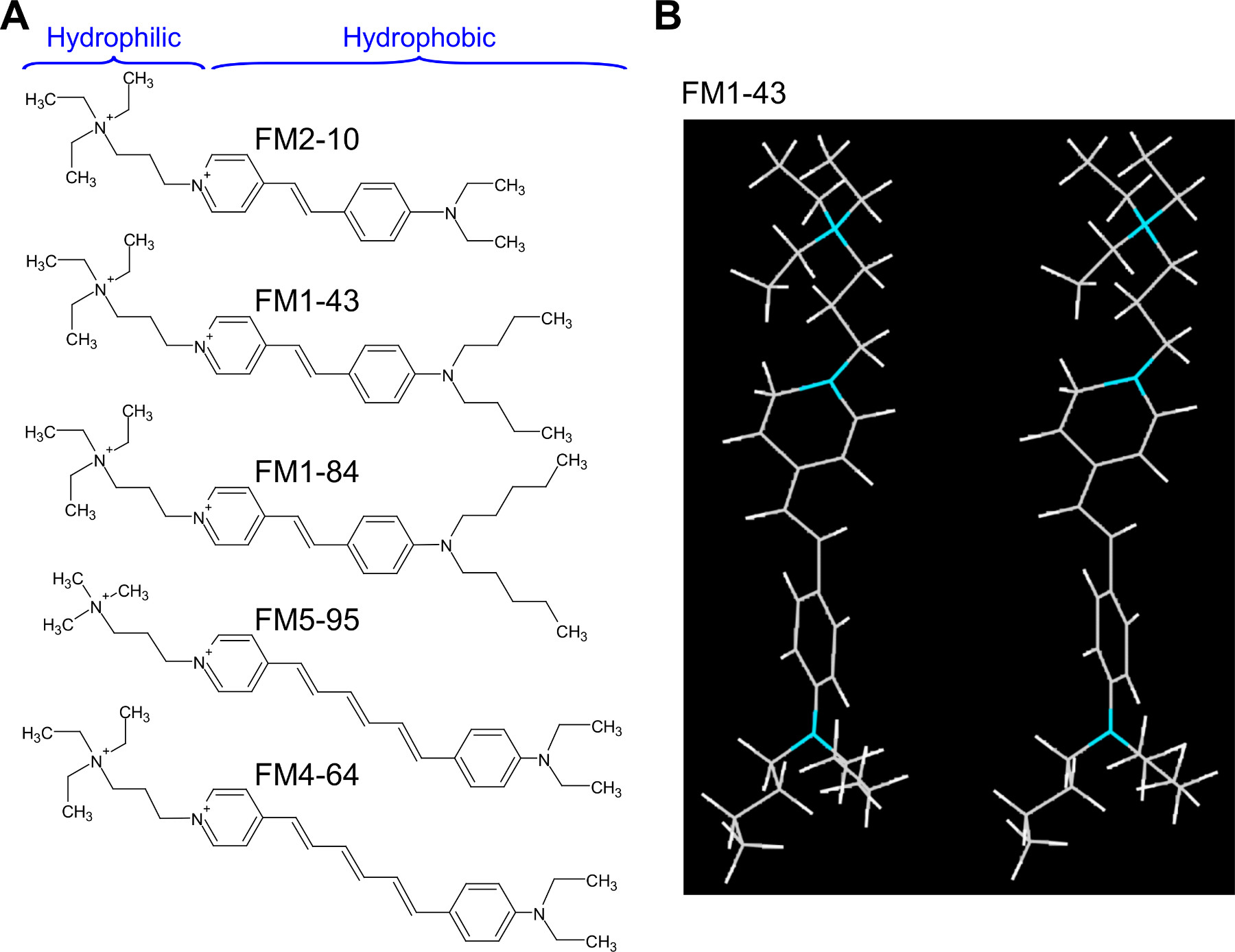
Figure 1. Structures of FM dyes. A. FM dyes shares some common structural features. The hydrophilic groups stay in aqueous solution and the hydrophobic tails allow the FM dyes to be partitioned into membrane. FM2-10, FM1-43 and FM1-84 show green emission, while FM5-95 and FM4-64 show red-shifted emission. B. Stereoview of FM1-43, the most commonly used FM dye. Hydrophilic group is facing up and hydrophobic tails are facing down. Nitrogen atoms are colored blue. For another view of FM1-43, see Schote and Seelig63.
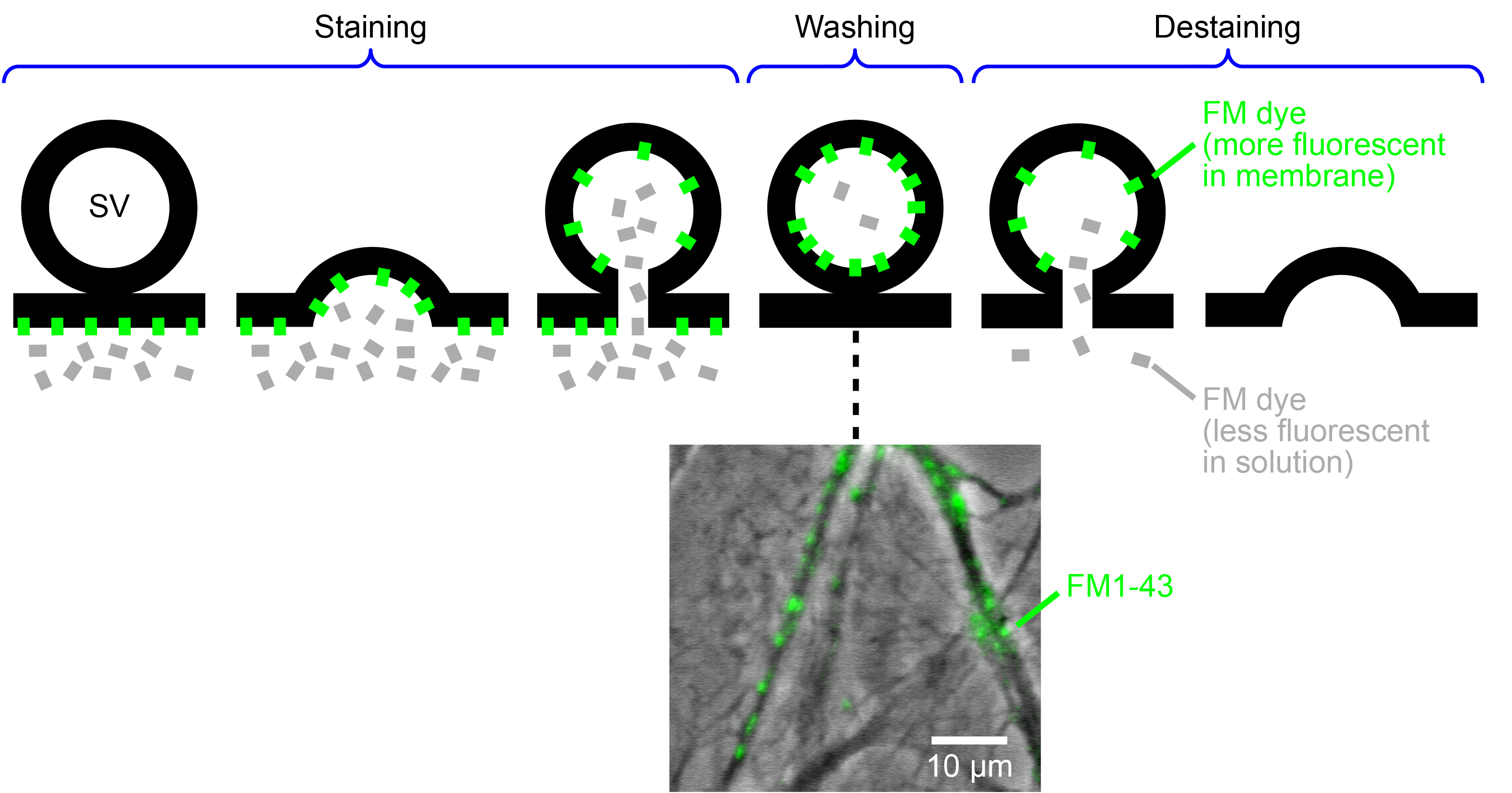
Figure 2. Basic scheme of FM dye usage. After application to neurons, the FM dye inserted into the plasma membrane becomes fluorescent (green), whereas that in the aqueous solution is much less fluorescent (gray). A synaptic vesicle (SV) is loaded (stained) by the FM dye, when it undergoes endocytosis, typically following exocytosis. Washing out the FM dye in extracellular solution enables only the stained vesicles to be fluorescent. Subsequently, a synaptic vesicle is unloaded (destained) when it undergoes exocytosis and therefore releases the FM dye. An image below illustrates an exemplar staining of cultured hippocampal neurons with FM1-43 (an overlay of fluorescence and phase contrast images). It is of note that, in the protocol described in this paper, the fluorescent puncta represent presynaptic nerve terminals (diameters ~1 μm in typical central neurons) with clusters of stained synaptic vesicles, not the individual synaptic vesicles (diameters ~40 nm). For simplicity, this scheme represents a general idea of exo-endocytosis of synaptic vesicles. It can include multiple forms of exo-endocytosis, such as the full-collapse fusion followed by clathrin-mediated endocytosis, the transient kiss-and-run exo-endocytosis, and the bulk endocytosis64.
Please click here to view a larger version of this figure.
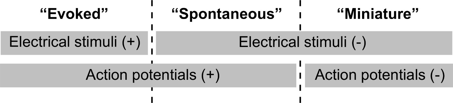
Figure 3. Three types of synaptic activities studied by FM imaging. Evoked synaptic activity typically requires external, electrical stimuli. Spontaneous synaptic activity occurs in the absence of electrical stimuli. Miniature synaptic activity occurs spontaneously without electrical stimuli and without action potentials: usually the action potential generation is suppressed by a blocker of voltage-dependent Na+ channel, tetrodotoxin. Additional activities include the evoked activity when exocytosis is stimulated by high-K+ solution (continuous depolarization), ionomycin (continuous increase in cytoplasmic Ca2+ concentration), and hypertonic solution containing sucrose (eliciting exocytosis of vesicles in the readily releasable pool), all of which will not require action potential firing for synaptic vesicles to undergo exocytosis. Note that, in some studies, the spontaneous activity is broadly defined to encompass miniature activity as well.

Figure 4. Representative result of destaining. A. Cultured hippocampal neurons were stained by spontaneous activity with 2.5 μM FM4-64 for 10 min at 37 °C (step 2.3), and washed (Protocol 3). Neurons were imaged during destaining with spontaneous activity (step 5.3), and three rounds of evoked activities (step 5.1) (continuous curve, average of n = 25 nerve terminals). Vertical bars represent SEM. Y-axis represents the absolute FM intensity and “0” represents the intensity when a camera shutter is closed. The total amount of evoked destaining is indicated (ΔFMEvoked). B. An expanded view of initial phase of destaining in panel A (continuous curve, SEM bars omitted for clarity). During a 60 sec observation, the detaining was mainly due to spontaneous activity (ΔFMSpont) but was partially due to photobleaching (ΔFMPB). The photobleaching time course of FM4-64 (thick dotted curve) was determined in a separate experiment by imaging fixable FM4-64 (Protocol 6). The rate was 2-3% over 2 min (a single exponential function with a time constant of 5,736 sec, determined by curve fitting over 9 min)19. The photobleaching curve was drawn as an exponential function with an initial value equivalent to that of the measured intensity of FM4-64. See supplementary Figure S5 in Kakazuet al.19 for the photobleaching over the longer period (9 min), and with variable excitation intensity and exposure time. Please click here to view a larger version of this figure.
Discussion
We have described protocols for staining and destaining synaptic vesicles in response to evoked, spontaneous and miniature synaptic activity, and for imaging during the destaining phase. In addition to the existing protocols, we have included a new protocol of observing the FM destaining based on miniature synaptic activity. Using these protocols, we previously identified abnormalities in cultured neurons from a mouse model of the movement disorder dystonia. In comparison to their counterparts in wild-type mice, those neurons underwent accelerated synaptic vesicle exocytosis in a Ca2+-dependent manner when stimulated by high activity20. Those neurons also showed more frequent miniature synaptic activity, as confirmed by patch-clamp electrophysiological recordings of neurotransmitter release19.
The critical aspect of the protocol for using FM dyes in such analyses is to assess and minimize photobleaching. Subtle changes in the FM fluorescence intensity can be reliably assessed if the changes caused by photobleaching are small in comparison to those triggered by synaptic activity. Reduced photobleaching also has the potential to suppress or eliminate cytotoxicity. Photobleaching can be reduced by minimizing the exposure of fluorophores to excitation light, and there are at least two components of equipment to do this in live-imaging experiments. One important component is a sensitive detector of photons, e.g. an EMCCD camera. This makes it possible to minimize the duration and intensity of the exposure without negatively affecting the detection of fluorescence emission. An associated component is the light source system that limits the exposure of the specimen to excitation light only when the detector acquires images. This is easily achieved by an LED light41 that allows for efficient control of the timing of light exposure (ON / OFF takes much less than 1 msec). The exposure can be triggered only during image capture, by digital output from the camera (e.g. "Fire" terminal in Andor camera). Additional advantages of using the LED include: the ability to control light intensity without using neutral density filters, long-term stability of the light intensity, and the absence of mechanical vibrations which would interfere with precise handling of glass pipettes as in patch-clamp recording.
In general, structurally different dyes photobleach at different rates under the same imaging conditions. It would thus be ideal to evaluate the extent of photobleaching of the fluorophore used in a particular experiment. For FM dyes in live nerve terminals, it is technically challenging to assess the photobleaching rate independent of synaptic activity, due to spontaneous or miniature synaptic activity. A decrease in fluorescence intensity during such activity could be due to the biological loss of FM dyes (exocytosis), photobleaching of FM dyes, or both. Fortunately, the structures of the fixable FM dyes are designed to be almost identical to those of the nonfixable FM dyes. In the protocol described here, fixable FM dyes were loaded into synaptic vesicles by the same methods as nonfixable FM dyes, whereas the synaptic activity was blocked afterwards by chemical fixation of the specimen. Notably, the rate of photobleaching measured using this system was as low as 2-3% over 2 min when the imaging conditions were the same as those for live-cell imaging19.
FM dyes can be used with different protocols to explore diverse aspects of synaptic vesicle recycling. Different synaptic activities during staining and destaining can be combined in various ways, depending on the experimental aims and specific features of synaptic vesicle recycling to be assessed. The selection of antagonists also depends on the purpose of the experiments. Furthermore, FM imaging can be carried out during the staining phase as well as during the destaining phase9,18,42. It should also be borne in mind, however, that FM dyes can have unexpected effects, such as blocking the muscarinic acetylcholine receptors43, and permeating the mechanotransducer channels44, store-operated Ca2+ channels45, and ATP receptors46. High concentrations of FM dyes can potentially modify the efficiency of synaptic vesicle exocytosis itself47. Thus we recommend caution in designing the experiments and interpreting the results regarding synaptic vesicle recycling. Complementary methods to consider will include uptake into synaptic vesicles of antibodies whose epitopes are intra-lumenal domains of vesicle proteins22,48. They also include expressing pH-sensitive GFP variants targeted to vesicle lumen49-51, and uptake of pH-sensitive antibody conjugates52-54 both of which detect the intra-vesicular pH changes accompanying exo-endocytosis.
Once such unwanted effects are excluded, FM dyes have wide applications. For example, they can be used to address whether the same synaptic vesicle pools are shared for spontaneous and evoked vesicle releases17,55, to what extent the efficiency of synaptic vesicle recycling can be regulated56,57, and what effects does a prior state (rest or stimulated) exert on the later state of vesicle recycling based on, e.g. spontaneous and miniature synaptic activity. FM dyes can also be used to evaluate synaptic vesicle recycling at the ultrastructural level, by correlating observations from light and electron microscopy by the FM photoconversion method30,58-62. FM dye can be used to simultaneously monitor synaptic functions and intracellular Ca2+ concentration5. In addition to the labeling of synaptic vesicles, the FM dyes and other fluorescent fluid-phase markers such as fluorescent dextran7 can be used to monitor the bulk endocytosis that is triggered by intense neuronal activity. In conclusion, the applications of FM dyes provide an invaluable source of information regarding synaptic vesicle recycling and additional synaptic functions.
Disclosures
The authors declare no competing financial interests.
Acknowledgements
The authors thank the members of the Harata lab for helpful discussions throughout the execution of this work. This work was funded by grants from the American Heart Association, the Dystonia Medical Research Foundation, the Edward Mallinckrodt, Jr. Foundation, the National Science Foundation, and the Whitehall Foundation to N.C.H.
Materials
| Name | Company | Catalog Number | Comments |
|---|---|---|---|
| Pulse generator | AMPI | Master-8 | |
| Isolated stimulator |  Digitimer Digitimer | DS3 | |
| Inverted microscope | Nikon | Eclipse TS100 | This is used for assessing the cell morphology at low magnification. |
| Inverted microscope | Nikon | Eclipse TiE | This is used for high-resolution fluorescence and transmitted light imaging, with minimal focus drift. |
| Objective lens | Nikon | Water-immersion lens is recommended. Oil-immersion lens is usable unless an imaged structure is deep from the coverslip surface (e.g. >10 μm). | |
| Filter cube | Nikon | 77032509 | 490/20-nm ex, 510-nm dclp, 520-nm-LP em for FM1-43 |
| Filter cube | Nikon | 77032809 | 490/420 nm ex, 510 nm dclp, 650 nm LP em for FM4-64. |
| EMCCD camera | Andor Technology | iXon EM+ DU-860 | This EMCCD camera is used for high-sensitivity detection of fluorescence. |
| Liquid recirculating chiller | Solid State Cooling Systems | Oasis 160 | This is used for continuously perfusing the camera with chilled water for maintaining a temperature of -80°C, and thereby reducing noise. |
| LED | CoolLED-Custom Interconnect | 490 nm | This light source is used for rapid on/off control of fluorescence excitation. |
| Image acquisition software | Andor Technology | Solis | |
| Imaging chamber | Warner Instruments | RC-21BRFS | |
| Fast perfusion system | Warner Instruments | SF-77B | |
| CNQX | Tocris Bioscience | 1045 | |
| D,L-AP5 | Tocris Bioscience | 0106 | |
| Tetrodotoxin | Tocris Bioscience | 1069 | Caution: toxic reagent. Handle with care. |
| FM1-43 | Invitrogen | T35356 | |
| Aldehyde-fixable FM1-43 (FM1-43FX) | Invitrogen | F35355 | |
| FM4-64 | Invitrogen | T13320 | |
| Aldehyde-fixable FM4-64 (FM4-64FX) | Invitrogen | F34653 | |
| Ionomycin | Sigma-Aldrich | I0634 | |
| Hanks’ balanced salt | Sigma-Aldrich | H2387 | |
| Minimum Essential Medium | Invitrogen | 51200-038 | This solution does not contain phenol red that will interfere with fluorescence imaging. |
| Paraformaldehyde | Electron Microscopy Sciences | 15710 | Caution: toxic reagent. Handle with care. |
| Sucrose | Sigma-Aldrich | S7903 |
References
- Betz, W. J., Bewick, G. S. Optical analysis of synaptic vesicle recycling at the frog neuromuscular junction. Science. 255, 200-203 (1992).
- Gaffield, M. A., Betz, W. J. Imaging synaptic vesicle exocytosis and endocytosis with FM dyes. Nat. Protoc. 1, 2916-2921 (2006).
- Hoopmann, P., Rizzoli, S. O., Betz, W. J. Imaging synaptic vesicle recycling by staining and destaining vesicles with FM dyes. , Cold Spring Harb Protoc. 77-83 (2012).
- Newton, J., Murthy, V. Measuring exocytosis in neurons using FM labeling. J. Vis. Exp. , (2006).
- Evans, G. J., Cousin, M. A. Simultaneous monitoring of three key neuronal functions in primary neuronal cultures. J. Neurosci. Methods. 160, 197-205 (2007).
- Cousin, M. A. Use of FM1-43 and other derivatives to investigate neuronal function. Curr. Protoc. Neurosci. 2, (2008).
- Clayton, E. L., Cousin, M. A. Quantitative monitoring of activity-dependent bulk endocytosis of synaptic vesicle membrane by fluorescent dextran imaging. J. Neurosci. Methods. 185, 76-81 (2009).
- Cheung, G., Cousin, M. A. Quantitative analysis of synaptic vesicle pool replenishment in cultured cerebellar granule neurons using FM dyes. J. Vis. Exp. , (2011).
- Welzel, O., Tischbirek, C. H., Kornhuber, J., Groemer, T. W. Pool-independent labelling of synaptic vesicle exocytosis with single vesicle resolution in rat hippocampal neurons. J. Neurosci. Methods. 205, 258-264 (2012).
- Winterer, J., Stanton, P. K., Muller, W. Direct monitoring of vesicular release and uptake in brain slices by multiphoton excitation of the styryl FM 1-43. Biotechniques. 40, 343-351 (2006).
- Kay, A. R. Imaging FM dyes in brain slices. Cold Spring Harb. Protoc. , (2007).
- Verstreken, P., Ohyama, T., Bellen, H. J. FM 1-43 labeling of synaptic vesicle pools at the Drosophila neuromuscular junction. Methods Mol. Biol. 440, 349-369 (2008).
- Amaral, E., Guatimosim, S., Guatimosim, C. Using the fluorescent styryl dye FM1-43 to visualize synaptic vesicles exocytosis and endocytosis in motor nerve terminals. Methods Mol. Biol. 689, 137-148 (2011).
- Guatimosim, C., von Gersdorff, H. Optical monitoring of synaptic vesicle trafficking in ribbon synapses. Neurochem. Int. 41, 307-312 (2002).
- Joselevitch, C., Zenisek, D. Imaging exocytosis in retinal bipolar cells with TIRF microscopy. J. Vis. Exp. , 1305(2009).
- Griesinger, C. B., Richards, C. D., Ashmore, J. F. FM1-43 reveals membrane recycling in adult inner hair cells of the mammalian cochlea. J. Neurosci. 22, 3939-3952 (2002).
- Sara, Y., Virmani, T., Deak, F., Liu, X. An isolated pool of vesicles recycles at rest and drives spontaneous neurotransmission. Neuron. 45, 563-573 (2005).
- Wilhelm, B. G., Groemer, T. W., Rizzoli, S. O. The same synaptic vesicles drive active and spontaneous release. Nat. Neurosci. 13, 1454-1456 (2010).
- Kakazu, Y., Koh, J. Y., Iwabuchi, S., Gonzalez-Alegre, P., Harata, N. C. Miniature release events of glutamate from hippocampal neurons are influenced by the dystonia-associated protein torsinA. Synapse. 66, 807-822 (2012).
- Kakazu, Y., Koh, J. Y., Ho, K. W., Gonzalez-Alegre, P., Harata, N. C. Synaptic vesicle recycling is enhanced by torsinA that harbors the DYT1 dystonia mutation. Synapse. 66, 453-464 (2012).
- Kavalali, E. T., Klingauf, J., Tsien, R. W. Activity-dependent regulation of synaptic clustering in a hippocampal culture system. Proc. Natl. Acad. Sci. U.S.A. 96, 12893-12900 (1999).
- Willig, K. I., Rizzoli, S. O., Westphal, V., Jahn, R., Hell, S. W. STED microscopy reveals that synaptotagmin remains clustered after synaptic vesicle exocytosis. Nature. 440, 935-939 (2006).
- Nosyreva, E., Kavalali, E. T. Activity-dependent augmentation of spontaneous neurotransmission during endoplasmic reticulum stress. J. Neurosci. 30, 7358-7368 (2010).
- Ryan, T. A., Smith, S. J. Vesicle pool mobilization during action potential firing at hippocampal synapses. Neuron. 14, 983-989 (1995).
- Ryan, T. A., Smith, S. J., Reuter, H. The timing of synaptic vesicle endocytosis. Proc. Natl. Acad. Sci. U.S.A. 93, 5567-5571 (1996).
- Harata, N., et al. Limited numbers of recycling vesicles in small CNS nerve terminals: implications for neural signaling and vesicular cycling. Trends Neurosci. 24, 637-643 (2001).
- Klingauf, J., Kavalali, E. T., Tsien, R. W. Kinetics and regulation of fast endocytosis at hippocampal synapses. Nature. 394, 581-585 (1998).
- Richards, D. A., Bai, J., Chapman, E. R. Two modes of exocytosis at hippocampal synapses revealed by rate of FM1-43 efflux from individual vesicles. J. Cell Biol. 168, 929-939 (2005).
- Kay, A. R., et al. Imaging synaptic activity in intact brain and slices with FM1-43 in C. elegans, lamprey, and rat. Neuron. 24, 809-817 (1999).
- Darcy, K. J., Staras, K., Collinson, L. M., Goda, Y. An ultrastructural readout of fluorescence recovery after photobleaching using correlative light and electron. 1, 988-994 (2006).
- Moulder, K. L., Jiang, X., Taylor, A. A., Benz, A. M., Mennerick, S. Presynaptically silent synapses studied with light microscopy. J. Vis. Exp. , (2010).
- Pyle, J. L., Kavalali, E. T., Choi, S., Tsien, R. W. Visualization of synaptic activity in hippocampal slices with FM1-43 enabled by fluorescence quenching. Neuron. 24, 803-808 (1999).
- Harata, N. C., Choi, S., Pyle, J. L., Aravanis, A. M., Tsien, R. W. Frequency-dependent kinetics and prevalence of kiss-and-run and reuse at hippocampal synapses studied with novel quenching methods. Neuron. 49, 243-256 (2006).
- Piedras-Renteria, E. S., et al. Presynaptic homeostasis at CNS nerve terminals compensates for lack of a key Ca2+ entry pathway. Proc. Natl. Acad. Sci. U.S.A. 101, 3609-3614 (2004).
- Rosenmund, C., Stevens, C. F. Definition of the readily releasable pool of vesicles at hippocampal synapses. Neuron. 16, 1197-1207 (1996).
- Pyle, J. L., Kavalali, E. T., Piedras-Renteria, E. S., Tsien, R. W. Rapid reuse of readily releasable pool vesicles at hippocampal synapses. Neuron. 28, 221-231 (2000).
- Kawano, H., et al. Long-term culture of astrocytes attenuates the readily releasable pool of synaptic vesicles. PLoS ONE. 8, (2012).
- Harata, N., Katayama, J., Takeshita, Y., Murai, Y., Akaike, N. Two components of metabotropic glutamate responses in acutely dissociated CA3 pyramidal neurons of the rat. Brain Res. 711, 223-233 (1996).
- Kira, T., Harata, N., Sakata, T., Akaike, N. Kinetics of sevoflurane action on GABA- and glycine-induced currents in acutely dissociated rat hippocampal neurons. Neuroscience. 85, 383-394 (1998).
- Harata, N., Katayama, J., Akaike, N. Excitatory amino acid responses in relay neurons of the rat lateral geniculate nucleus. Neuroscience. 89, 109-125 (1999).
- Albeanu, D. F., Soucy, E., Sato, T. F., Meister, M., Murthy, V. N. LED arrays as cost effective and efficient light sources for widefield microscopy. PLoS ONE. 3, e2146(2008).
- Neves, G., Lagnado, L. The kinetics of exocytosis and endocytosis in the synaptic terminal of goldfish retinal bipolar cells. J. Physiol. 515, 181-202 (1999).
- Mazzone, S. B., et al. Fluorescent styryl dyes FM1-43 and FM2-10 are muscarinic receptor antagonists: intravital visualization of receptor occupancy. J. Physiol. 575, 23-35 (2006).
- Gale, J. E., Marcotti, W., Kennedy, H. J., Kros, C. J., Richardson, G. P. FM1-43 dye behaves as a permeant blocker of the hair-cell mechanotransducer channel. J. Neurosci. 21, 7013-7025 (2001).
- Li, D., Herault, K., Oheim, M., Ropert, N. FM dyes enter via a store-operated calcium channel and modify calcium signaling of cultured astrocytes. Proc. Natl. Acad. Sci. U.S.A. 106, 21960-21965 (2009).
- Crumling, M. A., et al. P2X antagonists inhibit styryl dye entry into hair cells. Neuroscience. 161, 1144-1153 (2009).
- Zhu, Y., Stevens, C. F. Probing synaptic vesicle fusion by altering mechanical properties of the neuronal surface membrane. Proc. Natl. Acad. Sci. U.S.A. 105, 18018-18022 (2008).
- Thiagarajan, T. C., Lindskog, M., Tsien, R. W. Adaptation to synaptic inactivity in hippocampal neurons. Neuron. 47, 725-737 (2005).
- Miesenböck, G., De Angelis, D. A., Rothman, J. E. Visualizing secretion and synaptic transmission with pH-sensitive green fluorescent proteins. Nature. 394, 192-195 (1998).
- Li, H., et al. Concurrent imaging of synaptic vesicle recycling and calcium dynamics. Front. Mol. Neurosci. 4, 34(2011).
- Raingo, J., et al. VAMP4 directs synaptic vesicles to a pool that selectively maintains asynchronous neurotransmission. Nat. Neurosci. 15, 738-745 (2012).
- Martens, H., et al. Unique luminal localization of VGAT-C terminus allows for selective labeling of active cortical GABAergic synapses. J. Neurosci. 28, 13125-13131 (2008).
- Hua, Y., Sinha, R., Martineau, M., Kahms, M., Klingauf, J. A common origin of synaptic vesicles undergoing evoked and spontaneous fusion. Nat. Neurosci. 13, 1451-1453 (2010).
- Hua, Y., et al. A readily retrievable pool of synaptic vesicles. Nat. Neurosci. 14, 833-839 (2011).
- Groemer, T. W., Klingauf, J. Synaptic vesicles recycling spontaneously and during activity belong to the same vesicle. 10, 145-147 (2007).
- Moulder, K. L., et al. A specific role for Ca2+-dependent adenylyl cyclases in recovery from adaptive presynaptic silencing. J. Neurosci. 28, 5159-5168 (2008).
- Crawford, D. C., Jiang, X., Taylor, A., Moulder, K. L., Mennerick, S. Differential requirement for protein synthesis in presynaptic unmuting and muting in hippocampal glutamate terminals. PLoS ONE. 7, (2012).
- Henkel, A. W., Lubke, J., Betz, W. J. FM1-43 dye ultrastructural localization in and release from frog motor nerve terminals. Proc. Natl. Acad. Sci. U.S.A. 93, 1918-1923 (1996).
- Harata, N., Ryan, T. A., Smith, S. J., Buchanan, J., Tsien, R. W. Visualizing recycling synaptic vesicles in hippocampal neurons by FM 1- 43 photoconversion. Proc. Natl. Acad. Sci. U.S.A. 98, 12748-12753 (2001).
- Opazo, F., Rizzoli, S. O. Studying synaptic vesicle pools using photoconversion of styryl dyes. J. Vis. Exp. , (2010).
- Schikorski, T. Monitoring rapid endocytosis in the electron microscope via photoconversion of vesicles fluorescently labeled with FM1-43. Methods Mol. Biol. 657, 329-346 (2010).
- Hoopmann, P., Rizzoli, S. O., Betz, W. J. FM dye photoconversion for visualizing synaptic vesicles by electron microscopy. Cold Spring Harb. Protoc. 2012, 84-86 (2012).
- Schote, U., Seelig, J. Interaction of the neuronal marker dye FM1-43 with lipid membranes. Thermodynamics and lipid ordering. Biochim. Biophys. Acta. 1415, 135-146 (1998).
- Harata, N. C., Aravanis, A. M., Tsien, R. W. Kiss-and-run and full-collapse fusion as modes of exo-endocytosis in neurosecretion. J. Neurochem. 97, 1546-1570 (2006).
Reprints and Permissions
