The Tomato/GFP-FLP/FRT method involves visualizing mosaic photoreceptor cells in living Drosophila. It can be used to follow individual photoreceptor cell fates in the retina for days or weeks. This method is ideal for studies of retinal degeneration and neurodegenerative diseases or photoreceptor cell development.
Method Article
The Tomato/GFP-FLP/FRT Method for Live Imaging of Mosaic Adult Drosophila Photoreceptor Cells
In This Article
Summary
Abstract
The Drosophila eye is widely used as a model for studies of development and neuronal degeneration. With the powerful mitotic recombination technique, elegant genetic screens based on clonal analysis have led to the identification of signaling pathways involved in eye development and photoreceptor (PR) differentiation at larval stages. We describe here the Tomato/GFP-FLP/FRT method, which can be used for rapid clonal analysis in the eye of living adult Drosophila. Fluorescent photoreceptor cells are imaged with the cornea neutralization technique, on retinas with mosaic clones generated by flipase-mediated recombination. This method has several major advantages over classical histological sectioning of the retina: it can be used for high-throughput screening and has proved an effective method for identifying the factors regulating PR survival and function. It can be used for kinetic analyses of PR degeneration in the same living animal over several weeks, to demonstrate the requirement for specific genes for PR survival or function in the adult fly. This method is also useful for addressing cell autonomy issues in developmental mutants, such as those in which the establishment of planar cell polarity is affected.
Introduction
The Drosophila retina is composed of about 800 ommatidial units (Figure 1A), which are precisely organized, with a clearly defined axis of polarity (Figure 1B). Each unit contains 20 cells: eight photoreceptor cells (PRs) and 12 accessory cells, including cone cells, pigment cells and bristle cells 1,2. Two classes of PR are distinguished on the basis of the type of rhodopsin (Rh) they express. The six outer PRs (R1-6) express Rh1 and are arranged in a trapezoid pattern (Figure 1C). In the center of the trapezoid, the two inner PRs (R7/R8) express four possible types of rhodopsin (Rh3, Rh4, Rh5 or Rh6) and are organized such that R7 lies on top of R8 3.
More than 2,500 genes are involved in the morphogenesis of the Drosophila eye 4, which has proved a very powerful model for studies of a wide panel of processes, including eye development, PR recruitment, differentiation, planar cell polarity, morphogenesis, survival, apoptosis and visual transduction 5-9.
Researchers working on the Drosophila eye have, over many years, developed techniques for imaging the retina and carrying out systematic genetic screens. The easiest way to image the adult retina is to look at the cornea in an immobilized animal (Figure 1A). The structure of the cornea can be visualized precisely by scanning electron microscopy and was used in a large-scale genetic screen by the URCFG consortium (http://www.bruinfly.ucla.edu)10. This is a very effective approach for visualizing global morphological changes in corneal structure, such as eye roughness or glossiness, often induced by mutations in genes controlling early steps in development or cell viability. However, global visualization of the cornea is not sufficient to identify the factors regulating PR rhabdomere morphogenesis or adult PR viability and function. Such analyses require a more thorough investigation of PR integrity, based on phase-contrast microscopy of tangential semi-thin resin sections of the retina 11-13. This technique is suitable for mosaic analyses, in which mutant PRs can be identified by their lack of red pigment 14,15.
Mosaic mutant clones can also be visualized in whole-mount dissections of the pupal or adult retina, on the basis of their lack of green fluorescent protein (GFP) signal on fluorescence microscopy 16,17. These two techniques are very useful, but both are labor- and time-consuming and are therefore not suitable for large-scale screening. We and others have developed the use of cornea neutralization techniques for the imaging of PRs producing fluorescent proteins 18,19, to facilitate rapid PR analyses. With this technique, mutant clones can be identified on the basis of the autofluorescence of the red (w+) pigment in mosaic adult Drosophila clones. However, this method does not provide the single-cell resolution required to address cell autonomy issues 19. We overcame this problem by developing the Tomato/GFP-FLP/FRT method, which combines the imaging of fluorescent proteins by cornea neutralization with mitotic recombination 20. This method allows the high-throughput, rapid and precise identification of mutant PRs in mosaic clones, at single-cell resolution (http://www.ens-lyon.fr/LBMC/ApoDrosoDatabase/). It is suitable for use in kinetic analyses, for following individual PRs over a period of weeks in living Drosophila. We describe here the Tomato/GFP-FLP/FRT method and tips for its use in simple, kinetic analyses of mosaic eyes.
Protocol
1. Crossing with Tomato/GFP-FLP/FRT Lines
The generation of flies carrying the mutant clone requires a single cross between a FRT-carrying mutant line and the corresponding Tomato/GFP-FLP/FRT fly line.
This work utilized the BruinFly collection of FRT-recombined mutations (http://www.bruinfly.ucla.edu). Four Tomato/GFP-FLP/FRT lines can be used, carrying FRT Rh1-tdtomatoninaC on each arms of the 2nd and 3rd chromosomes combined with source of flipase (ey-FLP) and expression of GFP in all outer PRs (Rh1-Gal4 UAS-GFP) 20:
- 2L arm: line #43345, P{ry[+t7.2]=rh1-GAL4}1, P{ry[+t7.2]=ey-FLP.N}2, w[*]; P{w[+mC]=ninaE-tdTomato-ninaC}2L P{ry[+t7.2]=neoFRT}40A; P{w[+mC]=UAS-GFP-ninaC}3
- 2R arm: line #43346, P{ry[+t7.2]=rh1-GAL4}1, P{ry[+t7.2]=ey-FLP.N}2, w[*]; P{ry[+t7.2]=neoFRT}42D P{w[+mC]=ninaE-tdTomato-ninaC}2R; P{w[+mC]=UAS-GFP-ninaC}3
- 3L arm: line #43347, P{ry[+t7.2]=rh1-GAL4}1, P{ry[+t7.2]=ey-FLP.N}2, w[*]; P{w[+mC]=UAS-GFP-ninaC}2; P{w[+mC]=ninaE-tdTomato-ninaC}3L P{ry[+t7.2]=neoFRT}80B/TM6B, Tb[1]
- 3R arm: line #43348, P{ry[+t7.2]=rh1-GAL4}1, P{ry[+t7.2]=ey-FLP.N}2, w[*]; P{w[+mC]=UAS-GFP-ninaC}2; P{ry[+t7.2]=neoFRT}82B P{w[+mC]=ninaE-tdTomato-ninaC}3R/TM6B, Tb[1]
These four lines are available at the Bloomington Drosophila stock center. They provided a source of FLP in the developing eye for the generation of mitotic clones 21. The wild-type chromosome carries an FRT sequence and a rh1-tdTomatoninaC construct encoding the red fluorescent protein tdTomato, as a marker for wild-type and heterozygous outer PRs. In this construct, the last 41 amino acids of the p174 isoform of the ninaC (neither inactivation nor activation C) gene was appended in-frame to the C-terminus of the tdTomato sequence. The C-terminal tail of the p174 ninaC isoform is responsible for the rhabdomeral localization of the protein 22 and permits better live visualization of red fluorescent PRs using the cornea neutralization technique. The rh1 promoter is the minimal 234 bp (-152+82) rh1 promoter. The green fluorescent marker GFP is expressed by all the outer PRs, due to the presence of the rh1-Gal4 (3kb promoter) and UAS-GFPninaC constructs. Crosses between a fly stock carrying a mutation x on the FRT chromosome (FRT-x) and the Tomato/GFP-FLP/FRT lines yields a progeny with the following genotype (example of mutation on 2L): rh1-Gal4, ey-FLP; FRT40A, rh1-tdTomatoninaC/FRT40A-x; UAS-GFPninaC. In these flies, homozygous mutant cells are generated by mitotic recombination in the developing eye disc and divide to generate a clone of homozygous mutant PRs (Figure 2A). Homozygous mutant cells can be identified because they express only the green fluorescent protein (GFP), whereas the surrounding wild-type and heterozygous cells express both tdTomato and GFP (Figures 2B and B").
2. Preparing Drosophila for Photoreceptor Visualization
For PR visualization by the cornea neutralization technique, immobilize flies onto agarose plates.
- Prepare a wash bottle filled with 4 °C distilled water and place it on ice.
- Dissolve regular agarose in water (1.2-1.5% agarose in 100 ml of water) by heating in a microwave. Place the flask in a water bath at 55 °C and allow the agarose solution to cool to this temperature. Keep the agarose at 55 °C until use.
- Anesthetize the flies with CO2 for at least 1 min.
- Pour the warm agarose solution (at 55 °C) into a Petri dish (35 x 10 mm, 1.37 x 0.39 in) and place immediately the anesthetized Drosophila onto the agarose. For beginners, starting with 10 flies per Petri dish is reasonable. Bigger Petri dish (60 x 15 mm, 2.362 x 0.59 in) can also be used.
- Under a dissecting microscope, orient the Drosophila on its side with forceps, push one wing into the agarose, such that the wing and half of the body are embedded in the agarose (Figures 3A-3A""). Stick the other wing onto the surface of the agarose. Note: If the flies are not embedded deeply enough in the agarose, the fly will be free to move its head during imaging of the PR, and this will result in fuzzy images. The orientation of the eye will also be disturbed. It may be difficult to plunge the Drosophila into the agarose, due to either the concentration of the agarose or its temperature. It is easier to embed flies in more concentrated agarose, but if the agarose is too concentrated, the fly may sink. If the agarose is too cool, it may be difficult to embed the fly, but too high a temperature may damage the cornea.
- Put the Petri dish on ice and allow the agarose to solidify.
- With forceps, orient the head such that one eye is exposed to the immersion objective (Figure 3A' and 3A""). Generally, an eye may be considered to be well oriented under the dissecting microscope when the pseudopupil (visualized as a black spot) is in the middle of the eye. An optimal orientation of the eye maximizes the width of the ommatidial field in which PRs are focused. The aim of the orientation step is to find the region of the eye with the widest field of focused PRs, usually in the center of the eye. This step is also useful for removing any legs or aristae covering the eye and preventing its visualization.
- Cover the fly with ice-cold water and leave the Petri dish on ice until visualization. Ice-cold water keeps the flies anesthetized.
3. Visualizing Photoreceptors under the Microscope
For the visualization of PRs through the cornea, this protocol uses an upright microscope equipped with a water objective with a long working distance (W N-Achroplan 40X/0.75).
- Place the Petri dish covered with ice-cold water on a glass slide on the stage of the microscope (Figures 3 B-C). The Petri dish can be glued to the glass slide, making it possible to move it about smoothly with the micrometer screws.
- Plunge the immersion objective into the water of the Petri dish (Figures 3B'-3C'). Position the head of the fly under the excitation beam (start with the GFP filter, for example). Move the stage up and down until the beam converges on the eye. When the eye is at the right level, it tends to reflect the excitation light.
- Look through the eyepiece for fluorescence. When the eye is positioned in the center of the field of view, focus below the cornea to visualize the fluorescent photoreceptors.
Note 1: When looking through the eyepiece, it may be uneasy to distinguish the body parts of the fly, particularly the distal part of the abdomen and the head, for example. In such cases, you should look directly at the stage to reposition the Drosophila.
Note 2: Classical fluorescence or confocal microscopy may be used. Confocal microscopy gives images with less background and a wider field of focused PRs than classical microscopy. An example set-up is an LSM510 confocal microscope (Zeiss) with a 40X water objective. Better results may be obtained by opening the pinhole of the confocal microscope wider than is usually recommended for the objective. For example, use a value of 204, rather than the default 98 for the aperture of the pinhole.
Note 3: Higher levels of w+ pigmentation of the eye reduce the background fluorescence.
4. Time-course Visualization of Photoreceptor Cells
Care is required when following the fate of an individual PR within the same eye over a period of time.
- Reduce the temperature of the agarose to 45 °C, to maximize Drosophila survival. The agarose solidifies very quickly at this temperature. Only one Drosophila should be used per Petri dish, to ensure that flies are not mixed up during observations.
- Systematically position the flies on the same side, for viewing of the same eye. This can be achieved by taking the anesthetized fly from its vial by one wing and placing the Drosophila on the agarose. Always orient the eye in the same way if possible, making it easy to find PR clones that have been viewed before.
- Identify the clones on the basis of their shape under the confocal microscope. The field of view is often located next to the clone of interest. In such cases, the eye should be reoriented under water, to center the field of view on the clone of interest, based on the polarity of the eye (Figure 4).
5. Recovering Flies after Visualization
- Remove the water from the Petri dish.
- Gently pull the Drosophila out of the agarose with forceps.
- Dry the Drosophila on a tissue.
- Return the Drosophila to a vial, ensuring that it does not get stuck in the food. Allowed the fly to come round at 25 °C.
Results
The Tomato/GFP-FLP/FRT method can be used to study the effect of a mutation or of ectopic expression on the development and survival of PRs in the Drosophila retina. It is rapid, making it ideal for screening purposes, as recently demonstrated 20. The presence of mutant PRs next to wild-type PRs in the same eye makes it easy to detect defects associated with mutant PRs and to address the cell autonomy issue.
The developmental defects observed in our analysis affected various processes, including PR recruitment, morphogenesis and planar cell polarity (PCP) establishment (Figure 5). Seven in absentia is required for PR recruitment, particularly for R7 one of the inner PR. The loss of Seven in absentia results in the loss of the inner PR and some outer PRs (Figure 5A). Grainy head (grh) is involved in PCP establishment and some ommatidia are inverted in grh mutant clones (Figure 5B). This method makes it possible to identify the mutant PRs in mosaic ommatidia, facilitating identification of the PRs in which the gene is required for the correct acquisition of PCP. We have shown that grh is required for the correct acquisition of PCP in R3 precursors 20. Indeed, we observed that, in the inverted ommatidia, R4, which originates abnormally from the R3 precursor, was always mutant. Crumbs (Crbs) is an apical membrane protein required for rhabdomere morphogenesis 23. The loss of Crbs in the Crbs11A22 mutant results in irregular, larger or smaller PRs (Figure 5C).
This method can also be used to follow the fate of single PRs during adulthood (Figure 6). It is ideal for neurodegeneration studies, because a group of homozygous mutant PRs can be viewed and the same group can be found in the same eye of the same fly over a period of days or weeks. It is therefore possible to determine which PRs survive and which are doomed to die, because each clone has a unique shape that can be recognized when the eye is positioned under the fluorescence microscope. As the retina is polarized, it is possible to orient the retina to find the same clone again, on the basis of its shape. We used this method to show that fatpk10307 mutation induces progressive PR degeneration in adulthood (Figure 6, 24). This visualization method can also be used to analyze cell autonomy. In fatpk10307 mosaic retina, PR loss was restricted to fatp mutant PRs, the wild-type PRs being unaffected. The requirement of fatp for PR viability is therefore cell autonomous. We were also able to rescue the fatp mutant PR by reexpressing wild-type fatp with a rh1-Gal4 driver and a UAS-fatp construct (Figure 7). The possibility of carrying out such rescue experiments is one of the advantages of the Tomato/GFP-FLP/FRT method over clonal analysis based on histological sections of resin-embedded retina. Indeed, clonal detection with the Tomato/GFP-FLP/FRT method is not affected by the use of P(UAS, w+) transgenic constructs, whereas the red pigmentation associated with the mini-white gene (w+) covers the eye with red pigment, masking mosaic clones in histological sections.
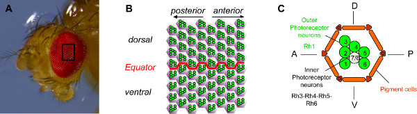
Figure 1. Organization of the Drosophila eye. (A) Photograph of a Drosophila eye taken with a stereomicroscope. The Drosophila eye is composed of approximately 800 ommatidia. (B) Schematic diagram of a field of 64 ommatidia in the middle of the eye. The six outer photoreceptor neurons (PRs) of each ommatidium, shown in green, are organized into a stereotypical trapezoid that points to the nearest poles of the eye. They consequently point in opposite directions in the ventral and the dorsal part of the eye and are mirror image according to the border between the two parts, which is called the equator. (C) Schematic diagram of an ommatidium. Each ommatidium contains eight PRs. PRs R1 to R6, shown in green, are the outer PRs. They express the photosensitive molecule rhodopsin1 (Rh1) and are the equivalent of mammalian rod cells. The inner PRs, R7 and R8, are shown in gray (R7 sits on top of R8). The inner PRs express rhodopsin 3, 4, 5 or 6 and are the equivalent of mammalian cone cells. For simplicity, only the rhabdomere, the light-sensitive part of the PR, is shown for each PR. Click here to view larger figure.
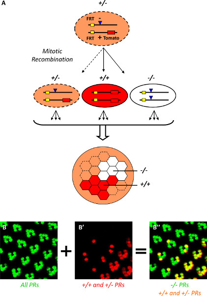
Figure 2. Generating and visualizing mosaic PR clones in the Drosophila eye with the Tomato/GFP-FLP/FRT method. (A) Schematic diagram of the generation of mosaic clones in the Tomato/GFP-FLP/FRT method. Mosaic clones are generated by mitotic recombination, due to the expression of the flipase (under control of the eyeless promoter) during eye development and the FRT sequences. After recombination of the FRT sequence and cell division, homozygous mutant, homozygous wild-type and heterozygous cells can be generated from a heterozygous cell. These cells divide again to form mosaic clones of PRs in the adult eye. The identification of mutant cells is facilitated by inserting a construct expressing the red fluorescent protein tdTomato into the wild-type chromosome, to label wild-type and heterozygous cells. (B-B'') Visualization of mosaic PR clones with the Tomato/GFP-FLP/FRT method. The Tomato/GFP-FLP/FRT method combines mitotic recombination, cornea neutralization and confocal microscopy. All PRs express the green fluorescent protein GFP, which facilitate the visualization of the PRs using cornea neutralization and confocal microscopy (B). PRs can be visualized at a single-cell level. Mutant mosaic clones are generated by mitotic recombination and can be identified by the absence of the red fluorescent protein tdTomato (B'). Consequently, on merged images, homozygous PRs appear in green, whereas wild-type and heterozygous PRs are yellow (B''). Click here to view larger figure.
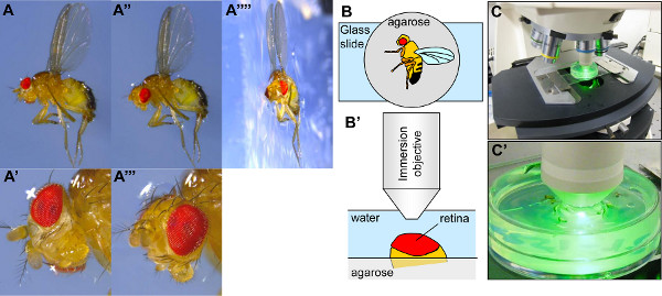
Figure 3. Setting up the Drosophila for visualization by cornea neutralization. (A-A'''') Photographs of a Drosophila immobilized on a plate filled with agarose. In panel A'''', a piece of agarose containing the fly has been cut to take a profile photograph. The Drosophila is half-embedded in the agarose, with the right wing within the agarose and the left wing stuck on the agarose surface (A, A'', A''''). When the Drosophila is embedded in the agarose, its head is frequently poorly positioned for the visualization of the eye (A, A'). The head must therefore be reorientated with forceps such that the middle of the eye is pointing upwards (A'', A'''). (B-B') Schematic diagram showing a Drosophila immobilized on an agarose plate and the positioning of the Drosophila under the objective of the microscope. (C-C') Photographs of the setting of the Drosophila on the stage of the microscope under its objective. Click here to view larger figure.
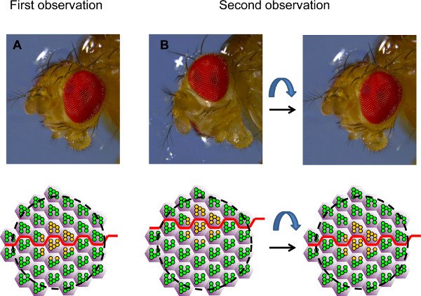
Figure 4. Orientation of the eye for time-course analysis. The figure shows photographs of a Drosophila head in two different orientations and the corresponding fields of ommatidia viewed by cornea neutralization, represented as schematic diagrams. (A) During the first observation, the eye is placed such that a clone of heterozygous or wild-type PRs (in yellow) is viewed in the middle of the field, at the level of the equator (red line). (B) During the second observation, the eye is often not in exactly the same orientation. Thus, the same group of PRs can be found again, but it is not possible to view the exact same field (on the left). The eye must be reoriented for the same field to be viewed again. The position of the equator can be used as a guide. In this example, we know that the observed field is too ventral and that the eye must therefore be turned ventrally to place the equator in the middle of the observed field again, as in the first observation.
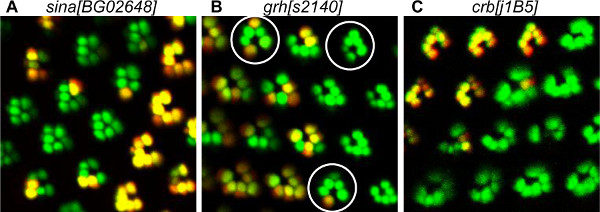
Figure 5. Examples of PR development defects detected by the Tomato/GFP-FLP/FRT method. (A) Visualization of mosaic sina[BG02648] mutant PRs, showing the loss of inner PRs. In the mutant ommatidia, outer PRs are clustered together due to the absence of the inner PR R7. Indeed, sina is known to be required for inner PR recruitment. (B) Visualization of mosaic grh[s2140] mutant PRs showing polarity defects. The mosaic mutant ommatidia, surrounded by a circle, are pointing in the opposite direction from wild-type ommatidia, indicating a dorso-ventral inversion. A detailed study with the Tomato/GFP-FLP/FRT method showed that grh was required in the R3 precursor for the correct acquisition of ommatidium polarity 20. (C) Visualization of mosaic crb[j1B5] mutant PRs, showing deformed homozygous mutant PRs. Crb is known to be required for rhabdomere morphogenesis.
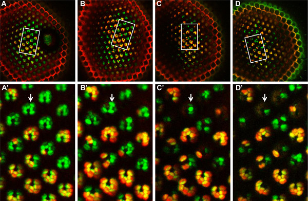
Figure 6. Time-course analysis of mosaic fatp[k10307] mutant PRs with the Tomato/GFP-FLP/FRT method, over 14 days. A mosaic fatp[k10307] mutant retina is observed on day 1 (A, A'), day 4 (B, B'), day 8 (C, C') and day 14 (D, D') after hatching. The same group of mosaic PRs can be found at each time point, in the observed field (white rectangle, A, B, C, D). In this field of PRs (A', B', C', D'), homozygous mutant PRs are labeled in green, whereas heterozygous and homozygous wild-type PRs are labeled in yellow. From day 4 (B') onwards, the homozygous mutant PRs begin to disappear, indicating that the fatp[k10307] mutation induces a progressive degeneration of these PRs. Click here to view larger figure.
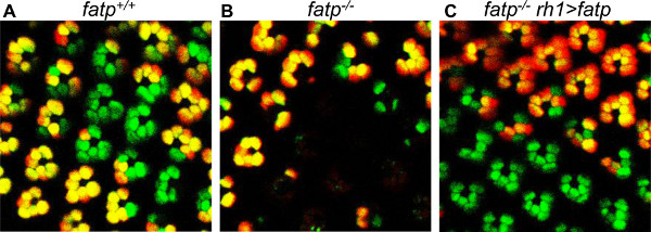
Figure 7. Rescue of fatp[k10307] mutant PRs with a mini-white-carrying fatp-expressing construct. The mosaic PRs are visualized with the Tomato/GFP-FLP/FRT method in 15-day-old flies. (A) Visualization of mosaic control PRs. (B) Visualization of mosaic fatp mutant PRs, showing the loss of mutant PRs. (C) Visualization of mosaic fatp mutant PRs in a fly reexpressing fatp in outer PR (rh1>fatp). The mutant PRs are rescued. The presence of a mini-white gene and the associated red eye pigmentation in rh1>fatp conditions does not alter Tomato or GFP fluorescence or clonal detection.
Discussion
The Drosophila eye has been widely used to decipher the signaling pathways regulating development, proliferation and survival. In the early 1990s, a large number of genetic screens were carried out to identify the pathways required during the initial phases of PR development 25-27. The efficiency of genetic screens was increased greatly by the mitotic recombination technique, which makes it possible to test loss-of-function mutations in mosaic clones 21. Hence, the role of embryonic lethal mutations could be systematically tested in homozygous mutant clones in the eyes of flies that were otherwise heterozygous. Most mosaic screenings have focused on the early PR recruitment, differentiation, axonal projection or morphogenesis occurring in the developing larvae or pupae 10,25-31. To date, only one mosaic screen has investigated the mechanisms regulating adult PR function, by monitoring the visual response via electroretinogram recordings 32. With the Tomato/GFP-FLP/FRT method and the possibility of performing time course analysis in living mutant animals, we have designed a new method for identifying the factors regulating adult PR viability and function 20,24.
The Tomato/GFP-FLP/FRT method has several major advantages over the classical histological sectioning of resin-embedded eyes. First, this method is much faster, cheaper and easier to perform than the histological method, provided that a fluorescence microscope equipped with an appropriate water dipping objective is available (see Table of Reagents and Equipment). Second, the Tomato/GFP-FLP/FRT method can be used in conjunction with transgene expression, such as the expression of P(UAS, w+), carrying a mini-white transgene. In contrast to histological sections, in which w+ is used for clonal detection, in the Tomato/GFP-FLP/FRT system, P(UAS, w+) does not interfere with clonal detection based on Tomato fluorescent protein. Using P(UAS-fatp, w+) transgenic flies, we were able to visualize the rescue of fatp mutant PRs (Figure 7). Third, a key pitfall of all types of clonal analysis, including that based on Tomato/GFP-FLP/FRT, is the ambiguity of the genotype of lost PRs. Indeed, it may be unclear whether a PR is absent because of the lack of a specific gene function or because of a cell non-autonomous effect, particularly at the border of the mutant clone. The Tomato/GFP-FLP/FRT method gets around this problem for degeneration by making it possible to follow wild-type and mutant PRs in the same animal over periods of several weeks. We were able to follow the fate of individual PRs by kinetic analyses on different fatp mutant flies (Figure 6). The same clone can be recognized in a given animal at different times, because of the precise shape of the surrounding wild-type clones. We were able to show unambiguously that all the missing PRs were mutant for fatp, indicating that the role of fatp mutants in PRs is cell-autonomous. Thus, by monitoring the loss of PRs in a kinetic analysis, it is possible to determine the genotype of PRs in models of adult-onset degeneration. Finally, the Tomato/GFP-FLP/FRT method has proved very powerful for the identification of factors regulating the establishment of PCP 20. The possibility of scoring a large number of mosaic ommatidia for a polarity phenotype without the need for sections, facilitates rapid determination of the PR requirement for the establishment of PCP. Nevertheless, finer analysis of PR integrity would require traditional sectioning followed by phase-contrast or electronic microscopies.
In conclusion, the Tomato/GFP-FLP/FRT method opens up new possibilities for efficient mosaic screening to identify factors regulating PR developmental processes and adult PR functions, such as the visual response and viability.
Disclosures
The authors have nothing to disclose.
Acknowledgements
PLATIM and Droso-Tools facilities of the UMS3444 Biosciences, Lyon, France. BM's research was supported by grants from the Fondation pour la Recherche Médicale from the CNRS (ATIP) and the ANR-12-BSV1-0019-01. PD was supported by Retina France association and the Ecole Normale Supérieure of Lyon (France). CL was supported by La Ligue Nationale contre le Cancer association.
Materials
| Name | Company | Catalog Number | Comments |
|---|---|---|---|
| Reagent | |||
| Agarose | Euromedex | D5-E | Regular agarose is used to immobilize the flies |
| Petri dish | BD Falcon | 353001 | 35 x 10 mm, 1.37 x 0.39 inch |
| Cold-ice water | To maintain the flies anesthetized | ||
| Equipment | |||
| Dissecting microscope | Dutcher | SMZ645 | |
| Water Bath | Julabo | 9550102 | To keep the agarose solution at warm temperature |
| 40X Water objective | Zeiss | 420967-9900-000 | Water dipping objective for the confocal microscope |
References
- Hardie, R. C. Sensory physiology 5. Ottoson, D. 5, 1-79 (1985).
- Wolff, T., Ready, D. F. Cell death in normal and rough eye mutants of Drosophila. Development. 113, 825-839 (1991).
- Rister, J., Desplan, C. The retinal mosaics of opsin expression in invertebrates and vertebrates. Dev Neurobiol. 71, 1212-1226 (2011).
- Halder, G., Callaerts, P., Gehring, W. J. Induction of ectopic eyes by targeted expression of the eyeless gene in Drosophila. Science. 267, 1788-1792 (1995).
- Mollereau, B., Domingos, P. M. Photoreceptor differentiation in Drosophila: from immature neurons to functional photoreceptors. Dev Dyn. 232, 585-592 (2005).
- Mollereau, B. Cell death: what can we learn from flies? Editorial for the special review issue on Drosophila apoptosis. Apoptosis. 14, 929-934 (2009).
- Charlton-Perkins, M., Brown, N. L., Cook, T. A. The lens in focus: a comparison of lens development in Drosophila and vertebrates. Mol Genet Genomics. 286, 189-213 (2011).
- Jenny, A. Planar cell polarity signaling in the Drosophila eye. Curr Top Dev Biol. 93, 189-227 (2010).
- Montell, C. Drosophila visual transduction. Trends Neurosci. 35, 356-363 (2012).
- Chen, J., et al. Discovery-based science education: functional genomic dissection in Drosophila by undergraduate researchers. PLoS Biol. 3, e59(2005).
- Tomlinson, A., Ready, D. F. Neuronal differentiation in the Drosophila ommatidium. Dev Biol. 120, 366-376 (1987).
- Mendes, C. S., et al. ER stress protects from retinal degeneration. Embo J. 28, 1296-1307 (2009).
- Jenny, A. Preparation of adult Drosophila eyes for thin sectioning and microscopic analysis. J Vis Exp. , (2011).
- Mollereau, B., et al. Two-step process for photoreceptor formation in Drosophila. Nature. 412, 911-913 (2001).
- Domingos, P. M., et al. Spalt transcription factors are required for R3/R4 specification and establishment of planar cell polarity in the Drosophila eye. Development. 131, 5695-5702 (2004).
- Mendes, C. S., et al. Cytochrome c-d regulates developmental apoptosis in the Drosophila retina. EMBO Rep. 7, 933-939 (2006).
- Domingos, P. M., et al. Regulation of R7 and R8 differentiation by the spalt genes. Dev Biol. 273, 121-133 (2004).
- Mollereau, B., et al. A green fluorescent protein enhancer trap screen in Drosophila photoreceptor cells. Mech Dev. 93, 151-160 (2000).
- Pichaud, F., Desplan, C. A new visualization approach for identifying mutations that affect differentiation and organization of the Drosophila ommatidia. Development. 128, 815-826 (2001).
- Gambis, A., Dourlen, P., Steller, H., Mollereau, B. Two-color in vivo imaging of photoreceptor apoptosis and development in Drosophila. Dev Biol. 351, 128-134 (2011).
- Golic, K. G. Site-specific recombination between homologous chromosomes in Drosophila. Science. 252, 958-961 (1991).
- Porter, J. A., Hicks, J. L., Williams, D. S., Montell, C. Differential localizations of and requirements for the two Drosophila ninaC kinase/myosins in photoreceptor cells. J Cell Biol. 116, 683-693 (1992).
- Johnson, K., Grawe, F., Grzeschik, N., Knust, E. Drosophila crumbs is required to inhibit light-induced photoreceptor degeneration. Curr Biol. 12, 1675-1680 (2002).
- Dourlen, P., et al. Drosophila fatty acid transport protein regulates rhodopsin-1 metabolism and is required for photoreceptor neuron survival. PLoS Genet. 8, e1002833(2012).
- Mlodzik, M., Hiromi, Y., Weber, U., Goodman, C. S., Rubin, G. M. The Drosophila seven-up gene, a member of the steroid receptor gene superfamily, controls photoreceptor cell fates. Cell. 60, 211-224 (1990).
- Gaul, U., Chang, H., Choi, T., Karim, F., Rubin, G. M. Identification of ras targets using a genetic approach. Ciba Found Symp. 176, 85-92 (1993).
- Wassarman, D. A., Therrien, M., Rubin, G. M. The Ras signaling pathway in Drosophila. Curr Opin Genet Dev. 5, 44-50 (1995).
- Janody, F., et al. A mosaic genetic screen reveals distinct roles for trithorax and polycomb group genes in Drosophila eye development. Genetics. 166, 187-200 (2004).
- Legent, K., Steinhauer, J., Richard, M., Treisman, J. E. A screen for X-linked mutations affecting Drosophila photoreceptor differentiation identifies Casein kinase 1alpha as an essential negative regulator of wingless signaling. Genetics. 190, 601-616 (2012).
- Newsome, T. P., Asling, B., Dickson, B. J. Analysis of Drosophila photoreceptor axon guidance in eye-specific mosaics. Development. 127, 851-860 (2000).
- Berger, J., et al. Systematic identification of genes that regulate neuronal wiring in the Drosophila visual system. PLoS Genet. 4, e1000085(2008).
- Bayat, V., et al. Mutations in the mitochondrial methionyl-tRNA synthetase cause a neurodegenerative phenotype in flies and a recessive ataxia (ARSAL) in humans. PLoS Biol. 10, e1001288(2012).
Reprints and Permissions
