We present a preparation for visualizing and manipulating calcium signaling in native, intact microvascular endothelium. Endothelial tubes freshly isolated from mouse resistance arteries supplying skeletal muscle retain in vivo morphology and dynamic signaling within and between neighboring cells. Endothelial tubes can be prepared from microvessels of other tissues and organs.
Method Article
Isolation of Microvascular Endothelial Tubes from Mouse Resistance Arteries
In This Article
Summary
Abstract
The control of blood flow by the resistance vasculature regulates the supply of oxygen and nutrients concomitant with the removal of metabolic by-products, as exemplified by exercising skeletal muscle. Endothelial cells (ECs) line the intima of all resistance vessels and serve a key role in controlling diameter (e.g. endothelium-dependent vasodilation) and, thereby, the magnitude and distribution of tissue blood flow. The regulation of vascular resistance by ECs is effected by intracellular Ca2+ signaling, which leads to production of diffusible autacoids (e.g. nitric oxide and arachidonic acid metabolites)1-3 and hyperpolarization4,5 that elicit smooth muscle cell relaxation. Thus understanding the dynamics of endothelial Ca2+ signaling is a key step towards understanding mechanisms governing blood flow control. Isolating endothelial tubes eliminates confounding variables associated with blood in the vessel lumen and with surrounding smooth muscle cells and perivascular nerves, which otherwise influence EC structure and function. Here we present the isolation of endothelial tubes from the superior epigastric artery (SEA) using a protocol optimized for this vessel.
To isolate endothelial tubes from an anesthetized mouse, the SEA is ligated in situ to maintain blood within the vessel lumen (to facilitate visualizing it during dissection), and the entire sheet of abdominal muscle is excised. The SEA is dissected free from surrounding skeletal muscle fibers and connective tissue, blood is flushed from the lumen, and mild enzymatic digestion is performed to enable removal of adventitia, nerves and smooth muscle cells using gentle trituration. These freshly-isolated preparations of intact endothelium retain their native morphology, with individual ECs remaining functionally coupled to one another, able to transfer chemical and electrical signals intercellularly through gap junctions6,7. In addition to providing new insight into calcium signaling and membrane biophysics, these preparations enable molecular studies of gene expression and protein localization within native microvascular endothelium.
Introduction
In this protocol, we describe the isolation of endothelial cell tubes from the mouse SEA. The SEA arises from the internal thoracic artery and supplies oxygenated blood to the anterior abdominal musculature. Our working with the SEA as a model is attributable to it providing relatively long, unbranched microvessel segments that are well suited for studying intercellular signaling events underlying the role of the endothelium in governing and coordinating smooth muscle cell relaxation. For those interested in tissues other than skeletal muscle, we have found this technique to be readily adapted for obtaining endothelial tubes from microvessels of brain, gut and the lymphatic system.
The key features of this method are that intact lengths (1-3 mm) of microvascular endothelium are isolated, secured against a glass coverslip in a flow chamber in which they are superfused with PSS, and imaged for Ca2+ signaling7-9 or impaled for electrical signaling6,8. Within the flow chamber, the upper half of a tube is exposed to the superfusion solution and responds to experimental interventions, while the bottom half (in contact with the coverslip) is protected and remains quiescent. In addition to studying both intra- and intercellular Ca2+ signaling events, endothelial tubes can be used for quantitative real-time PCR to assess gene expression6,10, immunohistochemistry to investigate protein expression6,10, and electrophysiological studies to define biophysical properties of electrical conduction6,11.
There are several advantages to the endothelial tube preparation for studying endothelial function. First is that the isolation process provides a simple distinction between two cell populations: endothelial cells (which remain in tube formation) and smooth muscle cells (individually dissociated; typically "C" shaped when relaxed). This allows for selective sampling and study of unique properties underlying respective cells' contribution to vessel function. Second, this model enables the resolution of signaling events intrinsic to the endothelium, independent from the influence of the blood flow, surrounding smooth muscle, nerves, and parenchyma. Third, the endothelium is studied while freshly isolated, thereby avoiding alterations in gene or protein expression associated with cell culture.
Protocol
1. Preparation of Equipment: Flow Chamber, Pipettes, and Solutions
- Flow chamber: Use a flow chamber with a 24 mm x 50 mm glass coverslip on the bottom to superfuse the isolated endothelial tubes (see Figure 1A). Before assembling the chamber, wash the glass coverslips with both 3% HCl and 70% ethanol, rinse with ultrapure water, and dry with nitrogen gas.
- Pipettes: Three different types of pipettes are used during the protocol: cannulation, trituration, and pinning. Both the cannulation and pinning pipettes can be made before the day of the experiment, but the trituration pipette should be made the day of as it requires knowledge of the diameter of the vessel to size the lumen appropriately.
- Cannulation pipettes: In order to flush red blood cells from the lumen, pull borosilicate glass capillary tubes using a micropipette puller to have long, tapered ends. Break the ends with forceps to an outer diameter ~30% less than that of the arteries (~50-80 µm), and fire polish the tip to be smooth (see Figure 1B). It is helpful to also angle the tips of the pipettes to approximately 45°.
- Trituration pipettes: To mechanically dissociate the smooth muscle cells and adventitia from the endothelial tubes, make triturating pipettes from borosilicate glass capillary tubes. Prepare capillary tubes as above, and break the end with forceps. Tip: Use a glass scoring device to make it easier to break the thicker wall of the trituration pipette glass cleanly. Break the pipette to the internal diameter determined on the day of the experiment, approximately 10-20 µm larger than that of the outer diameter of the vessel from which endothelial tubes will be isolated. Fire polish the broken ends until smooth (see Figure 1C).
- Pinning pipettes: To stabilize the endothelial tube within the flow chamber, make pinning pipettes to secure the endothelial tube against the coverslip bottom of the chamber. Pull glass capillary tubes in the same manner as the cannulation pipettes. Then break the ends with forceps to a diameter of ~100 µm, and fire polish until the tips are sealed, rounded and smooth (see Figure 1D). Note: If the tip of the pinning pipette is not completely sealed, fluid will enter the lumen and may confound experimental results.
- Solutions: Prepare all solutions in ultrapure (18.2 MΩ) H2O and sterilize them through a 0.2 µm filter. Make sure the osmolality of the solutions is between 285-300 mOsm. Solutions remain good for approximately two weeks when stored at 4 °C.
- Dissection solution: The salt solution used during the dissection of the superior epigastric artery is composed of (in mmol/L): 137 NaCl, 5 KCl, 1 MgCl2, 10 N-2-hydroxyethylpiperazine-N'-2-ethanesulfonic acid (HEPES), 10 glucose, 0.01 sodium nitroprusside (SNP), and 0.1% bovine serum albumin (BSA) with a pH of 7.4. The NO donor SNP is included to help relax smooth muscle cells, thus making them easier to cannulate the artery and to facilitate smooth muscle cell removal during trituration.
- Dissociation solution: The salt solution used during the dissociation of the endothelial tube is composed of (in mmol/L): 137 NaCl, 5 KCl, 1 MgCl2, 10 HEPES, 10 glucose, 2 CaCl2, and 0.1% BSA, with a pH of 7.4. To a 1 ml aliquot of this buffer, add: 0.62 mg/ml papain, 1.5 mg/ml collagenase and 1.0 mg/ml dithioerythritol. Since enzymes from different suppliers can have varying levels of enzymatic activity, the product numbers of those used for this work are included for reference in the table of materials. Note: During the protocol both dissociation buffer and dissociation buffer with enzymes are used at different steps.
- Superfusion solution: The physiological salt solution (PSS) used for superfusing the isolated endothelial tube is composed of (in mmol/L): 137 NaCl, 5 KCl, 1 MgCl2, 10 HEPES, 10 glucose and 2 CaCl2, with a pH of 7.4.
- Prepare buffers and solutions before beginning surgical procedures. Remove the solutions from 4 °C and aliquot 50 ml of Dissection buffer into a 200 ml Erlenmeyer flask and place it on ice. Additionally, aliquot 50 ml of Dissociation buffer without enzymes and place it on the bench top to equilibrate to room temperature. Remove the superfusion solution from 4 °C and allow it to equilibrate to room temperature on the bench top.
2. Animal Preparation
Make sure that all procedures and protocols involving animals are in accord with the National Institutes of Health Guide for the Care and Use of Laboratory Animals. The procedures described here were approved by the Animal Care and Use Committee of the University of Missouri.
- Use male or female mice at least 9 weeks old and study each individually. Anesthetize a mouse with pentobarbital sodium (60 mg/kg) via intraperitoneal (i.p.) injection. Tip: Diluting the pentobarbital to 10 mg/ml in sterile saline before injection reduces the possibility of overdose.
- Anesthesia compromises temperature regulation, so keep the mouse warm immediately after the first injection of pentobarbital. Using a metal carrier (ventilated aluminum basket) on top of a heating plate (calibrated to ~37 °C) works well to keep the temperature of the mouse stable. Alternatively, use a heat lamp, taking care to position it at an appropriate distance from the mouse. Monitor the mouse every 5-10 min until the appropriate level of anesthesia is achieved (lack of withdrawal to toe or tail pinch). A supplementary dose may be required after 15-20 min. It is best to proceed slowly and with caution to avoid overdose, especially with obese, aged, or animals otherwise stressed.
- Remove the hair from the ventral side by carefully shaving it with small pet hair clippers. Particular attention should be taken to avoid trauma to the animal. It is helpful to remove all hair from an area spanning the armpits to the pubic area. Remove any loosely clinging hair with a fresh alcohol swab.
- Position the mouse on its back, lying with both fore- and hind limbs spread out to expose as much of the abdominal wall as possible. If necessary, use laboratory tape to secure the legs in a spread-eagle manner.
3. Isolation of the Superior Epigastric Artery
- Make a small incision through the skin just above the area of the pubic region. Ensure that only the skin is being cut while avoiding damage to the underlying abdominal muscle; extend the incision laterally in each direction out to respective hindlimbs. Continue the incision rostrally along the ventral midline to the top of the rib cage and then extend the incision laterally in each direction to respective forelimbs.
- Gently lift the skin and use scissors to sever the connective tissue holding the skin to the underlying muscle, thereby exposing the entire surface of the abdominal musculature (see Figure 3A). Irrigate the exposed muscle with room temperature 0.9% saline solution.
- Due to the small size of the SEA, position the animal underneath a stereomicroscope for further procedures. Lift the fat pad located at the bottom of the sternum with forceps, and make an incision through it. Continue the incision along the bottom rib, making sure not to cut too deeply into the underlying tissue. The SEA should be visible; take care to avoid damaging it. Once the incision is complete, irrigate the exposed tissue with room temperature 0.9% saline solution.
- After the top layer of skeletal muscle has been retracted, the SEA and underlying muscle are easily identified. Tip: Note the length of the SEA in vivo before isolating to extend endothelial tubes (which shorten along their axis upon isolation) to approximate their original length. Using forceps and scissors, carefully excise the thin muscle layer underneath the SEA.
- Using angled forceps, pass a length of 6-0 silk suture underneath the SEA and ligate the artery and its adjacent vein to keep it pressurized and blood retained within the vessel lumen to facilitate visualization during subsequent isolation and cleaning.
- Repeat the SEA ligation procedure on the contralateral side.
- Once both SEAs have been ligated, make an incision along the midline of the abdominal muscles to separate respective sides.
- Extend the incision laterally in each direction as done for the skin, then continue the incision vertically along the outer edge to completely separate the abdominal muscle from the body. Avoid damage to vasculature fed by the SEA to maintain the pressurized lumen.
- Cut the SEA above the ligation to maintain the seal, and place the isolated muscle and artery in a 50 ml beaker containing 10 ml of 4 °C dissection buffer.
- Repeat the procedure for the other side of the abdomen and incubate the tissues in the dissection buffer for 10 min.
- Place the abdominal muscle containing the SEA in a chilled (4 °C) dissection chamber containing dissection buffer (see Figure 3B). The dissection chamber consists of a Petri dish with a layer of Sylgard (polydimethylsiloxane [PDMS]) at the bottom.
- Using insect pins (0.15 mm), stretch the isolated SEA and abdominal muscle out to approximate in vivo lengths (noted above) and secure it to the Sylgard. Tip: Orienting the muscle such that the thin layer facing the peritoneum is on top makes the SEA readily visible using transillumination.
- Utilizing fine microdissection instruments and working from the upstream site of ligation towards the downstream end, clear the SEA from its paired vein and the surrounding tissue until the first major branch site (1-2 cm). Then excise the SEA by cutting it just above the branch site and just below the ligation.
- To remove blood retained within the vessel lumen, cannulate the SEA and flush it with ice cold dissection buffer. Using a piece of silastic tubing, attach the back end of a cannulation pipette to a 5 ml syringe containing ice cold dissection buffer and secure the micropipette in a micromanipulator to position its tip within the dissection chamber to cannulate the SEA.
- Once all of the erythrocytes are flushed out, remove the SEA from the cannulation pipette, and lift the pipette out of the dissection dish.
- Cut the SEA into smaller segments each 1-3 mm in length. These shorter segments facilitate the isolation of endothelial tubes.
4. Isolation of the Endothelial Tube
- Fill a 12 mm x 75 mm glass culture tube halfway with ice cold dissection buffer. Using angled forceps, transfer the arterial pieces into the culture tube and place on ice. At this time, combine the digestion enzymes with Dissociation buffer to a final volume of 1 ml in a separate 12 mm x 75 mm culture tube (see 1.3.2 Solutions) and preheat the solution to 37 °C with a heating block.
- While the dissociation buffer and enzymes are warming up, carefully aspirate the dissection buffer from the culture tube leaving a small volume containing the vessel segments.
- Gently add room temperature dissociation buffer without enzymes to wash away remaining dissection buffer. Tip: Add the dissociation buffer slowly such that the arterial pieces remain on the bottom of the culture tube to save time waiting for them to resettle. Raise the buffer temperatures over multiple steps from ice (4 °C), to room temperature (~24 °C), to 37 °C to minimize shock.
- Carefully aspirate the dissociation buffer from the culture tube, again being sure to leave a small volume containing the vessel segments.
- Remove the preheating dissociation buffer with enzymes from the heating block, transfer the 1 ml of enzyme solution to the culture tube containing the vessel segments, and place the culture tube back in the heating block. Incubate at 37 °C for 30 min.
- During the incubation with enzymes, prepare the trituration pipette, backfill it with mineral oil and secure it on a microsyringe that is mounted in a micromanipulator. Then retract the microsyringe plunger to fill the pipette with 2 nl of dissociation buffer and position the pipette tip over the flow chamber.
- At the end of the 30 min incubation, remove the culture tube from the heating block and carefully aspirate the buffer as above.
- Wash the arterial segments with 4 ml of room temperature dissociation buffer. Note: It is no longer necessary to keep the vessel segments at the bottom of the culture tube; disturbing the arterial pieces actually helps to ensure that residual enzymes are effectively removed.
- Transfer one vessel segment to the flow chamber by gently aspirating it with a 1 ml micropipette, and place it into the flow chamber with 1 ml of the dissociation buffer.
- Utilizing the micromanipulator containing the microsyringe, position the tip of the trituration pipette near one end of the vessel segment.
- Aspirate the vessel segment (500 nl withdrawn) into the triturating pipette slowly (225 nl/sec), so as not to cause mechanical strain to the ECs, and then eject it back into the chamber. If the digestion was successful, the smooth muscle cells and adventitia will be dissociated from the endothelial tube.
- Repeat the trituration until all smooth muscle cells are dissociated. Be aware that excessive trituration (4x or more) can damage the ECs and render them unresponsive to agonists. Tip: using the trituration pipette, move the adventitia away from the endothelial tube as soon as possible to keep it from entangling the tube. Note: Using the trituration pipette, the isolated endothelial tube can be aspirated and transferred to a separate chamber.
- Position the endothelial tube in the center of the flow chamber, aligned along the direction of flow, and remove the triturating pipette.
- Secure pinning pipettes in micromanipulators mounted at either end of the flow chamber, and position their tips at respective ends of the endothelial tube.
- Lower a pinning pipette over one end of the tube to press it against the bottom of the chamber. Position the second pinning pipette in the same way at the opposite end of the tube. Note: Placement of the pinning pipettes is a trade-off between securing the tube (placing them farther from the end of the tube) and allowing for more imaging/experimental area (placing them closer to the end of the tube). Locating them ~50 µm from each end of the tube works well. Just as the pinning pipette touches the tube, slowly retract it along the axis of the tube, thereby extending it to approximate in vivo length; then press the pipette against the chamber bottom to secure the tube.
- Begin the flow of superfusion solution, and allow the endothelial tube to rest for at least 20 min to equilibrate and adhere (see Figure 3C). Utilize a peristaltic pump to maintain constant fluid flow (superfusion) across the endothelial tube. Note: Pinning pipettes can be removed once the endothelial tube has adhered to the bottom of the flow chamber (~25 min) or left in place to ensure the endothelial tube does not become dislodged.
5. Dye Loading and Calcium Imaging
- To visualize intracellular calcium responses, load the endothelial tube with fluo-4 AM dye at room temperature (~24 °C). Stop fluid flow in the bath, and add fluo-4 AM to the chamber at a final concentration of 10 µM. Wait 10 min to allow the dye to enter cells, then superfuse the endothelial tube with fresh PSS for 20 min to ensure extracellular dye is removed and that intracellular dye has been fully de-esterified.
- Perform calcium imaging. For this work, an upright microscope (see Figure 2) equipped with a spinning disc confocal imaging system was used at ambient room temperature. Excite the fluorescent calcium dye with a 491 nm laser and acquire images (at 500-550 nm emission) using an intensified digital camera.
Results
Calcium responses can be initiated in freshly isolated endothelial tubes using acetylcholine (ACh). In this movie, an endothelial tube loaded with 10 µM fluo-4 AM is stimulated with superfusion of 1 µM ACh (Movie 1). Click here to view movie. The dye is excited at 491 nm and emission is recorded from 500-550 nm using an intensified charge-coupled device camera. Image stacks are acquired at 120 frames/sec for a 25 sec period and can then be averaged (e.g. to 40 frames/sec). Representative fluorescence images over time are shown in Figure 4.

Figure 1. Flow chamber and pipettes used during isolation of endothelial tubes. A) The flow chamber assembled with a 24 mm x 50 mm glass coverslip as the base. B) Examples of a pulled (left) and a pulled, broken, polished and angled (right) cannulation pipette. This pipette is used to flush erythrocytes from the lumen of the artery following dissection. Scale bar = 200 µm. C) Examples of a pulled (left) and a pulled, broken and polished (right) trituration pipette. The internal diameter of this pipette is determined by the exterior diameter of the artery. Scale bar = 200 µm. D) Examples of a pulled (left) and a pulled, broken and polished (right) pinning pipette. The tip of this pipette is rounded off and completely sealed to secure the endothelial tube in the flow chamber. Scale bar = 200 µm. Click here to view larger image.
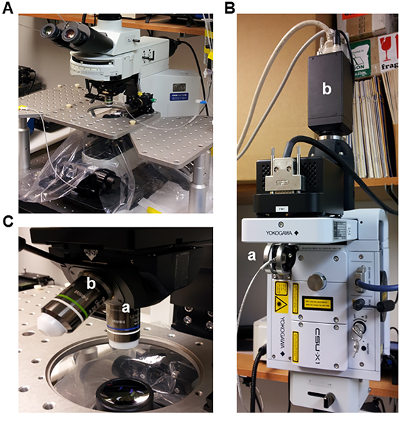
Figure 2. Spinning disc confocal microscope for imaging of Ca2+ signals. A) Upright microscope used for calcium imaging. B) (a) Confocal spinning disc unit with an (b) intensified charge coupled device camera. C) Immersion objectives (a) 63X (NA = 1) and (b) 20X (NA = 0.5) used for imaging. Click here to view larger image.

Figure 3. Isolation of the abdominal muscle and superior epigastric artery. A) Anesthetized mouse with abdominal muscle exposed and irrigated with room temperature 0.9% saline solution. Red arrows mark the sites under fat pads at which respective SEAs enter each rectus abdominis muscle (i.e. where vessels should be ligated). Scale bar = 1 cm. B) Isolated abdominal muscle (unilateral) and SEA pinned out in a dissection chamber for microdissection. Red arrow indicates the first major bifurcation site of the SEA (i.e. the point at which cleaning of the vessel is stopped). Scale bar = 1 cm. C) Isolated endothelial cell tube from an SEA. Differential interference contrast image of an endothelial tube following 1 hr incubation at room temperature. The endothelial tube was superfused with Superfusion buffer at 3 ml/min. Scale bar = 50 µm. Click here to view larger image.

Figure 4. Calcium fluorescence in an endothelial cell tube. Representative fluorescent images of an endothelial tube in response to stimulation with 1 µM acetylcholine. A) Fluorescence before stimulation. B) endothelial tube fluorescence at the time of acetylcholine administration, t=0 sec. C) endothelial tube fluorescence at t=5 sec. D) endothelial tube fluorescence at t=10 sec. E) endothelial tube fluorescence at t=15 sec. F) endothelial tube fluorescence at t=20 sec. Scale bars = 40 µm. Click here to view larger image.
Discussion
Here we describe the isolation of endothelial tubes from the SEA and the use of this preparation to visualize Ca2+ signaling events within its constitutive ECs. This procedure was adapted from one originally developed to isolate endothelial tubes from arterioles of the hamster cremaster muscle8. Utilizing minor variations of the techniques presented here, we have isolated endothelial tubes from a variety of vascular beds, including: feed arteries of the hamster retractor muscle and cheek pouch arterioles10, mouse superior epigastric artery6,7,9, mouse mesenteric and cerebral arteries and lymphatic microvessels (unpublished observations; references are included for isolating mesenteric vessels12 and cerebral vessels13). Isolating endothelial tubes from new vascular beds may require modifications to the original protocol. If isolation is unsuccessful, it is best to begin by altering digestion times: Increase the digestion time if endothelial tubes are difficult to isolate from smooth muscle cells and adventitia and reduce digestion time if the endothelial tube does not remain intact. If altering digestion times is unsuccessful, adjusting the concentration of digestion enzymes or the digestion temperature should be tried. Another option is to reduce the internal diameter of the trituration pipette, which increases shear force in removing smooth muscle cells. Increasing the velocity of fluid ejection can also be tried for the same effect but is more likely to damage the preparation. It should be recognized that it may not be possible to isolate intact endothelial tubes from vessels in which endothelial cells are not well connected to each other by junctional proteins.
Dissociation of smooth muscle cells following enzymatic digestion was initially performed using a hand-style pipettor8. We have refined the trituration procedure using a microsyringe system, which has enabled consistent isolation of longer endothelial tubes (up to 3 mm)6. When using this system, the trituration pipette is backfilled with mineral oil to provide a continuous fluid column between the piston controlling fluid movement and PSS containing the vessel segment. The incompressibility of fluid along with the constant driving force of the microsyringe piston results in constant shear as the vessel is forced through the pipette tip. In contrast, hand techniques often used for dissociating cells (e.g. squeezing rubber bulb at the end of a Pasteur pipette) involves the compression of air, which introduces variability in the driving pressure and, thereby, shear exerted at the pipette tip.
There are multiple advantages to the tube preparation for studying endothelial function in microvessels. The first is that the isolation process provides distinct homogenous native cellular populations of ECs and smooth muscle cells. Since the ECs remain physically connected as tubes, they are distinct from individual smooth muscle cells, which retain a "C" configuration if they remain relaxed during trituration. Thus respective cell types are readily distinguished, e.g. when obtaining samples for molecular techniques such as real-time PCR and immunohistochemistry10. Since multiple tubes are isolated from a single vessel (or from bilateral vessels as done for the SEA), molecular data and functional data can be obtained from the same vessels of a given animal. Thus molecular expression can be correlated with the functional behavior of microvascular endothelium. Further, both intra- and intercellular signaling events intrinsic to microvascular endothelium can be resolved independent from the influence of hemodynamic forces (pressure, flow), vasoactive agents carried in the blood stream (e.g. hormones), or from surrounding smooth muscle cells (e.g. via myoendothelial coupling14-16), nerves17, or tissue parenchyma18.
Importantly, since the endothelium is freshly isolated from designated microvessels, there is no alteration of phenotype that is otherwise associated with culturing ECs19-21. For example, cultured ECs lose muscarinic receptor expression and thereby alter their calcium signaling profiles. Further, electrophysiological properties of ECs can change in culture22. Because individual ECs remain coupled through functional gap junction channels, the endothelial tube presents an ideal model for studying the conduction of electrical and Ca2+ signals between cells6,7. It should also be recognized that the dissociated smooth muscle cells are readily studied with patch-clamp techniques for complementary data underlying microvascular function23.
A key limitation to the endothelial tube model is the instability of the preparation with increasing temperature. While our preparations from the SEA have proven stable and healthy at ambient room temperature (~24 °C) and for several hours at 32 °C, morphological and functional degradation occur in less than an hour at 37 °C9. A second important limitation of the endothelial tube is the loss of myoendothelial junctions and their inherent signaling domains that are integral to EC function in the intact vessel wall14-16. It should also be recognized that, while longer tubes enable intercellular signaling to be studied over relatively great distances6, preparation of tubes is complicated as they get longer because it becomes more difficult to fully dissociate surrounding smooth muscle cells and adventitia. We have found that tubes longer than 1-2 mm are also more difficult to position and secure in the flow chamber. In contrast, while shorter tubes (e.g. <1 mm) enable Ca2+ and electrical signals to be studied8, they are difficult to maintain during superfusion in the flow chamber. Finally, even with optimal isolation of tubes bilaterally, there is insufficient material for traditional quantification of protein expression using Western blots, though immunolabeling provides an index of protein expression and localization. Despite such limitations, the endothelial tube represents a novel preparation for providing new insight into mechanisms of microvascular endothelial cell function in vivo.
Acknowledgements
The authors thank Dr. Pooneh Bagher and Dr. Erika Westcott for editorial comments in preparing the manuscript. This work was supported by the National Heart, Lung and Blood Institute of the National Institutes of Health under award numbers R37-HL041026 and R01-HL086483 to SSS and F32-HL107050 to MJS. This content is solely the responsibility of the authors and does not necessarily represent the official views of the National Institutes of Health.
Materials
| Name | Company | Catalog Number | Comments |
|---|---|---|---|
| Sodium Chloride | Fisher | S642-212 | |
| Potassium Chloride | Sigma | P9541 | |
| Magnesium Sulfate | Sigma | M2643 | |
| Calcium Chloride | Sigma | C1016 | |
| Sodium Nitroprusside | Sigma | 431451 | |
| Bovine Serum Albumin | USB Products | 9048-46-8 | |
| Papain | Sigma | P-4762 | |
| Collagenase | Sigma | C-8051 | |
| Dithioerythritol | Sigma | D-8255 | |
| Boroscilicate Glass (Cannulation and Pinning Pipettes) | Warner Instruments | G150-4 | |
| Boroscilicate Glass (Trituration Pipettes) | World Precision Instruments | 4897 | |
| Nanoliter Injector Microsyringe | World Precision Instruments | B203MC4 | Updated to Nanoliter 2010 from Nanoliter 2000 |
| Micro4 Controller | World Precision Instruments | SYS-MICRO4 | |
| 12 mm x 75 mm Culture Tubes | Fisher | 14-961-26 | |
| 3-Axis Micromanipulator | Siskiyou Corp | DT3-100 | Holding and positioning the pinning pipettes |
| Flow Chamber | Warner Instruments | RC-27N | |
| 100-1,000 µl Pipette | Eppendorf | 3120000062 | For transferring digested vessel segment to the flow chamber |
| 1 ml Pipette Tips | Fisher | 02-707-405 | For transferring digested vessel segment to the flow chamber |
| Upright Microscope | Olympus | BX51W1 | The actual microscope is up to the user |
| Spinning Disc Confocal System | Yokogawa | CSU-X1 | Confocal imaging is optional |
| XR/TURBO EX Camera | Stanford Photonics | XR/TURBO EX | Ideal for our needs; may vary with user |
| Piper Control Software | Stanford Photonics | Imaging software for TURBO EX camera | |
| Stereo Microscrope | Leica | MZ8 | may vary with user |
| Sylgard | Dow Corning | 184 | |
| Microdissection Scissors | Fine Science Tools | 15003-08 | |
| Dumont Forceps | Fine Science Tools | #5/45 | |
| Minutiens Insect Pins | Austerlitz | M size 0.15 mm | |
| GP Millipore Express PLUS Membrane | Millipore | SCGPT05RE | |
| Pipette Scoring Device | Austin Flameworks | Small Handheld Scoring Tool | austinflameworks.com |
| Compact Pet Trimmer | Wahl Clipper Corp. | Model 9966 | Clean well after each use to maintain life |
References
- Campbell, W. B., Gebremedhin, D., Pratt, P. F., Harder, D. R. Identification of epoxyeicosatrienoic acids as endothelium-derived hyperpolarizing factors. Circ. Res. 78, 415-423 (1996).
- Crutchley, D. J., Ryan, J. W., Ryan, U. S., Fisher, G. H. Bradykinin-induced release of prostacyclin and thromboxanes from bovine pulmonary artery endothelial cells. Studies with lower homologs and calcium antagonists. Biochim. Biophys. Acta. 751, 99-107 (1983).
- Kruse, H. J., Grunberg, B., Siess, W., Weber, P. C. Formation of biologically active autacoids is regulated by calcium influx in endothelial cells. Arterioscler. Thromb. 14, 1821-1828 (1994).
- Busse, R., et al. EDHF: bringing the concepts together. Trends Pharmacol. Sci. 23, 374-380 (2002).
- Ledoux, J., Bonev, A. D., Nelson, M. T. Ca2+-activated K+ channels in murine endothelial cells: block by intracellular calcium and magnesium. J. Gen. Physiol. 131, 125-135 (2008).
- Behringer, E. J., Socha, M. J., Polo-Parada, L., Segal, S. S. Electrical conduction along endothelial cell tubes from mouse feed arteries: confounding actions of glycyrrhetinic acid derivatives. Br. J. Pharmacol. 166, 774-787 (2012).
- Socha, M. J., Domeier, T. L., Behringer, E. J., Segal, S. S. Coordination of intercellular Ca2+ signaling in endothelial cell tubes of mouse resistance arteries. Microcirculation. 19, 757-770 (2012).
- Cohen, K. D., Jackson, W. F. Membrane hyperpolarization is not required for sustained muscarinic agonist-induced increases in intracellular Ca2+ in arteriolar endothelial cells. Microcirculation. 12, 169-182 (2005).
- Socha, M. J., Hakim, C. H., Jackson, W. F., Segal, S. S. Temperature effects on morphological integrity and Ca2+ signaling in freshly isolated murine feed artery endothelial cell tubes. Am. J. Physiol. Heart Circ. Physiol. 301, 773-783 (2011).
- Hakim, C. H., Jackson, W. F., Segal, S. S. Connexin isoform expression in smooth muscle cells and endothelial cells of hamster cheek pouch arterioles and retractor feed arteries. Microcirculation. 15, 503-514 (1908).
- Behringer, E. J., Segal, S. S. Tuning electrical conduction along endothelial tubes of resistance arteries through Ca2+-activated K+ channels. Circ. Res. 110, 1311-1321 (2012).
- Bagher, P., et al. Low intravascular pressure activates endothelial cell TRPV4 channels, local Ca2+ events, and IKCa channels, reducing arteriolar tone. Proc. Natl. Acad. Sci. U.S.A. 109, 18174-18179 (2012).
- Faraci, F. M., et al. Cerebral vascular effects of angiotensin II: new insights from genetic models. J. Cereb. Blood Flow Metab. 26, 449-455 (2006).
- Emerson, G. G., Segal, S. S. Electrical coupling between endothelial cells and smooth muscle cells in hamster feed arteries: role in vasomotor control. Circ. Res. 87, 474-479 (2000).
- Ledoux, J., et al. Functional architecture of inositol 1,4,5-trisphosphate signaling in restricted spaces of myoendothelial projections. Proc. Natl. Acad. Sci. U.S.A. 105, 9627-9632 (2008).
- Tran, C. H., et al. Endothelial Ca2+ wavelets and the induction of myoendothelial feedback. Am. J. Physiol. Cell Physiol. 302, 1226-1242 (2012).
- Nausch, L. W., et al. Sympathetic nerve stimulation induces local endothelial Ca2+ signals to oppose vasoconstriction of mouse mesenteric arteries. Am. J. Physiol. Heart Circ. Physiol. 302, 594-602 (2012).
- Duza, T., Sarelius, I. H. Increase in endothelial cell Ca2+ in response to mouse cremaster muscle contraction. J Physiol. 555, 459-469 (2004).
- Colden-Stanfield, M., et al. Bradykinin-induced increases in cytosolic calcium and ionic currents in cultured bovine aortic endothelial cells. Circ. Res. 61, 632-640 (1987).
- Brunner, F., Kukovetz, W. R. Radioligand binding to muscarinic receptors of bovine aortic endothelial cells. Br. J. Pharmacol. 102, 373-380 (1991).
- Tracey, W. R., Peach, M. J. Differential muscarinic receptor mRNA expression by freshly isolated and cultured bovine aortic endothelial cells. Circ. Res. 70, 234-240 (1992).
- Adams, D. J., Hill, M. A. Potassium channels and membrane potential in the modulation of intracellular calcium in vascular endothelial cells. J. Cardiovasc. Electrophysiol. 15, 598-610 (2004).
- Jackson, W. F., Huebner, J. M., Rusch, N. J. Enzymatic isolation and characterization of single vascular smooth muscle cells from cremasteric arterioles. Microcirculation. 4, 35-50 (1997).
Reprints and Permissions
