Here we demonstrate a technique for widespread neuronal transduction by intraventricular injection of adeno-associated virus into the neonatal mouse brain. This method provides a rapid and easy way to attain lifelong expression of virally-delivered transgenes.
Method Article
Intracerebroventricular Viral Injection of the Neonatal Mouse Brain for Persistent and Widespread Neuronal Transduction
* These authors contributed equally
In This Article
Summary
Abstract
With the pace of scientific advancement accelerating rapidly, new methods are needed for experimental neuroscience to quickly and easily manipulate gene expression in the mouse brain. Here we describe a technique first introduced by Passini and Wolfe for direct intracranial delivery of virally-encoded transgenes into the neonatal mouse brain. In its most basic form, the procedure requires only an ice bucket and a microliter syringe. However, the protocol can also be adapted for use with stereotaxic frames to improve consistency for researchers new to the technique. The method relies on the ability of adeno-associated virus (AAV) to move freely from the cerebral ventricles into the brain parenchyma while the ependymal lining is still immature during the first 12-24 hr after birth. Intraventricular injection of AAV at this age results in widespread transduction of neurons throughout the brain. Expression begins within days of injection and persists for the lifetime of the animal. Viral titer can be adjusted to control the density of transduced neurons, while co-expression of a fluorescent protein provides a vital label of transduced cells. With the rising availability of viral core facilities to provide both off-the-shelf, pre-packaged reagents and custom viral preparation, this approach offers a timely method for manipulating gene expression in the mouse brain that is fast, easy, and far less expensive than traditional germline engineering.
Introduction
Traditional methods for modifying neural gene expression require time-consuming and expensive germline manipulations. Alternative de novo approaches such as in utero electroporation or stereotaxic lentiviral injection yield faster results and are less costly but have the disadvantage of requiring complex surgical intervention1-3. Furthermore, transgene expression has a limited spatial range with these methods. Herein, we describe a fast, easy, and economical method for widespread neuronal manipulation via intraventricular injection of adeno-associated virus (AAV) into the neonatal mouse brain. The method was first described by John Wolfe and Marco Passini in 2001, where they suggested small particle size of AAV allowed it to diffuse within the cerebral spinal fluid as it passes from the lateral ventricles through the immature ependymal barrier and into the brain parenchyma4,5. Intraventricular injection of AAV within the first 24 hr after birth yields widespread viral transduction of neural subsets spanning every region of the brain, from the olfactory bulbs to the brain stem6,7. Virally-delivered transgenes are expressed and active within days of injection and persist for up to a year after transduction. Thus, this versatile manipulation enables studies ranging from early postnatal brain development to aging and degeneration in the adult.
In adapting the technique to our specific experimental needs, we have focused primarily on AAV8 serotype because it is the most efficient at transducing neurons6. We show that viral titer can be diluted to control the density of transduced neurons for experiments testing cell-intrinsic consequences of genetic manipulation. In addition, we demonstrate that two viruses could be co-injected to produce expression patterns that are biased towards distinct or overlapping sets of neurons, depending on the serotypes chosen for viral packaging. Our work expands the versatility of this technique for use in a broad range of experimental neuroscience settings.
Protocol
Perform all procedures and protocols involving animals in accordance with the National Institutes of Health Guide for the Care and Use of Laboratory Animals. The procedures described here were reviewed and approved by the Baylor College of Medicine Institutional Animal Care and Use Committee.
Replication-incompetent adeno-associated virus (AAV) vectors for transgene delivery in the rodent brain are approved for Biosafety Level 1 use. Refer to the CDC website for the US Government publication “Biosafety in Microbiology and Biomedical Laboratories (BMBL)” which details specific requirements regarding proper protection and virus handling procedures. Check with local veterinary and environmental safety staff to learn institutional specific requirements for procedures using viruses. Regulations regarding procedure rooms, quarantine, and identification of biohazard cages vary across institutions.
1. Prepare P0 Pups and Foster Mothers
- Provide high fat chow for all pregnant and nursing females to support the energy demands of breeding and lactation.
- In the evening, set up a breeding cage by adding one or two healthy females to the cage of a single resident male. The following morning, check the females for a mating plug and separate the females from the males if a plug is present.
- Use foster mothers to raise pups from a genetic background with poor maternal characteristics (for example, C57BL/6 or C3HeJ). Foster mothers accept pups from another litter if the transferred pups are born within four days of the foster mother’s own litter.
- Set up a separate mating cage using ICR or FVB females alongside the experimental breeders so that the two sets of females deliver at approximately the same time.
- Females will deliver in 19 ±1 days from the plug date, depending on the background strain. Three days prior to delivery (16 days after the plug date), place the pregnant female into a clean cage with fresh nesting material and a covered shelter.
- Check for newborn pups twice daily starting 2 days prior to the expected delivery date (17 days after the plug date). Examine the cage with minimum stress to the mothers by peeking through the bottom for the presence of pink newborns. Mice generally deliver pups in the morning but on rare occasions will deliver in afternoon.
- Plan to perform injections as soon as the pups are nursing (which will be evident by visible milk spots), or within 6 hr of birth, whichever comes first.
2. Prepare Equipment for Injection
- Prepare a 10 µl injection syringe with a 32 G needle for general P0 neonates. If performing stereotaxic injection, mount the syringe onto the manipulator arm.
- If using a stereotaxic frame for injections, cool the neonatal stage to 4-8 °C by adding 100% ethanol and dry ice to the reservoir at the front end of the block. Maintain the temperature above 1 °C to avoid frostbite of the pups.
3. Prepare Viral Dilutions for Injection
- Prepare a 1% trypan blue dye solution (20x stock).
- Prepare 5-25 µl aliquots of adeno-associated virus (AAV) stock inside a Class 2 biosafety cabinet. Place these aliquots at -80 °C for future use. Avoid additional freeze-thaw cycles as this causes the virus to lose transduction efficacy.
- Remove an aliquot of AAV from the -80 °C freezer and place on ice to thaw.
- Prepare the viral solution for injection by diluting the virus into ice-cold 1x phosphate buffered saline (PBS). Start with a ten-fold serial dilution (1010 to 108 viral particles/hemisphere) for initial optimization of the transduction pattern.
- Add 20x trypan blue to the AAV solution to a final concentration of 0.05%.
4. Staging the Newborn Pups for Injection
- Place a small aluminum plate on ice to cool. Place a dry task wipe on top of the plate to protect the pup’s skin from the cold metal. This serves as a flat cold surface for anesthetizing and injecting the pups.
- Prepare a warming pad suitable for keeping neonatal mice warm before and after injection.
- Once the newborn pups have begun to nurse, remove approximately half of them from the cage and leave the other half with their mother. Place the collected pups on the warming pad while awaiting injection.
NOTE: If the biological mother does not care for the pups, remove the entire litter from the cage immediately and place the pups onto a warming pad to await injection. This situation may arise with the first litter born to C57BL/6 females or with other inbred strains that have poor breeding characteristics. In this situation, milk spots will not be visible on newborn pups. - Transfer one pup from the warming pad onto the cold metal plate to induce hypothermia anesthesia. Wait 2-3 min for the pup to become fully anesthetized. Confirm anesthesia by very gently squeezing a paw and monitor for lack of movement or respiration.
5. Injection of AAV into Neonatal Mice
- Free-hand intracranial injection of AAV into neonatal mice:
- Load 5 µl of diluted AAV with 0.05% trypan blue into the injection syringe.
- Gently wipe the head of the anesthetized pup with a cotton swab soaked in 70% ethanol.
- Identify the injection sites at 2/5 of the distance from the lambda suture to each eye. An alternative injection site is located approximately 0.8-1 mm lateral from the sagittal suture, halfway between lambda and bregma. These landmarks are visible through the skin at P0.
- Mark the injection site with a non-toxic laboratory pen.
- Hold the syringe with the scale visible for monitoring the volume of solution dispensed.
- Ensure that the thumb can reach the top of the plunger. Then remove the thumb from the plunger while positioning the needle to avoid accidentally dispensing virus.
- Find a comfortable position to brace the arm for injection by putting the elbow on the bench and leaning the arm on the ice bucket.
- Lay the pup on its side with its head directly under the syringe. Turn the pup’s head so that the marked injection site is facing up, and gently but firmly hold this position with an open hand.
- Hold the syringe perpendicular to the surface of the skull and insert the needle at the marked injection site to a depth of approximately 3 mm. Inject the needle until the resistance decreases slightly, indicating that the needle has penetrated into the lateral ventricle. If a reference is needed, mark 3 mm from the tip of needle with non-toxic maker. Adjust the injection depth for pup size and strain based on dye targeting of the ventricles.
- Hold the syringe rigidly so that the plunger can be depressed without moving the needle farther into the brain. If the injection is displaced into the thalamus, viral spread will be severely limited.
- Begin slowly injecting virus while monitoring the volume dispensed from the syringe. Administer a maximum volume of 2 µl into each ventricle. If the needle is in the correct position, the dye will spread to fill the ventricle.
- Slowly withdraw the needle.
- Allow the first injection site to close before injecting the other hemisphere.
NOTE: Do not inject the same site more than once. Reinserting the needle forces virus that has already been injected to leak out and causes injury or death of the pup. - Inject the contralateral ventricle using the same procedure.
- Stereotaxic injection of AAV into neonatal mice:
- Load 5 µl of diluted AAV with 0.05% trypan blue into the injection syringe held by the stereotaxic manipulator.
- Gently place the pup’s head between the ear bars of the neonatal frame. Make sure the head is level in the Y-axis (front to back) by checking that the line between lambda and bregma is parallel to the stage. Make sure the head is level in the X-axis (side to side) by checking that an imagined line between the ears, or a line between the eyes, is parallel to the stage.
- Gently wipe the anesthetized pup’s head with a cotton swab soaked in 70% ethanol.
- Use the stereotaxic manipulator to position the syringe above lambda and then zero the X and Y coordinates. Move the stereotaxic arms to (X, Y) = (0.8, 1.5) mm for standard P0 pups.
NOTE: Pup size and stereotaxic coordinates may vary by strain and age. Adjust coordinates based on dye injections as needed to target the lateral ventricles. - Slowly lower the needle into the injection site. The surface of the skull will indent and then release once the needle has penetrated the skin. Retract the needle until the skull recovers its normal concave shape, but keep the bevel of the needle under the skin. Zero the Z coordinate at this point.
- Insert the needle until Z = -1.7 mm and then retract to -1.5 mm.
- Slowly inject the virus. Each hemisphere can accept up to 2 µl of solution.
- Keep the syringe in place for 30-60 sec after completing the injection and then slowly retract the needle over 1-2 min.
- Repeat for the contralateral hemisphere, using negative coordinates in the X-axis for the injection site.
NOTE: Alternative coordinates for injection are (X, Y, Z) = (0.8, 2.0, -1.5) mm and (1.2, 1.0, -1.5) mm. Perform the first injection at (0.8, 2.0), then move to (0.8, -2.0), (1.2, 1.0) and finally (1.2, -1.0). Inject 1 μl of solution at each site, for a total of 2 μl per hemisphere. Move quickly between injections to complete the procedure in less than 10 min.
6. Post-injection Care
- After completing injections into both hemispheres, place the pup back on the warming pad until its body temperature and skin color return to normal and the pup begins to move.
- If using the biological mother to nurse the injected neonates, return the injected pups to the biological mother after they recover normal movement. Place the remaining uninjected pups on the warming pad. Repeat the procedure until all mice have been injected.
- If using a foster mom to nurse the injected neonates, transfer all injected pups to the foster mother for care after they recover normal movement.
- Remove most or all of the pups belonging to the foster mother from the cage to ensure the success of the injected animals.
- If foster mother’s pups and injected pups have the same eye and skin color, mark pups from the foster mother with a laboratory pen so they can be distinguished later.
- Place the foster mother’s pups and some of their bedding together with the injected pups. Ensure that the injected pups acquire the scent of her biological offspring to improve acceptance of the new pups.
- Place only the injected pups back into the foster mother’s cage. Cull any remaining pups from the foster mother.
- Ensure that the mother is attending to and nursing the injected pups within 10 min of placing them into the cage. If the mother does not collect the pups within this time, transfer pups to another foster mother.
- Label the cage with a biohazard card to show that it contains virally-injected animals. House the cage in an approved area for 72 hr.
- After the 72 hr quarantine period, transfer the mother and pups (but no bedding or other cage items) to a clean cage. Perform this transfer inside a Level 2 biosafety cabinet to avoid exposure to any virally-contaminated bedding. Return the clean cage with the injected animals to the regular mouse facility.
- Place the dirty cage into a biohazard bag, sterilize by autoclaving, and then return the cage to the vivarium.
7. Cleanup
- Place the remaining virus solution at 4 °C for future use. The virus retains transduction efficiency for at least 2 months when stored at 4 °C8.
- Disinfect the injection needle and syringe with 2% bleach followed by repeated rinses in deionized water.
- Clean the working area with 2% bleach followed by 70% ethanol.
- Collect all disposable items including pipette tips, tubes, mask, and gloves in a plastic biohazard bag. Dispose of waste materials as required by local law and institutional policies (i.e. autoclave or incinerate).
8. Prepare Mouse Brains for Imaging
- Place an injected mouse into a carbon dioxide euthanasia chamber at 3-4 weeks post-injection. Follow institutional guidelines for proper euthanasia procedures. Do not perfuse animals with paraformaldehyde as this will quench the fluorescence label.
- Remove the whole brains from the skull and fix for 3-12 hr in 4% paraformaldehyde in PBS at 4 °C.
- Transfer the fixed brain to 30% sucrose for cryoprotection. Incubate at 4 °C until the brain sinks to the bottom.
- Collect a bucket of finely crushed dry ice. Cut the brain in half down the midline. Bury the brain halves in the dry ice to freeze.
- Cool the microtome stage by adding dry ice and 100% ethanol.
- Secure the brain to the stage with the midline down using PBS, and allow to freeze.
- Section through the brain at 30-45 µm thickness using a freezing-sliding microtome.
- Place sections into 24-well plates containing antifreeze solution (250 ml glycerol, 300 ml ethylene glycol, 500 ml 0.1 M phosphate buffer, pH 7.4). Cover plates with tin foil and store at -20 °C for future use.
- Collect sections from 1-4 wells and wash 3x in PBS.
- Mount sections onto microscope slides and coverslip. Allow slides to dry in a dark location.
- Image slides using appropriate filters for native YFP or tdTomato to check the virus transduction pattern.
Results
Successful intraventricular viral injection yields widespread and robust neuronal expression. Here we evaluated viral transduction using YFP or tdTomato fluorescent genes under the control of chicken beta actin promoter (CBA promoter). These constructs were packaged into AAV8 and injected into the lateral ventricles of neonatal (P0) ICR mice. High viral titers (1010 particles per hemisphere) resulted in dense labeling of the olfactory bulb, striatum, cerebral cortex, hippocampus and cerebellum (Figure 1A, left). Labeling was also apparent in cerebellar Purkinje neurons, but notably absent from cerebellar granule neurons which are not yet present in large numbers at P0. Injection of fewer viral particles (107) resulted in sparse labeling and lower intensity expression (Figure 1A, right). We commonly use AAV8 within a concentration range between 107 and 1010 particles per hemisphere as an easy and reliable way to control the degree of transgene mosaicism produced by injection (Figure 1B). We have also adapted the technique to express multiple transgenes by co-injecting two or more viruses. Co-injection of two viruses packaged into the same serotype (i.e. both AAV8) biases the resulting transduction to overlapping neuronal populations, so that many cells express both transgenes (Figure 2A). At lower concentrations, the expression patterns become more independent, and the percentage of co-labeled cells is decreased (Figure 2B). Non-overlapping expression can also be achieved by co-injecting different AAV serotypes. Co-injection of AAV1 and AAV8 encoding distinct fluorescent reporters produces a largely independent pattern of dual-mosaicism (Figure 2C).
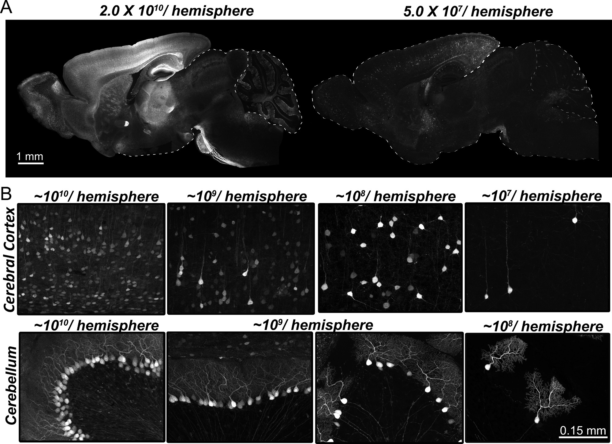
Figure 1. Viral titer can be used to control the density of viral transduction. (A) A broad range of transgene mosaicism can be achieved by diluting the viral solution from 1010 down to 107 particles/hemisphere. Representative images of sagittal sections show two distinct transduction patterns based on viral titer. Images were taken from mice harvested 3 weeks after injection with AAV8-YFP to deliver 2.0 x 10¹⁰ (left) or 5.0 x 10⁷ (right) particles/hemisphere. (B) Higher magnification images of cortex (upper row) and cerebellum (lower row) show the spectrum of transgene mosaicism achieved by serial dilution of AAV8. Transduction is visualized by native YFP fluorescence. Exposure times for whole brain images were determined by the cortex and hippocampus, which fluoresce most brightly, while exposure times for the magnified panels were adjusted to show patterns within each region. Please click here to view a larger version of this figure.
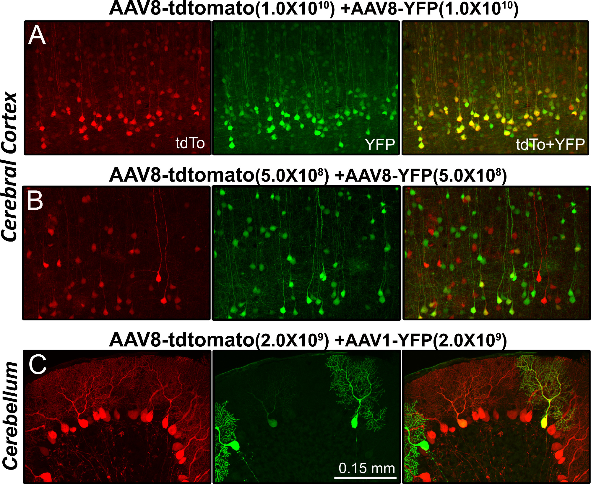
Figure 2. Titer and serotype independently control the degree of viral co-expression. Representative images show the transduction pattern in cerebral cortex (A, B) and cerebellum (C) 3 weeks after viral injection. (A) Co-injection of two AAV8 viruses containing different fluorescent reporters (tdTomato or YFP) produced extensive transduction throughout the brain. The majority of transduced cells expressed both transgenes. (B) Co-injection of the same viruses at lower titers produced sparse expression of each protein, in which fewer cells were transduced by both viruses. (C) Co-injection of AAV packaged into different serotypes (AAV8 and AAV1), even at moderately high titers, produced a largely non-overlapping pattern of viral expression. The pattern of each fluorescent protein was nearly identical to its expression when injected alone, indicating that transduction of one AAV is independent of the other. TdTomato fluorescence shown in red, YFP in green. Transduction is visualized by native fluorescence. Please click here to view a larger version of this figure.
Discussion
We have described a versatile procedure for manipulating neuronal gene expression using AAV as a vehicle for widespread delivery into the neonatal mouse brain. Compared with other methods of neuronal transgenesis such as in utero electroporation1 or stereotaxic intracranial injection2,3, neonatal viral injection is relatively easy and simple. The basic procedure can be performed in minutes with only an ice bucket and a microliter syringe. Optimal survival and transgene expression can be attained by attending to a few technical details, the most important being the quality of viral stocks, the timing and accuracy of injection, and the post-natal care.
The quality of viral preparation is critical for successful transduction. Bad viral preparations will significantly diminish neuronal infectivity and produce significant astrocytic transduction, possibly by phagocytosis of non-infectious particles. Many universities have core laboratories on-site that specialize in viral packaging, and large facilities at the University of North Carolina and the University of Pennsylvania offer high quality off-the-shelf reagents in a variety of serotypes at reduced cost. These facilities also provide custom packaging for vectors that are not available as pre-packaged stocks. Once received into the laboratory, remember that AAV particles can be stable for years at -80 ºC, but are very sensitive to temperature fluctuations. For this reason, aliquots should be stored at -80 °C and should not be refrozen once thawed.
For highest transduction efficiency, it is critical to inject virus as soon as possible after pups are delivered. Based on our studies with AAV8 and those of our collaborators, delaying the injections not only diminishes the spread of virus from the ventricle, but will also bias the transduction from neurons to astrocytes6,7. Therefore we recommend injecting on the day of birth for optimal neuronal expression. At this age - before the cerebrospinal fluid-brain barrier has matured9 - viral particles injected into the lateral ventricle will diffuse throughout the ventricular system and then follow the flow of cerebrospinal fluid into the brain4. Consequently, precise targeting of the lateral ventricle is critical to maximize viral spread. Accurate targeting also minimizes tissue damage from the rapid injection of a relatively large volume of fluid. Therefore, we strongly recommend repeated practice until targeting the lateral ventricles becomes reliable and reproducible. Note that the coordinates provided in this protocol are appropriate for general P0 pups and should be adjusted for the strain and age of experimental pups. Initially, injections should be performed with dye solutions, such as trypan blue or India ink, so the targeting and spread can be visualized by harvesting the brain immediately after injection. If the lateral ventricles have been successfully targeted, the dye will spread throughout the ventricular chambers and be visible all the way from the olfactory bulb to the cerebellum. A stereotaxic device is recommended if the ventricles cannot be reliably targeted by free-hand injection.
If the injections are performed correctly, the proportion of mice lost to injection will be negligible. Instead, the survival rate after injection is most affected by maternal care. Start with healthy animals. The mother’s health is indicated by her behavior and her coat. She should be well-groomed and clean. The litter size can also be an indication of well-being. Healthy young females should produce litters of 6 - 8 pups for C57BL/6, or 10 - 16 pups for ICR. Healthy pups should be pink and wiggly. If pups do not look well or have no milk spot on their belly, they should be transferred to a new foster female immediately. We recommend ICR or FVB for fostering because they produce ample milk and readily accept new pups into their litter. It is imperative that disruptions to the mother are minimized before and after the injections as stress may cause her to reject or cannibalize the pups. Reduce stressors by limiting noise, light, and other disturbances and keeping the cage in a quiet place. Open the cage as little as possible when checking for newborn pups and in the first few days after injection, although it is essential to monitor the mother’s attentiveness to the pups once they are replaced. The mother should display innate behaviors such as bunching the pups in one place and sitting atop the pile of offspring. If the mother does not respond immediately when the pups are returned to the cage, try again to mix the pups with dirty bedding and nesting material from her cage ensure they have acquired her scent. Check again later in the day that the pups still have milk spots on their bellies. If possible, do this by looking into the cage from underneath rather than opening the cage from above. The first days are most critical for survival, but also when the female will be most sensitive to disruption.
When done carefully, intraventricular AAV injection provides a fast and easy means of manipulating neuronal gene expression in vivo without the cost and time of traditional germline transgenesis. The native mosaicism of viral transduction can be harnessed to control the density of expression, making it an ideal approach for experiments to separate cell-intrinsic and cell-extrinsic consequences of transgenesis. In addition, two viruses can be co-injected making it possible to express multiple proteins in either overlapping or distinct neuronal populations. These manipulations highlight the flexibility of the approach, but we believe we’ve only just scratched the surface of its potential. The growing availability of off-the-shelf reagents will make it easier to develop viral injection for new experimental needs, while the emergence of hybrid serotypes with distinct tropisms may expand the cellular repertoire that can be targeted10,11.
Disclosures
Authors declare that they have no competing financial interests.
Acknowledgements
This research was supported by the Robert A. and Rene E. Belfer Family Foundation, NIA R21 AG038856 (JLJ), BrightFocus Foundation Alzheimer’s Disease research grant A2010097 (JLJ), and NIA Biology of Aging Training grant T32 AG000183 (support for SDG).
Materials
| Name | Company | Catalog Number | Comments |
|---|---|---|---|
| ICR outbred mice | Harlan | Hsd:ICR (CD-1) | This strain is also known as CD-1 |
| FVB inbred mice | The Jackson Laboratory | 1800 | 5-6 weeks of age |
| Nestlets | Lab Supply | NESTLETS | |
| Shepherd shacks | Lab Supply | SS-mouse | |
| High fat rodent chow | Purina Mills | PicoLab Mouse diet 20, #5058 | This is our standard breeder chow |
| High fat rodent chow (alternative) | Harlan Laboratories | Teklad Global 19% protein rodent diet #2019S | If low phytoestrogen, autoclavable diet is needed |
| Injection syringe | Hamilton | 7653-01 | 10 ml syringe |
| Injection needles | Hamilton | 7803-04, RN 6PK PT4, 0.375" | 32 G, for standard P0 injections |
| Metal plate for cryoanesthesia | McMaster Carr | 8975K439 | Raw aluminum plate, 6” x 12”, 0.25” thick, will need to be cut into 3 equal pieces and edges sanded by local machine shop |
| Small animal stereotaxic device with digital readout | David Kopf Instruments | Model 940 | |
| Universal syringe holder with needle support foot | David Kopf Instruments | Model 1772-F1 | |
| Neonatal frame | Stoelting | 51625 | Officially called a mouse and neonatal rat adaptor |
| Biohazard disposal bags with sterile indicator | VWR | 14220-030 | Important! – Check with local veterinary and environmental safety staff to learn your institute’s protocol for biohazard waste disposal |
References
- De Vry, J., et al. In vivo electroporation of the central nervous system: a non-viral approach for targeted gene delivery. Progress in neurobiology. 92, 227-244 (2010).
- Sun, B., Gan, L. Manipulation of gene expression in the central nervous system with lentiviral vectors. Methods Mol Biol. 670, 155-168 (2011).
- Puntel, M., et al. Gene transfer into rat brain using adenoviral vectors. Curr Protoc Neurosci. 4, (2010).
- Passini, M. A., Wolfe, J. H. Widespread gene delivery and structure-specific patterns of expression in the brain after intraventricular injections of neonatal mice with an adeno-associated virus vector. J Virol. 75, 12382-12392 (2001).
- Passini, M. A., et al. Intraventricular brain injection of adeno-associated virus type 1 (AAV1) in neonatal mice results in complementary patterns of neuronal transduction to AAV2 and total long-term correction of storage lesions in the brains of beta-glucuronidase-deficient mice. J Virol. 77, 7034-7040 (2003).
- Kim, J. Y., et al. Viral transduction of the neonatal brain delivers controllable genetic mosaicism for visualising and manipulating neuronal circuits in vivo. Eur J Neurosci. 37, 1203-1220 (2013).
- Chakrabarty, P., et al. Capsid serotype and timing of injection determines AAV transduction in the neonatal mice brain. PloS one. 8, (2013).
- Croyle, M. A., Cheng, X., Wilson, J. M. Development of formulations that enhance physical stability of viral vectors for gene therapy. Gene therapy. 8, 1281-1290 (2001).
- Bauer, H. C., Bauer, H. Neural induction of the blood-brain barrier: still an enigma. Cellular and molecular neurobiology. 20, 13-28 (2000).
- Cearley, C. N., et al. Expanded repertoire of AAV vector serotypes mediate unique patterns of transduction in mouse brain. Molecular therapy : the journal of the American Society of Gene Therapy. 16, 1710-1718 (2008).
- Grimm, D., et al. In vitro and in vivo gene therapy vector evolution via multispecies interbreeding and retargeting of adeno-associated viruses. J Virol. 82, 5887-5911 (2008).
Reprints and Permissions
Request permission to reuse the text or figures of this JoVE article
Request Permission