Demyelinating diseases can be modeled in animals by focal application of lysolecithin into the CNS. A single injection of lysolecithin into mouse spinal cord produces a lesion that spontaneously repairs over time. The goal is to study factors involved in de- and remyelination, and to test agents for enhancing repair.
Method Article
Experimental Demyelination and Remyelination of Murine Spinal Cord by Focal Injection of Lysolecithin
Opens in a new tab
In This Article
Summary
Loading...
$$\rightleftharpoonup{xx}$$
$$\longleftharp{xx}$$,
$$\longrightharp{xx}$$,
Abstract
Loading...
$$\rightleftharpoonup{xx}$$
$$\longleftharp{xx}$$,
$$\longrightharp{xx}$$,
Multiple sclerosis is an inflammatory demyelinating disease of the central nervous system characterized by plaque formation containing lost oligodendrocytes, myelin, axons, and neurons. Remyelination is an endogenous repair mechanism whereby new myelin is produced subsequent to proliferation, recruitment, and differentiation of oligodendrocyte precursor cells into myelin-forming oligodendrocytes, and is necessary to protect axons from further damage. Currently, all therapeutics for the treatment of multiple sclerosis target the aberrant immune component of the disease, which reduce inflammatory relapses but do not prevent progression to irreversible neurological decline. It is therefore imperative that remyelination-promoting strategies be developed which may delay disease progression and perhaps reverse neurological symptoms. Several animal models of demyelination exist, including experimental autoimmune encephalomyelitis and curprizone; however, there are limitations in their use for studying remyelination. A more robust approach is the focal injection of toxins into the central nervous system, including the detergent lysolecithin into the spinal cord white matter of rodents. In this protocol, we demonstrate that the surgical procedure involved in injecting lysolecithin into the ventral white matter of mice is fast, cost-effective, and requires no additional materials than those commercially available. This procedure is important not only for studying the normal events involved in the remyelination process, but also as a pre-clinical tool for screening candidate remyelination-promoting therapeutics.
Introduction
Loading...
$$\rightleftharpoonup{xx}$$
$$\longleftharp{xx}$$,
$$\longrightharp{xx}$$,
Multiple sclerosis (MS) is a chronic demyelinating disease of the central nervous system (CNS) characterized by immune cell infiltration and plaques containing lost myelin, oligodendrocytes, axons and neurons. Most patients have a disease course consisting of inflammatory relapses accompanied by a wide range of neurological symptoms, followed by periods of remission. Over half of these patients eventually transition to a secondary progressive stage with no apparent relapses but continual neurological decline. It is believed that this progressive deterioration is due to axonal damage and loss, contributed in part by chronic demyelination. Strategies to restore lost myelin are thus considered a promising treatment approach to delay disease progression and perhaps reverse neurological symptoms.
Remyelination is an endogenous repair response in the CNS whereby new myelin sheaths are generated from recruited oligodendrocyte precursor cells (OPCs) that differentiate into myelin-forming oligodendrocytes. Remyelination has been shown in animal models to be quite robust1-3, however, its efficiency declines with age4. Indeed, remyelination occurs in humans although it is incomplete in the majority of MS patients5. All currently available medications for MS primarily target the aberrant immune component of the disease and, while effective at reducing relapses, do not markedly delay disease progression. The next generation of therapeutic strategies for the management of MS will integrate advances in immunomodulation with the enhancement of endogenous remyelination in order to prevent both relapses and progression6.
One method to study de- and remyelination in the CNS involves the direct injection of the detergent lysophosphatidylcholine (lysolecithin) into the spinal cord white matter1,3,7. This procedure produces a well characterized demyelinating injury consisting principally of macrophage/microglial infiltration and activation8,9, reactive astrogliosis, perturbation of axonal homeostasis/axonal injury, and OPC proliferation and migration10. The lesion predictably evolves over the period of a few weeks and is eventually capable of fully remyelinating. This method has been particularly useful in studying the choreography of events involved in de- and remyelination. Further, it has been adopted as a tool for pre-clinical testing of candidate therapies to accelerate repair following a demyelinating insult.
Protocol
Loading...
$$\rightleftharpoonup{xx}$$
$$\longleftharp{xx}$$,
$$\longrightharp{xx}$$,
NOTE: The animals used in this procedure were cared for in accordance with the Canadian Council on Animal Care (CCAC) guidelines. Ethics were approved by the Animal Care Committee of the University of Calgary.
1. Prepare Syringe for Injection
- Dissolve lysolecithin to a 1% solution in phosphate-buffered saline (PBS; pH 7.4) and store at -20 °C in small aliquots (75 μl). Thaw a vial to RT.
NOTE: if lysolecithin is undissolved, sonicate the tube in an ultrasonic cleaner (40 kHz) for approximately 30 min to form a uniform solution. - Handle the pre-pulled glass capillary with extreme care to avoid damaging the delicate tip. Unscrew the nut of a 10 μl injecting syringe and thread it onto the flat end of the capillary followed by 2 ferrules, ensuring that the mating ends of the ferrules align and the capillary is snug in the conical ferrule (Figure 1). Rinse the syringe with isopropyl alcohol and remove the plunger. Once dry, screw the needle assembly hand tight onto the injecting syringe.
- Attach the metal hub needle to the priming syringe. Pierce a rubber disc with the needle and slide it down to the base. Fill the priming syringe with lysolecithin. Gently depress the priming syringe until liquid is visible at the tip of the needle. This ensures air bubbles will not be introduced into the injecting syringe.
- Insert the metal hub needle of the priming syringe into the injecting syringe. Making a firm seal with the rubber disc, slowly depress the priming syringe until the capillary fills to the tip with solution. Carefully remove the priming syringe while simultaneously depressing to fill the barrel of the injecting syringe without introducing air bubbles. Insert the plunger into the injecting syringe and ensure solution flows from the tip of the capillary as the plunger is gently depressed.
NOTE: If any air bubbles are visible in the capillary or injecting syringe, the syringe preparation must be repeated from the beginning. Continuous fluid in the injecting apparatus is critical to ensure accurate injection volumes. - Attach the completed injecting syringe to the arm of a stereotactic micromanipulator. This completed apparatus will be able to inject 15-20 animals before needing to be refilled.
- Discard the remaining lysolecithin from the priming syringe. Withdraw and depress isopropyl alcohol several times, and detach the metal hub needle. Wait several hours for the remaining isopropyl alcohol in the priming syringe to evaporate before filling again.
2. Prepare Animal for Surgical Procedure
NOTE: This procedure is described for female C57BL/6 mice, aged 8-10 weeks.
- Anesthetize the animal with an intraperitoneal injection of ketamine (200 mg/kg) and xylazine (10 mg/kg) or per institutional animal care regulations. Plan for the animal to be under anesthesia for approximately 1 hr if using injectable anesthesia.
- Test that the animal is deeply anesthetized by firmly pinching the foot. A properly anesthetized animal will not respond to the pinch.
- Using clippers, shave a 2-3 cm2 area on the dorsal side of the animal, close to the ears. Be careful not to damage the ears.
- Wipe the area clean with 70% ethanol applied to a gauze pad. Ensure all clipped hair has been removed from the area. Disinfect the area with iodine.
- Apply petroleum jelly to the eyes to prevent drying throughout the procedure.
- Keep the animal in a heated recovery chamber until ready to begin the procedure.
3. Perform the Surgical Procedure
NOTE: Ensure adequate aseptic technique for all steps of the procedure. This includes proper use of gloves, hairnets, masks, and drapes. All tools should be sterilized before coming in contact with the animal.
- Move the animal to a stereotactic frame, dorsal side up, elevated at the mid section by folded paper towels to exaggerate the curvature of the spine. Fasten the arms and tail with surgical tape and secure the head with a tooth clamp. Stabilizing ear bars are not necessary for this procedure.
- Use a scalpel to make a 3 cm midline incision, starting just below the ears and cutting in the caudal direction.
- Locate the divide between the 2 large adipose structures and use fine forceps in each hand to pull these apart. Spread retractors to open the surgical field.
- Under a surgical microscope, locate the prominent outgrowth process of the T2 vertebra (Note: this feature is characteristic of the C57BL/6 mouse strain). Perform a blunt dissection with closed spring scissors through the overlaying musculature to better visualize T2. Using the forceps, feel for the hard surfaces of T3 and T4 to confirm proper anatomical location.
- Using spring scissors, make shallow lateral cuts (2-3 mm deep) of the connective tissue between T3 and T4. Due to natural spacing between vertebrae in the upper thoracic portion of the mouse vertebral column, a laminectomy is not necessary to reveal the spinal cord. Be mindful that too deep a cut will pierce and damage the cord.
NOTE: A small degree of bleeding is common during this step. If this occurs, hold a sponge spear into the area until the bleeding subsides (30-60 sec). - Visualize the spinal cord. It will be covered with a thick layer of visible dura if this meningeal layer was not yet cut while exposing the cord. A prominent blood vessel runs caudal/rostral through the approximate midline of the spinal cord.
NOTE: This vasculature should not be used as a landmark for the midline. Instead, adequate lighting should reveal the grey-white matter boundaries flanking the dorsal column, and these should be used to estimate the midline. - If the dura remains intact, make gentle lateral scrapes with a 32 G metal needle until it is cleared. The goal is to remove the dura while not cutting the remaining underlying meninges, which are not as thick and harder to see.
NOTE: Release of cerebrospinal fluid indicates a breach of the arachnoid and while this can occur without mechanical damage to the tissue, the accumulated cerebrospinal fluid should be removed with a sponge spear to better visualize the surface of the cord. - Move the injecting syringe in place and slowly lower it until the tip of the capillary just barely touches the spinal cord immediately lateral of either side of the midline. Lock the arm in place.
- Use the graded measurements of the Z-direction stereotactic arm to make a baseline position measurement. From this reading, subtract 1.3 mm. Use a quick and shallow downward motion to pierce the tissue and then carefully lower the capillary until the new measurement is reached. Optional: if desired, lesions in the dorsal column can be produced by the same piercing motion at the midline and a depth of 0.3 mm (see discussion for more information).
NOTE: These values are specific for injecting between T3 and T4. If opting to perform the injection at any other location in the spinal cord, these values must be derived from any available mouse brain atlas. - Use the micromanipulator to depress lysolecithin into the ventral spinal cord white matter. Make 1 rotation of the micromanipulator every 5 sec for 2 min, resulting in a final volume of 0.5 μl. Leave the capillary in place for 2 additional min to prevent backflow of solution, and then carefully remove the capillary.
- Tie a single suture in the muscle/adipose tissue overlaying the spinal column. Use a non-interrupted suture to close the skin. Apply more iodine to the incision site.
- Place the animal in a heated recovery chamber until it recovers, then return it to its cage. Apply analgesics post-operatively as per institutional animal care regulations. Additional post-operative care is usually not necessary as the animals are fully ambulatory and capable of self-feeding and drinking as soon as they recovery from anesthesia.
- Repeat the procedure for the remaining animals.
NOTE: With proficiency, the operation can be completed in 10-15 min per animal, particularly with the help of a second person tying sutures. The same glass capillary can be used for approximately 15-20 surgeries before the tip becomes blunt and should be replaced. Control surgeries can be performed identically as described, with an injection of PBS into the spinal cord instead of lysolecithin. We do not recommend cleaning the capillaries for future use.
4. Tissue Processing and Analysis
- Sacrifice the animals with an intraperitoneal overdose of ketamine (500 mg/kg) and xylazine (25 mg/kg) at desired time points. Lesions typically evolve in the following manner: 1-3 days, active demyelination; 3-7 days, OPC recruitment; 7-10 days, oligodendrocyte differentiation; 10-21 days, active remyelination2,10,11.
- To prepare tissue for histology, first perform a transcardial perfusion12 with 20 ml RT PBS followed by 20 ml ice-cold 4% paraformaldehyde in PBS. For preparation of resin embedded tissue for semi- or ultrathin sectioning, see step 4.9.
- Remove the spinal cord using curved bone scissors to cut through each vertebrae starting at the cervical end of the spinal column and working down to the lower thoracic level. Fix the spinal cords O/N in 4% paraformaldehyde in PBS at 4 °C. Switch the cords to 30% sucrose in PBS at 4 °C for at least 72 hr.
- Identify the injection site as an abnormality on the dorsal surface of the spinal cord. Cut a 3 mm piece of tissue (with the injection site in the center) and align the pieces in cryomolds containing optimal cutting temperature (OCT) compound. Suspend the underside of the cryomolds in a chilled mixture of 2-methylbutane and dry ice until the OCT is completely frozen. Store at -80 °C until ready to section.
- Section the spinal cords on a cryostat at a width of 20 μm onto microscope slides and allow to air dry O/N before storing the slides at -20 °C.
- Detect lesion location and size using a eriochrome cyanine histological stain of myelin13. Perform all steps at RT. Air-dry slides for 30 min. Place slides in clearing agent for 1 min followed by rehydration in graded ethanol solutions (100%, 95%, 90%, 70%, 50%, water) for 1 min each.
- Place slides in eriochrome cyanine solution for 15 min, followed by a 1 min wash with water. Differentiate in 0.5% ammonium hydroxide for 10 sec, followed by a 1 min wash with water. Dehydrate in graded ethanol solutions (reverse order as described above) for 1 min each, coverslip with mounting media, and image on a bright-field microscope.
- Use immunohistochemistry to visualize axons and myelin. Air-dry slides for 30 min. Dehydrate with graded ethanol (water, 50%, 70%, 90%, 95%, 100%) for 2 min each at RT, then reverse order back to water. This step results in greater resolution of individual myelin rings14.
- Block non-specific antibody interactions using 10% goat serum and 0.25% triton X-100 in PBS for 60 min at RT. Dilute primary antibodies (mouse anti-SMI312 1:2,000, rabbit anti-MBP 1:1,000) in PBS and incubate on slides O/N at 4 °C.
- Wash 5 times for 5 min with PBS at RT. Dilute secondary antibodies (Alexa 488 anti-rabbit 1:500, Alexa 546 anti-mouse 1:500) in PBS and incubate on slides for 60 min at RT. Wash 5 times for 5 min with PBS at RT. Mount slides with coverslips using gelvatol and image on a fluorescent microscope.
- Use immunohistochemistry to visualize cells of the oligodendrocyte lineage. Follow the procedure for step 4.7, omitting the dehydration/rehydration step, and using 10% horse serum instead of goat serum. Use the following primary antibody dilutions: goat anti-PDGFRα 1:100, mouse anti-CC1 1:200, rabbit anti-Olig2 1:200. Use the following secondary antibody dilutions: Alexa 488 anti-goat 1:500, Alexa 594 anti-mouse 1:500, Alexa 647 anti-rabbit 1:500.
- To prepare tissue for semi- or ultrathin sectioning, first perform a transcardial perfusion12 with 20 ml RT PBS followed by 20 ml ice-cold 4% paraformaldehyde/1% glutaraldehyde in PBS. Remove spinal cord as described in step 4.3. Fix O/N in 4% paraformaldehyde/1% glutaraldehyde in PBS at 4 °C.
- Identify the injection site as described in step 4.4. Cut a 1 mm piece of tissue containing the injection site. Additional 1 mm blocks on either side of the lesion can be prepared as well if desired.
- In a fume hood, add 10 times volume 1% osmium tetroxide/1.5% potassium ferrocyanide to each block on ice for 60 min.
NOTE: osmium tetroxide is a highly toxic substance and should be handled with extreme care. All materials in contact with osmium tetroxide should be placed in corn oil (twice volume as osmium tetroxide solution) for neutralization. - Wash cords 3 times with 0.2 M cacodylate for 10 min at RT, discarding in corn oil. Dehydrate with graded ethanol solutions (water, 50%, 70%, 90%, 2 times with 95%, 2 times with 100%, 2 times with propylene oxide) for 10 minutes at RT.
- Embed cords in resin as per manufacturer’s instructions.
- Cut blocks on an ultramicrotome and mount sections onto water droplets on microscope slides. Place slides on a hot plate (approximately 50 °C) until dry.
- Visualize myelin by adding a few drops of 1% toluidine blue/2% borax solution onto the slides for 10-15 sec at 50 °C. Rinse with water, dry, and coverslip with mounting media. Image sections on a bright-field microscope.
Results
Loading...
$$\rightleftharpoonup{xx}$$
$$\longleftharp{xx}$$,
$$\longrightharp{xx}$$,
Focal injection of lysolecithin into the ventral white matter produces a discrete demyelinating lesion that is detectable over a distance of approximately 3 mm (Figure 2). Immunohistochemical staining of the lesion core for myelin (MBP) and axons (SMI312) shows axons that have been stripped of myelin at 7 days (Figure 3). By 14 days, many axons are surrounded by MBP-positive rings, which suggests the occurrence of remyelination. Staining for cells of the oligodendrocyte lineage (PDGFRα, Olig2, CC1), there is a significant increase in both the total number of cells at 14 days compared to 7 days, as well as the distribution of mature oligodendrocytes compared to OPCs (Figure 4). Consistent with this finding, semithin sections stained with toluidine blue reveal the presence of thin myelin sheaths at 14 days that are rarely detected at 7 days (Figure 5), indicating that these are remyelinated internodes.
The procedure is highly reproducible between animals. Variation occurs when heavy breathing alters the stationary position of the capillary—this is usually not an issue with adequate sedation. Damage to axons appears to be minimal, except in the very center of the lesion, which has been described since the earliest use of the model1. We believe this to be mechanical injury from the glass capillary, as it is also observable in PBS injected controls. Nevertheless, variability tends to be small, and we and others using a similar procedure have detected differences between experimental conditions with as few as 4 animals per group15.
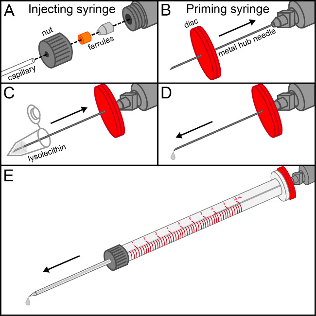
Figure 1. Assembly of the injecting syringe. (A) The nut of the injecting syringe is threaded onto the flat end of the glass capillary, followed by the 2 ferrules such that their mating ends interlock. Once the capillary is firmly snug in the conical ferrule, the assembly is screwed hand tight onto the end of the injecting syringe. (B) Piece the center of a rubber disc with the metal hub needle attached to the priming syringe and slide it down to the base. (C) Withdraw lysolecithin solution into the priming syringe. (D) Gently depress the priming syringe until the first drop of lysolecithin is visible at the tip of the needle. (E) Insert the priming syringe into the barrel of the injecting syringe, making a firm seal with the rubber disc. Gently depress the solution until it runs to the end of the capillary. Carefully withdraw the priming syringe while maintaining pressure on the plunger to remove the metal hub needle without introducing air bubbles into the injecting syringe. Please click here to view a larger version of this figure.
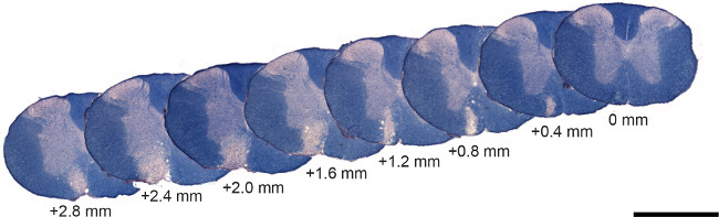
Figure 2. Representative lysolecithin lesion stained with eriochrome cyanine. Serial sections (spaced 400 μm apart) of a characteristic lysolecithin lesion at 14 days stained with eriochrome cyanine to visualize myelin (blue). Note that demyelination is restricted to the ventral white matter and that the lesion spans approximately 3 mm in the rostral/caudal direction. Scale bar = 1 mm. Please click here to view a larger version of this figure.
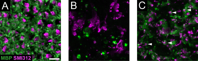
Figure 3. Axons and myelin in the lysolecithin model. (A) A sham (PBS) injected spinal cord at 7 days shows healthy axons that are surrounded by myelin rings. (B) A lysolecithin lesion at 7 days shows denuded axons as well as degraded myelin. (C) At 14 days, a proportion of axons (examples denoted with white arrowheads) are associated with the reappearance of myelin rings. Scale bar = 10 μm. Please click here to view a larger version of this figure.
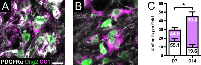
Figure 4. Oligodendrocyte lineage cells in the lysolecithin model. (A, C) At 7 days, there is a greater representation of PDGFRα+ OPCs (white bars) relative to CC1+ oligodendrocytes (magenta bars); the numbers within the bars represent percentages. Olig2 was probed to confirm staining of oligodendrocyte lineage cells. (B, C) At 14 days, there is a significant increase in the total number of oligodendrocyte lineage cells compared to 7 days (p <0.05, two-tailed t-test, n = 4 per group). There is also a significant increase in the distribution of oligodendrocytes to OPCs at 14 days compared to 7 days (p <0.0001, Fisher’s exact test). Scale bar = 10 μm. Each field is captured at an original magnification of 60X. Values are mean ± SD. * signifies p <0.05. Please click here to view a larger version of this figure.
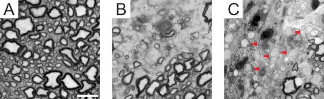
Figure 5. Remyelination in the lysolecithin model. (A) A cross-sectional toluidine blue stained semithin section of the ventral white matter in a healthy mouse shows axons over a wide range of caliber with respective myelin thickness. (B) At 7 days, a lack of myelin sheaths is observed adjacent to an unaffected area (bottom right). (C) At 14 days, thinly myelinated sheaths (examples denoted with red arrowheads) appear throughout the lesion, indicative of remyelinated segments. Scale bar = 10 μm. Please click here to view a larger version of this figure.
Discussion
Loading...
$$\rightleftharpoonup{xx}$$
$$\longleftharp{xx}$$,
$$\longrightharp{xx}$$,
A number of animal models have been developed to study MS, most recognizably the experimental autoimmune encephalomyelitis (EAE) model. In EAE, rodents are immunized against a fragment of a myelin peptide and undergo inflammatory lesion development manifesting in ascending paralysis. While this model has been useful for pre-clinical testing of immunomodulatory MS drugs, it is not ideal for studying remyelination for three main reasons: Firstly, the location of inflammatory lesions is somewhat random, and locating lesions when processing tissue for semi- or ultrathin sections can be challenging. The second is that remyelination occurs over a specific time course, and the exact age of a single EAE lesion cannot be known without continual non-invasive magnetic resonance imaging. The third is that remyelination is a naturally occurring phenomenon in rodents, and evidence of remyelination following drug treatment in EAE may not be a primary result of the drug, but instead a secondary phenomenon of reducing inflammation.
Another common method of producing demyelination is achieved by introducing the copper chelator cuprizone in the diet. This results in widespread demyelination, most notably in the corpus callosum. There are limitations in studying the corpus callosum as a site of remyelination for the following reasons: Firstly, axon diameters (and thus myelin thickness) are smaller than other CNS regions, and thus thinly remyelinated sheaths can be indistinguishable from those that were never demyelinated. Secondly, because the mouse corpus callosum contains >70% unmyelinated axons16, it can be unclear whether a remyelinated segment is true repair of damaged myelin or de novo myelin synthesis in the adult, which occurs normally17.
It is of our belief that the best model for studying remyelination is the direct injection of toxins, either lysolecithin, ethidium bromide, or others, into the caudal cerebral peduncles18 or the spinal cord white matter. The former location is achieved only by precise 3-dimension stereotactic injection, and is limited to larger rodents (rats) due to the small size of the cerebellar peduncles. This excludes the extensive resource of transgenic mice in studying de- and remyelination. The spinal cord, however, contains many large white matter tracts that are easily accessible surgically. Spaces between vertebrae in the rostral thoracic segment allows for exposure of the spinal cord without the need for a laminectomy, which is a necessary step in caudal thoracic surgical procedures. An advantage of specifically targeting the ventral white matter is that the axons are uniformly larger than the dorsal white matter, making quantification of remyelination a less ambiguous task—similar to the challenges associated with the corpus callosum. Additionally, the ventral white matter makes up a much larger target area to inject; several hundred microns laterally in the dorsal region would place the capillary outside the column, while the same deviation ventrally would still produce a prominent demyelinating lesion. Some protocols inject lysolecithin into both the dorsal and ventral columns of the same animal19. This can increase both the likelihood of proper capillary placement and the number of quantifiable lesions in fewer animals. While the current data presented is from 8-10 week old animals at time of operation, we have also had success using the same procedure on 8-10 month old mice, where remyelination is described as being markedly slower4.
Quantification of remyelination is not a trivial undertaking. A central dogma posits that remyelinated segments are shorter in length and thinner on average than their healthy counterparts, and thus g-ratio calculations (axon diameter divided by axon + myelin diameter) of cross-sectional semi- or ultrathin sections have become standard procedure. However, it is known that remyelinated segments thicken over time2 and a recent study using a transgenic reporter of remyelinating oligodendrocytes suggests that many internodes eventually become indistinguishable from control20. Quantifying the number of mature oligodendrocytes within the lesion is an indirect way to measure repair, as oligodendrocytes are capable of making a wide number of internodes, and a significant proportion of remyelination—depending on the model used—can occur from Schwann cells3. Of course, as remyelination has been linked to restoration of saltatory conduction21, the ultimate metric of repair would be functional recovery of neurological deficits. While remyelination has been linked to recovery of function in some species22,23, it has not become a standard procedure in murine lysolecithin studies. This is likely due to a lack of overt observable deficits from either dorsal or ventral lesions, compared to more robust demyelination models such as EAE and even cuprizone. We believe that functional deficits resulting from lysolecithin injection, and subsequent recovery with remyelination, will only be observable using sensitive tests of fine sensorimotor functioning.
A PubMed search of “remyelination” alongside either of the animal models listed above, albeit a brusque methodological approach, shows fewer search hits for lysolecithin (109) compared to EAE (188) and cuprizone (197). If our argument that lysolecithin demyelination is the superior approach for studying remyelination, why is it the least discussed? Perhaps an apprehension for using this method derives from a belief of technical difficulty in performing the surgical operation. In actuality, this procedure is fast, cost-effective, and is no more difficult than routine tissue dissection, requiring materials that are all commercially available. It is our hope that this protocol proves useful for those that wish to add this powerful model to their repertoire for studying the exciting and expanding field of myelin repair.
Disclosures
Loading...
$$\rightleftharpoonup{xx}$$
$$\longleftharp{xx}$$,
$$\longrightharp{xx}$$,
The authors have nothing to disclose.
Acknowledgements
Loading...
$$\rightleftharpoonup{xx}$$
$$\longleftharp{xx}$$,
$$\longrightharp{xx}$$,
This project was funded by a grant from the Multiple Sclerosis Society of Canada and the Alberta Innovates - Health Solutions CRIO Team program. MBK is a recipient of studentships from Alberta Innovates - Health Solutions and the Multiple Sclerosis Society of Canada. SKJ is funded by a graduate student support grant from the Alberta endMS Regional Research and Training Center of the Multiple Sclerosis Society of Canada. The authors wish to acknowledge Dr. Jan van Minnen and the Regeneration Unit in Neurobiology core facility for training and use of equipment.
Materials
| Name | Company | Catalog Number | Comments |
|---|---|---|---|
| spring scissors | Fine Science Tools | 15004-08 | |
| forceps | Fine Science Tools | 11254-20 | |
| retractor | Fine Science Tools | 17003-03 | |
| clippers | Philips | QG3330 | |
| heating recovery chamber | Peco Services | V1200 | |
| surgical tape | 3M | 1527-1 | |
| scalpel handle | Fine Science Tools | 10003-12 | |
| scalpel blade | Feather | No. 15 | |
| sponge spear | Beaver Visitec | 581089 | |
| 5-0 Vicryl sutures | Ethicon | J511G | |
| curved ToughCut spring scissors | Fine Science Tools | 15123-12 | |
| 32 G metal needle | BD | 305106 | |
| needle holders | Fine Science Tools | 12002-14 | |
| angled forceps | Fine Science Tools | 11251-35 | |
| cotton tipped applicator | Puritan | 806-WC | |
| gauze pads | Safe Cross First Aid | 3763 | |
| 10 μl syringe | Hamilton | 7635-01 | make sure to purchase the microliter, not gastight syringe |
| compression fitting | Hamilton | 55750-01 | contains the 2 ferrules and removable nut, but the nut that comes with the 10 μl syringe is a tighter fit |
| priming kit | Hamilton | PRMKIT | contains the priming syringe, removable hub needle and the rubber discs |
| pre-pulled glass capillaries | WPI | TIP10TW1 (pack of 10) | contains capillaries with 10 μm inner diameter. 30 μm inner diameter also work (TIP30TW1) |
| stereotactic frame | David Kopf instruments | Model 900 | |
| Ultrasonic cleaner | Fisher Scientific | FS-20 | |
| Tissue-Tek optimal cutting temperature (OCT) compound | VWR | 25608-930 | |
| Tissue-Tek intermediate cryomold | VWR | 25608-924 | |
| cryostat | Leica | CM1900 | |
| microscope slides | VWR | 48311-703 | |
| bright field microscope | Olympus | BX51 | |
| ultramicrotome | Leica EM UC7 | EM UC7 | |
| Lysophosphatidylchoine | Sigma | L1381 | |
| 2-methylbutane | Sigma | M32631 | |
| Triton x-100 | Sigma | X-100 | |
| goat serum | Sigma | G9023 | |
| Mouse anti-SMI312 antibody | Covance | SMI-312R | 1:2,000 dilution |
| Rabbit anti-MBP antibody | Abcam | AB40390 | 1:1000 dilution |
| Goat anti-PDGFRα antibody | R&D Systems | AF1062 | 1:100 dilution |
| Rabbit anti-Olig2 antibody | Millipore | AB9610 | 1:200 dilution |
| Mouse anti-CC1 antibody | Calbiochem | OP80 | 1:200 dilution |
| Alexa Fluor 488 goat anti-rabbit IgG | Life technologies | A-11008 | 1:500 dilution |
| Alexa Fluor 546 goat anti-mouse IgG | Life technologies | A-11003 | 1:500 dilution |
| Alexa Fluor 488 donkey anti-goat IgG | Jackson Immuno | 705-546-147 | 1:500 dilution |
| Alexa Fluor 594 donkey anti-mouse IgG | Jackson Immuno | 715-586-151 | 1:500 dilution |
| Alexa Fluor 647 donkey anti-rabbit IgG | Jackson Immuno | 711-606-152 | 1:500 dilution |
| PBS | Oxoid | BR0014 | |
| isopropyl alcohol | Sigma | 109827 | |
| ketamine | CDMV | ||
| xylazine | CDMV | ||
| iodine | West Penetone | 2021 | |
| Vaseline petroleum jelly | VWR | CA05971 | |
| paraformaldehyde | Sigma | P6148 | |
| sucrose | Sigma | S5016 | |
| Eriochrome Cyanine R | Sigma | 32752 | |
| sulfuric acid | Sigma | 320501 | |
| iron(III) chloride | Sigma | 157740 | |
| ammonium hydroxide | Sigma | 320145 | |
| Acrytol | Leica Biosystems | 3801700 | |
| Citrisolv | Fisher Scientific | 22-143-975 | |
| horse serum | Sigma | H0146 | |
| glutaraldehyde | Electron Micrscopy Sciences | 16220 | |
| osmum tetroxide | Electron Micrscopy Sciences | 19150 | highly toxic |
| potassium hexacyanoferrate (II) trihydrate | Sigma | P3289 | |
| corn oil | Sigma | C8267 | |
| polyvinyl alcohol | Sigma | P8136 | |
| glycerol | Sigma | G9012 | |
| cacodylic acid | Electron Micrscopy Sciences | 12300 | |
| propylene oxide | Electron Micrscopy Sciences | 20401 | |
| EMBED kit | Electron Micrscopy Sciences | 14120 | |
| Toluidine Blue O | Sigma | T3260 | |
| Sodium tetraborate decahydrate | Sigma | S9640 | |
| Recipes | |||
| Eriochrome cyanine solution | |||
| Ingredient | Amount to add | ||
| Eriochrome Cyanine R | 0.8 g | ||
| sulfuric acid | 400 ml 0.5% | ||
| iron(III)chloride | 20 ml 10% | ||
| water | 80 ml | ||
| *add eriochrome cyanine to sulfuric acid, followed by iron(III) chloride and water. Solution can be kept after use. Make fresh once per year. | |||
| Gelvatol | |||
| Ingredient | Amount to add | ||
| PBS | 140 mL | ||
| polyvinyl alcohol | 20 g | ||
| glycerol | 40 g | ||
| *mix well, place at 37 °C O/N, centrifuge at 1,960 x g 30 min, aliquot into 40 tubes |
References
Loading...
$$\rightleftharpoonup{xx}$$
$$\longleftharp{xx}$$,
$$\longrightharp{xx}$$,
- Blakemore, W. F., Eames, R. A., Smith, K. J., McDonald, W. I. Remyelination in the spinal cord of the cat following intraspinal injections of lysolecithin. J. Neurol. Sci. 33 (1-3), 31-43 (1977).
- Jeffery, N. D., Blakemore, W. F. Remyelination of mouse spinal cord axons demyelinated by local injection of lysolecithin. J. Neurocytol. 24 (10), 775-781 (1995).
- Blakemore, W. F. Invasion of Schwann-cells into the spinal-cord of rat following local injections of lysolecithin. Neuropathol. Appl. Neurobiol. 2 (1), 21-39 (1976).
- Shields, S. A., Gilson, J. M., Blakemore, W. F., Franklin, R. J. Remyelination occurs as extensively but more slowly in old rats compared to young rats following gliotoxin-induced CNS demyelination. Glia. 28 (1), 77-83 (1999).
- Patrikios, P., et al. Remyelination is extensive in a subset of multiple sclerosis patients. Brain. 129 (Pt 12), 3165-3172 (2006).
- Keough, M. B., Yong, V. W. Remyelination therapy for multiple sclerosis). Neurotherapeutics. 10 (1), 44-54 (2013).
- Hall, S. M. The effect of injections of lysophosphatidyl choline into white matter of the adult mouse spinal cord. J. Cell Sci. 10 (2), 535-546 (1972).
- Miron, V. E., et al. M2 microglia and macrophages drive oligodendrocyte differentiation during CNS remyelination. Nat. Neurosci. 16 (9), 1211-1218 (2013).
- Kotter, M. R., Setzu, A., Sim, F. J., Van Rooijen, N., Franklin, R. J. Macrophage depletion impairs oligodendrocyte remyelination following lysolecithin-induced demyelination. Glia. 35 (3), 202-212 (2001).
- Lau, L. W., et al. Chondroitin sulfate proteoglycans in demyelinated lesions impair remyelination. Ann. Neurol. 72 (3), 419-432 (2012).
- Fancy, S. P., et al. Dysregulation of the Wnt pathway inhibits timely myelination and remyelination in the mammalian CNS. Genes Dev. 23 (13), 1571-1585 (2009).
- Gage, G. J., Kipke, D. R., Shain, W. Whole animal perfusion fixation for rodents. J. Vis. Exp. (65), (2012).
- Skihar, V., et al. Promoting oligodendrogenesis and myelin repair using the multiple sclerosis medication glatiramer acetate. Proc. Natl. Acad. Sci. U.S.A. 106 (42), 17992-17997 (2009).
- Plemel, J. R., et al. Platelet-derived growth factor-responsive neural precursors give rise to myelinating oligodendrocytes after transplantation into the spinal cords of contused rats and dysmyelinated mice. Glia. 59 (12), 1891-1910 (2011).
- Chong, S. Y., et al. Neurite outgrowth inhibitor Nogo-A establishes spatial segregation and extent of oligodendrocyte myelination. Proc. Natl. Acad. Sci. U.S.A. 109 (4), 1299-1304 (2012).
- Sturrock, R. R. Myelination of the mouse corpus callosum. Neuropathol. Appl. Neurobiol. 6 (6), 415-420 (1980).
- Young, K. M., et al. Oligodendrocyte dynamics in the healthy adult CNS: evidence for myelin remodeling. Neuron. 77 (5), 873-885 (2013).
- Woodruff, R. H., Franklin, R. J. Demyelination and remyelination of the caudal cerebellar peduncle of adult rats following stereotaxic injections of lysolecithin, ethidium bromide, and complement/anti-galactocerebroside: a comparative study. Glia. 25 (3), 216-228 (1999).
- Mei, F., et al. Micropillar assays as a high-throughput screening platform for therapeutics in multiple sclerosis. Nat. Med. 20 (8), 954-960 (2014).
- Powers, B. E., et al. Remyelination reporter reveals prolonged refinement of spontaneously regenerated myelin. Proc. Natl. Acad. Sci. U.S.A. 110 (10), 4075-4080 (2013).
- Smith, K. J., Blakemore, W. F., McDonald, W. I. Central remyelination restores secure conduction. Nature. 280 (5721), 395-396 (1979).
- Jeffery, N. D., Blakemore, W. F. Locomotor deficits induced by experimental spinal cord demyelination are abolished by spontaneous remyelination. Brain. 120 (Pt 1), 27-37 (1997).
- Duncan, I. D., Brower, A., Kondo, Y., Curlee, J. F. Jr, Schultz, R. D. Extensive remyelination of the CNS leads to functional recovery. Proc. Natl. Acad. Sci. U.S.A. 106 (16), 6832-6836 (2009).
Reprints and Permissions
Request permission to reuse the text or figures of this JoVE article
Request Permission