In order to study brain reorganization under pathological conditions we used miniosmotic pumps for direct protein delivery into the brain circumventing the blood brain barrier. Tract tracers are then injected to study alterations in brain connectivity under the influence of the protein.
Method Article
Implantation of Miniosmotic Pumps and Delivery of Tract Tracers to Study Brain Reorganization in Pathophysiological Conditions
In This Article
Summary
Abstract
Pharmacological treatment in animal models of cerebral disease imposes the problem of repeated injection protocols that may induce stress in animals and result in impermanent tissue levels of the drug. Additionally, drug delivery to the brain is delicate due to the blood brain barrier (BBB), thus significantly reducing intracerebral concentrations of selective drugs after systemic administration. Therefore, a system that allows both constant drug delivery without peak levels and circumvention of the BBB is in order to achieve sufficiently high intracerebral concentrations of drugs that are impermeable to the BBB. In this context, miniosmotic pumps represent an ideal system for constant drug delivery at a fixed known rate that eludes the problem of daily injection stress in animals and that may also be used for direct brain delivery of drugs. Here, we describe a method for miniosmotic pump implantation and post operatory care that should be given to animals in order to successfully apply this technique. We embed the aforementioned experimental paradigm in standard procedures that are used for studying neuroplasticity within the brain of C57BL6 mice. Thus, we exposed animals to 30 min brain infarct and implanted with miniosmotic pumps connected to the skull via a cannula in order to deliver a pro-plasticity drug. Behavioral testing was done during 30 days of treatment. After removal the animals received injections of anterograde tract tracers to analyze neuronal plasticity in the chronic phase of recovery. Results indicated that neuroprotection by the delivered drug was accompanied with increase in motor fibers crossing the midline of the brain at target structures. The results affirm the value of these techniques for drug administration and brain plasticity studies in modern neuroscience.
Introduction
The delivery of proteins and pharmacological compounds into the brain are important strategies for studying mechanisms underlying brain diseases and evaluating candidate molecules for new treatments 1,2. In experimental neurosciences, the delivery of vectors such as plasmids or adenoviruses has become an important tool for studying long-term actions of proteins in the brain 3,4. Single injections of vectors present the advantage of a system which by itself will maintain highly stable levels of the therapeutic agent in the brain 4. However, for long term experiments with purified drugs systemic administration by intraperitoneal injection induces stress in mice or rats, and is not the best choice when a targeted brain response is needed, requiring also large doses of drug5. Miniosmotic pumps represent an ideal system for prolonged direct drug delivery into the brain by circumventing both low accessibility to the brain and also peaks of drug concentration, as the delivery of the drug happens directly into a targeted place in the brain and at a fixed flow rate determined by the pump model that is chosen2,6,7. Indeed, this system has allowed us to successfully study brain recovery after stroke by delivery of several drugs such as recombinant human erythropoietin (rhEpo) and vascular endothelial growth factor 6,7.
Brain plasticity is essential for the rewiring of connections in response to brain injuries. Plasticity is a broad concept that ranges from the formation or elimination of synaptic contacts, growth of dendritic spines and also elongation or retraction of long distance connections8,9. The brain was previously believed to not be capable of reconstructing connections after a lesion. However many approaches have shown that if properly stimulated it can reestablish connectivity 6,7,10. One technique that is particularly useful to study this is the use of tract tracers. Anterograde tract tracers are compounds that can enter neurons at the soma and then distribute all along the axons until these reach their target structures. Two examples are cascade blue (CB) and biotinylated dextran amine (BDA). Conversely, retrograde tract tracers, such as cholera toxin B (CTB) or fluorogold (FG) enter the neuron through the axon terminal and then distribute back to the soma thus revealing the site of origin of neurons targeting the injection site.
Here, we present the methods that we use for implantation of miniosmotic pumps for direct delivery of proteins or drugs that have potential effects on neural plasticity as well as the injection of BDA and FG to unveil input and output connections to the motor cortex. BDA will also be used as an example of a tract tracer used to demonstrate increased plasticity of axons emerging from the co after stroke under rhEpo treatment.
Protocol
Animal experiments were performed with government approval (G1361/13, AZ84-02.04.2013.A192 and G1362/13, AZ84-02.04.2013.A194; Bezirksregierung Düsseldorf) based on NIH Guidelines for the Care and Use of Laboratory Animals.
1. Preparation of Miniosmotic Pumps
- In a sterile environment (i.e., cell culture hood), obtain the pump, catheter, flow moderator, brain infusion cannula and the spacer discs to be used (Figure 1).
- For delivery of drugs directly into the ventricles use one spacer disc so that only the tip of the cannula needle is in contact with the ventricle. Hold the cannula upside down with a forceps. Add two drops of cyanoacrylate adhesive (C.A.) and introduce one spacer disc.
- Leave the cannula on a flat surface facing upwards so that the glue between the spacer disc and the cannula dries.
- Cut the catheter in sections of about 2.5 cm.
- For each pump, prepare a minimal of 300-350 µl of the solution of interest as it will be needed to completely fill the pump thus extruding all air inside of it to prevent the formation of bubbles within the catheter. Bubbles will impede the flow of the solution into the brain.
- Carefully connect the catheter to the flow moderator.
- Using a 2 ml syringe connected to the needle provided by the brain infusion kit, fill the pump until a little amount of solution escapes the pump, thus preventing air bubbles inside it.
- Carefully fill the flow moderator and catheter paying attention to prevent any bubbles remaining inside the catheter.
- Introduce the flow moderator inside the pump.
- Once filled, carefully connect the cannula to the end of the catheter. If it is observed that a bubble is formed remove the cannula and carefully refill the catheter with vehicle or drug solution and then reintroduce the cannula.
- Place the pump in a container with sterile saline solution and leave it at 37 °C O/N.
- Before implantation recheck for bubble formation. If necessary (i.e., bubbles are observed) remove the cannula and refill the tube. Reconnect the cannula. This should take only a few microliters of solution.
2. Implantation of Miniosmotic Pumps
Note: For these experiments animals were anesthetized by 1% isoflurane (30% O2, 70% N2O). However if this is not available, the use of intraperitoneal injections of anesthesia is also possible11.
- Locate the animal in the stereotactic device under anesthesia and cover the eyes with a protective ointment to prevent dryness while under anesthesia. Confirm anesthesia by observing a lack of response in the hindpaw when pressed with the fingers or with a forceps. Do not proceed until the animal is completely asleep.
- Cut the fur over the head either with scissors or a shaving machine. Cut as much as possible without damaging the skin.
- Clean the skin with 70% ethanol and disinfectant with antibacterial and fungicide properties.
- With a scalpel open a 1 cm incision slightly to the right of the midline and expose the skull.
- Clean the skull with a cotton swab. Soak it with 70% ethanol and pass it over the skull. This will induce the skull to dry.
- If the skull has light bleeding, use a cauterizer to eliminate any bleeding points. Blood prevents the C.A. from drying properly over the bone once the cannula is implanted.
- Use the stereotactic device to make a mark on the point of the skull where the cannula will be implanted. For ventricular infusions on the left hemisphere coordinates are -0.2 mm caudal, 0.9 mm lateral to bregma (Figure 2A).
- Gently drill over the skull making a 45° angle; this prevents the drill from accidentally going into the brain. Repeatedly drill for a few seconds and then check how deep the hole is. Stop drilling once the skull has been thinned but still not fully penetrated.
- Break the meninges with the tip of a sterile needle until full access to the brain is achieved. Do so gently so as to not damage the brain.
- Clean the skull with 70% ethanol using a cotton swab.
- Introduce a straight forceps under the skin of the animal in an antero-caudal direction. Use the forceps to open the space under the skin in the back of the animal where the pump will be implanted. Introduce the pump into the back of the animal leaving the catheter and cannula outside (Figure 2B). The pump will remain in this position until it is removed and does not need any kind of fixation.
- Carefully place four little drops of glue next to the needle in the cannula.
- Carefully introduce the needle through the skull without moving it sideways. Hold it in position for 15-30 sec until the cannula is completely attached (Figure 2C.1). Once in place, the cannula will reach 2.5 mm in the dorso ventral direction if one spacer disc has been used. If properly attached, the cannula will remain attached to the bone until the end of the experiment.
- Place one finger over the removable tab and then use the other hand to cut it off by its neck with a set of scissors (Figure 2C.2).
- Close the wound on the skin with a 5-0 suture and add a few drops of povidone-iodine solution (PVP-I) on top of the wound to prevent infection (Figure 2D).
- Move the animal into a new cage. Do not place operated animals with animals that have still not been operated. Keep animals that have been implanted with pumps alone in their cages during the time of drug administration.
- Do not leave mice unattended until they are awake and have regained sternal recumbency.
3. Pump Removal
Note: Usually the experiment will end at the end of the delivery time allowed by the pump, however it is possible to remove the pump in order to do secondary experiments as a follow up to the drug delivery. In order to do tract tracer injections it is thus necessary to remove the pump.
- Place the animal in the seterotactic device under anesthesia and cover the eyes with protective ointment to prevent dryness while under anesthesia. Confirm anesthesia by observing a lack of response in the hindpaw when pressed with the fingers or with a forceps. Do not proceed until the animal is completely asleep.
- Carefully open the skin by cutting through the incision done on the day of the pump implantation.
- With a surgical clamp hold the cannula and pull it out. It should be easily removed from the skull. As gentle bleeding is expected, stop it by placing a cotton swab and wait for 1-2 min.
- Pull the pump out by the catheter. Help it come out by pushing it, creating pressure over the skin.
- Close the wound again with a 5-0 suture and add a few drops of PVP-I.
- Move the animal into a new cage. Do not put operated animals with animals that have still not been operated. Animals that have undergone surgery can be put together.
- Do not leave mice unattended until they are awake and have regained sternal recumbency.
- Allow the animal to recover for 10 days before proceeding to tract tracer injection.
4. Pressure Tract Tracer Injection at 45° Angles on the Right Motor Cortex
- Place the animal on the stereotactic device under anesthesia and cover the eyes with a protective ointment to prevent dryness while under anesthesia. Confirm anesthesia by observing a lack of response in the hindpaw when pressed with the fingers or with a forceps. Do not proceed until the animal is completely asleep.
- Open the head wound.
- Clean the skull with 70% Ethanol using a cotton swab.
- Introduce a 5 µl glass syringe (26S ga) on the vertical holder of the device. Make sure the lower end of the glass holder is precisely at the holding piece. The syringe must not be too far below it as then it will be impossible to place it at 45° from the vertical position and perform the injection.
- Orient the syringe directly over bregma and then locate the desired coordinates. For an injection on the motor cortex coordinates are: Point #1: +0.5 mm rostral and 2.5 mm lateral with respect to bregma. Point #2: +1.34 mm rostral, 2.5 mm lateral with respect to bregma.
- Once coordinates have been located, indicate their position over the skull with a marker with a thin tip. Make only one dot in the skull as excessive ink release might hide the correct coordinate.
- Carefully drill the skull holding the drill at 45°. As in step 2.8, check the skull frequently so that it is not fully perforated in order to avoid damage to the brain caused by the drill. Use the tip of a syringe to ensure that all bone has been removed.
- Load 600 nl of tract tracer dilution into the syringe.
- Adjust the vertical position to 45° towards the right side of the animal. Under the microscope, place the tip of the syringe’s needle right in front of the hole.
- Move it in the vertical direction by 1.5 mm. Let the needle steady in this position for 30 sec to 1 min before doing pressure injections.
- Inject 300 nl of tracer dilution in three steps of 100 nl separated from each other by 30 sec. After the last injection, leave the syringe steady for 30 sec to 1 min in order to avoid the tracer to flow out of the brain.
- Slowly pull out the syringe, relocate it over the second hole and repeat the same process with the remaining 300 nl that are inside the syringe.
- After the second injection, remove the syringe and proceed to close the wound with a 5-0 suture and add a few drops of PVP-I.
- Move the animal into a new cage. Do not put operated animals with animals that have still not been operated. Animals that have undergone surgery can be put together. Do not leave mice unattended until they are awake and have regained sternal recumbency.
- Allow the animal to recover for 10 days before sacrifice.
5. Tract Tracer Observation
- Anesthetize the animal fully per institutional guidelines. Confirm anesthesia by observing a lack of response in the hindpaw when pressed with the fingers or with a forceps. Do not proceed until the animal is completely asleep.
- Perfuse the animals with 4% paraformaldehyde in PBS (pH 7) under standard protocols6.
- Remove the brain and post fix it by O/N immersion in 4% paraformaldehyde.
- Cryoprotect brains in sucrose at 30% until the brains sink. Then remove the brains and freeze them by 10 sec immersion in liquid nitrogen. Keep brains at -80 °C until sections are produced.
- Produce sections at 20 µm for retrograde tract tracer analysis and at 40 µm for anterograde tract tracer analysis by sectioning on a cryostate.
Note: FG labelled neurons can be observed as white cells under ultraviolet light excitation. BDA is detected by O/N incubation with an avidin-biotin-peroxidase complex and 3,3’ diaminobenzidine with addition of nickel at 0.4% to enhance contrast of the fibers6,7.
Results
We submitted animals to 30 min of middle cerebral artery occlusion by the intraluminal suture method inducing a lesion in the left striatum and then delivered rhEpo directly into the brain by means of miniosmotic pumps (Figure 1, Figure 3) during 30 days starting 3 days after stroke6. Figure 4 shows a schematic of the cortico spinal tract that was traced after CB and BDA injection and the area where tracers were injected. We showed an improvement of grip strength and motor performance (Figure 5) after 14 and 42 days of rhEpo delivery respectively. Delivery of BDA into the right motor cortex of animals that received a stroke on the left striatum, showed an increase in motor fibers crossing the mid line at the level of the red and facial nucleus (Figure 5), demonstrating successful staining of sprouting fibers as a consequence of pharmacological treatment with miniosmotic pumps. rhEpo treatment also increased neuronal survival, delayed diffuse astrocytosis, reduced glial scar formation and increased angiogenesis in the studied period6. By using this same technique for tract tracer injection we can successfully detect thalamic nuclei that are connected to the cortex by injection of the retrograde tract tracer FG (Figure 6).
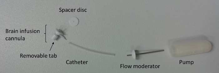
Figure 1. Components of the miniosmotic pump used in this protocol. The spacer disc, cannula and removable tab, catheter, flow moderator and miniosmotic pump can be observed. The aspect of the fully assembled pump can be seen in Figure 2A. Please click here to view a larger version of this figure.
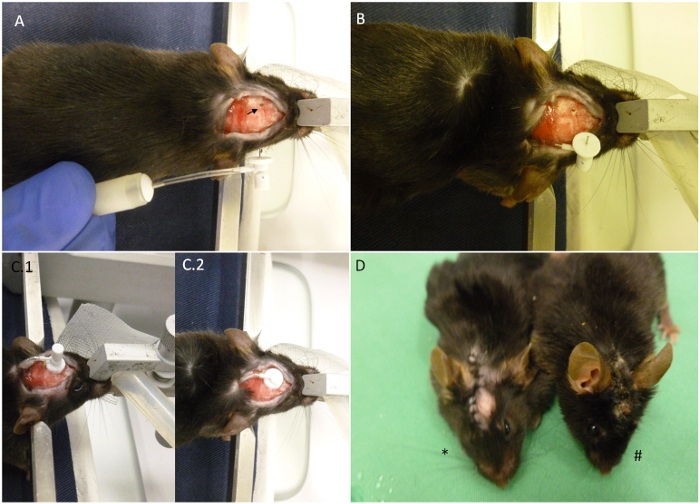
Figure 2. Summary of pump implantation key points. (A) The mouse is shown as placed on the stereotactic device with the fully constructed miniosmotic pump next to it. Arrow indicates the coordinates selected for implantation. (B) The pump has been introduced on the back of the animal and only the cannula remains on the exterior. The skull has already been drilled. (C) Aspect of the head after implantation. (C.1) The cannula is on position but the removable tab has not been cut. (C.2) The removable tab has been cut and stitching of the wound can now begin. (D) The asterisk shows a recently implanted animal as compared to an animal 30 days after implantation (#). When housed correctly, the wound should remain closed until the end of the procedure as shown on the image. Please click here to view a larger version of this figure.
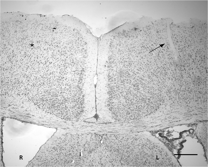
Figure 3. Nissl staining indicating the site of implantation on the cortex. A small incision can be observed on part of the left cortex (arrow). The width of the penetrated area is approximately 50 μm. The are no evident severe tissue alterations based on Nissl staining as compared to the corresponding contralateral area (*). R: Right hemisphere. L: Left hemisphere. Scale bar = 300 μm. Please click here to view a larger version of this figure.
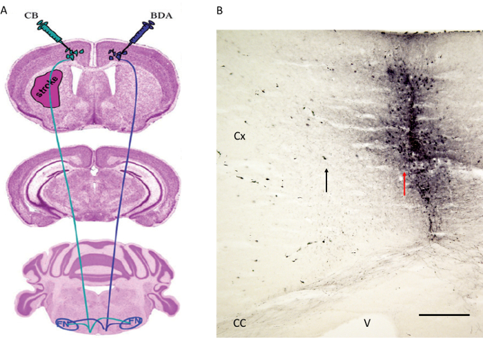
Figure 4. Tract tracer injection strategy as published before by Reitmeier et al.6,7. (A) Schematic indicating injection sites for the tract tracer BDA at the contralateral motor cortex whereas CB was injected in the motor cortex of the infarcted hemisphere. Fibers were followed to the red nucleus (not shown) and facial nucleus (see Figure 5). (B) The injection site of the anterograde tract tracer BDA next to the motor cortex is shown. Note the red arrow indicating the needle track whereas the black arrow shows a few cortical cells labelled with the BDA. Cx: Cortex. CC: Corpus callosum. V: Ventricle. FN: Facial nucleus. Scale bar in B = 200 µm. Figure 4A is reproduced with kind permission6. Please click here to view a larger version of this figure.
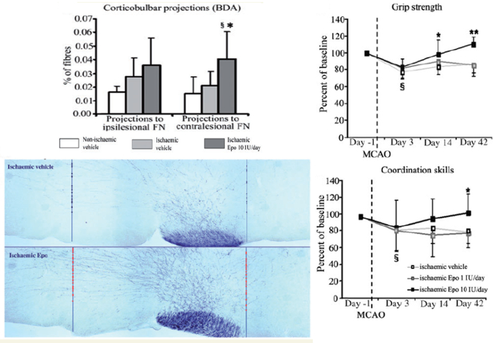
Figure 5. Recovery of an infarcted brain after delivery of rhEpo. (A) BDA injected in the contralesional motor cortex is then detected in corticobulbar fibers at the level of the facial nucleus (Bregma -5.8 mm to -6.3 mm). Intersection lines on each hemisphere were drawn parallel to the midline and fibers crossing each line in direction to the ipsilesional and contralesional hemisphere were counted and expressed as percentage of total labelled fibers in the corticospinal tract. Erythropoietin increased fiber crossings in direction to the contralesional facial nucleus Data are means +- SD. Data were analysed by one-way ANOVA followed by least significant differences tests, §P<0.05 compared with vehicle-treated non-ischaemic mice. (B) Motor behavior showed an improvement of hand grip strength and coordination in the rota rod test. Data are mean values +- SD. Data were analysed by two-way repeated measures ANOVA, followed by one-way ANOVA/least significant differences tests for each time-point. §P<0.05 compared with pre-ischaemic baseline; *P<0.05 compared with vehicle treated ischemic mice. Figures 5A and B are reproduced with kind permission6. Please click here to view a larger version of this figure.
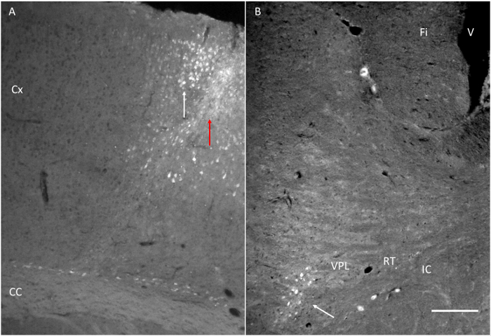
Figure 6. Target structures reached after injection of fluorogold (FG). (A) Injection site of FG next to the motor cortex as indicated in Figure 1. Note the red arrow indicating the needle track and the white arrow indicating a few labelled cells in the cortex. (B) FG injected near the motor cortex is detected in the VPL. Fi: Fimbria. IC: Internal capsule. RT: Thalamic reticular nucleus. VPL: Thalamic ventral posterolateral nucleus. Scale bar = 200 µm. Please click here to view a larger version of this figure.
Discussion
For many years, research on neurodegenerative conditions like ischemic stroke or traumatic brain injury has focused on development of neuroprotective therapies that aim to promote neuronal survival in the acute stroke phase. The vast majority of drug therapies that have been found to be effective in rodent models failed when translated to the clinic. Reasons for this therapeutic failure include but are not restricted to the lack of sustained drug effects resulting in persisting functional neurological recovery. It is thus important to develop strategies promoting brain remodeling in the longer run. Because the promotion of neuronal survival alone is not sufficient to allow successful stroke recovery, as suggested by the large number of unsuccessful neuroprotection trials, the stimulation of neuronal plasticity has recently obtained major interest in the field.
Means for drug delivery are intraperitoneal injection, tail intravascular injection, femoral injection, single stereotactic injection of vectors into the brain and continued constant delivery by miniosmotic pumps. The latter can include systemic delivery, if the pump does not have a cannula, or which can be organ-directed, as we have shown for delivery into the brain. With the exception of miniosmotic pumps and the use of viral vectors, all other strategies will induce fluctuating drug concentrations. For long term experiments it thus becomes necessary to submit the animal to the stress of receiving frequent injections. The BBB imposes an important impediment for the brain uptake of proteins or drugs from the blood, resulting in the need of huge protein or drug dosages in order to achieve therapeutic concentrations in the brain. For example Pellegrini et al. (2013) 5 delivered rhEpo by intraperitoneal injection at a dose equivalent to 75 IU/day for an animal of 30 g (750 IU/day for a 300 g rat). In comparison, the targeted delivery of rhEpo to the brain allowed us to use a much lower dose of only 10 IU/day in our study for successful stroke recovery, which enabled us to achieve recovery over a large time scale at a fixed rate of 0.25 µl/hr.
In this work we have shown the method of implantation of minipumps with a cannula connected to the skull in order to deliver the plasticity-promoting protein rhEpo directly into the ventricle, thus circumventing the BBB. By this method, rhEpo promoted neurological recovery in a number of ways, including reduction of infarct size, reduction of glial scar formation and induction of angiogenesis. rhEpo also promoted neuronal survival and increased projections from the contralesional motor cortex towards the denervated red nucleus and facial nuclei. The sprouting of the fibers was revealed by injection of the anterograde tract tracer BDA into the motor cortex (Figures 4A and 5A). A functional correlate to the sprouting of the fibers is provided by the improvement of motor skills (Figure 5B). Additionally, we have shown that the same approach for tract tracer injection can be applied to unveil thalamo-cortical connections by injection of the retrograde tract tracer FG (Figure 6B).
In the preparation of the miniosmotic pump, it is critical to consider the target point and the use of spacers. We use one spacer to reduce the length of the needle by 0.5mm as in this way the very tip of the needle is in contact with the ventricle at the given coordinates (-0.2 mm caudal, 0.9 mm lateral, 2.5 mm dorso ventral, with respect to bregma). However if deeper structures are the target of the research, then no spacers will be needed. Likewise, if a more external delivery point is desired (i.e., the cortex), then more spacer discs will be necessary. The catheter must be long enough so that the pump is not too close to the head, as it will impede movements of the mouse, but also not too long as once implanted excessive length may cause the catheter to bend, thus increasing the risk of cannula removal by the natural movement of the mouse. A section of 2 cm of catheter gives very good results in terms of mobility and stability of the implant (Figures 1 and 2). Incubation of the pump at 37 °C O/N allows the pump to immediately start pumping the drug into the brain at the moment of implantation.
In the miniosmotic pump implantation it is critical to assure that the skull is properly dried before implanting the cannula. Usually cleaning with 70% Ethanol will induce the bone to dry, but if continuous bleeding is found, touching the skull gently with a cauterizer will completely dry it. It is critical to assure that the introduction of the needle is as vertical and slow as possible. Once in position, and while the glue is drying, placing the finger on top of the cannula prevents it from moving sideways over the skull. Special care should be given to the wound and placement of the cannula. It is important that the incision is not performed exactly over the middle line of the skull but slightly to the right side. When closing the wound, if the incision was made at the middle line, the skin will be overstretched, thus increasing the risk of wound opening. Making the incision slightly to one side will allow the suture points to be away from the highest part of the cannula. As a consequence there will be less tension on the suture points and the wound will heal properly. Animals should be caged alone and checked every day, especially during the first 10-15 days after the implantation. In case of wound dehiscence, wounds have to be closed as soon as possible. If the cannula is removed or the animal presents an infection, the experiment has to be terminated. Re-implantation of the cannula is not recommended. It is very important for successful implantation to use adequate amounts of tissue adhesive (not too much!) as it degrades the bone and increases the risk of cannula removal. However using too little adhesive will also not hold the cannula attached to the bone. The miniosmotic pumps can carry drugs dissolved in a wide variety of substances, being the only limitation to this that the solvent is biocompatible. Additionally, given that the volume is small (200 µl) one must determine whether the concentration required for the experiment is suitable and will not cause precipitation inside the pump.
Tract tracing with either anterograde or retrograde tracers is a very well established technique to study brain connectivity and plasticity. Care must be given to the use stereotactic frames when injecting to ensure accuracy on targeting the brain area one wishes to study (i.e., to prevent injection on the corpus callosum when injecting the cortex).
For all surgical interventions and in order to reduce pain and inflammation, animals should be treated with 0.1 mg/kg Buprenorphine before the intervention and Caprofen at 4 mg/kg once a day for three days after the intervention.
In conclusion, this approach provides a proper tool for studying effect of proteins or pharmacological compounds in the injured brain, representing a method that is well suited for studies on brain plasticity.
Disclosures
Production and Open Access publication fees provided by DURECT Corporation, which produces the miniosmotic pumps used in this Article.
Acknowledgements
This work was supported by the Dr. Werner Jackstädt Foundation (to Eduardo Sanchez-Mendoza), the German Academic Exchange Service (DAAD; to Jeismar Carballo), the German Research Council (HE3173/2-1, HE3173/2-2, and HE3173/3-1; to Dirk M. Hermann), Heinz Nixdorf Foundation (to Dirk M. Hermann).
Materials
| Name | Company | Catalog Number | Comments |
|---|---|---|---|
| Alzet miniosmotic pump. Model 2004. | Alzet | 000298 | Drug container |
| Brain infusion kit 3 1-3 mm | Alzet | 0008851 | Drug brain delivery system |
| Loctite 454 Prism gel | Loctite | 45404 | Cyanoacrylate adhesive for cannula adhesion to the skull |
| 75N glass syringe | Hamilton | 87900/00 | Injection of tract tracers |
| Biotin Dextran Amine (10,000 MW) | Molecular probes | N-7167 | Anterograde tract tracer |
| Fluorogold | Fluorochrome, LLC. | Retrograde tract tracer | |
| Quintessential Stereotaxic Injector (QSI) | Stoelting | 53311 | Stereotactic device for coordinate determination, pump implantation and tract tracer injection. |
References
- Doeppner, T. R., et al. MicroRNA-124 protects against focal cerebral ischemia via mechanisms involving Usp14-dependent REST degradation. Acta Neuropathol. 126, 251-265 (2013).
- Hoyo-Becerra, C., et al. Rapid Regulation of Depression-Associated Genes in a New Mouse Model Mimicking Interferon-alpha-Related Depression in Hepatitis C Virus Infection. Mol Neurobiol. , (2014).
- Puntel, M., et al. Gene transfer into rat brain using adenoviral vectors. Curr Protoc Neurosci. Chapter 4, Unit 4.24(2010).
- Miao, J., et al. Overexpression of adiponectin improves neurobehavioral outcomes after focal cerebral ischemia in aged mice. CNS Neurosci Ther. 19, 969-977 (2013).
- Pellegrini, L., et al. Therapeutic benefit of a combined strategy using erythropoietin and endothelial progenitor cells after transient focal cerebral ischemia in rats. Neurol Res. 35, 937-947 (2013).
- Reitmeir, R., et al. Post-acute delivery of erythropoietin induces stroke recovery by promoting perilesional tissue remodelling and contralesional pyramidal tract plasticity. Brain. 134, 84-99 (2011).
- Reitmeir, R., et al. Vascular endothelial growth factor induces contralesional corticobulbar plasticity and functional neurological recovery in the ischemic brain. Acta Neuropathol. 123, 273-284 (2012).
- Hermann, D. M., Chopp, M. Promoting brain remodelling and plasticity for stroke recovery: therapeutic promise and potential pitfalls of clinical translation. Lancet Neurol. 11, 369-380 (2012).
- Overman, J. J., et al. A role for ephrin-A5 in axonal sprouting, recovery, and activity-dependent plasticity after stroke. Proc Natl Acad Sci U S A. 109, E2230-E2239 (2012).
- Wolf, W. A., Martin, J. L., Kartje, G. L., Farrer, R. G. Evidence for Fibroblast Growth Factor-2 as a Mediator of Amphetamine-Enhanced Motor Improvement following Stroke. PLoS One. 9, e108031(2014).
- Arras, M., Autenried, P., Rettich, A., Spaeni, D., Rulicke, T. Optimization of intraperitoneal injection anesthesia in mice: drugs, dosages, adverse effects, and anesthesia depth. Comp Med. 51, 443-456 (2001).
Reprints and Permissions
