We report a Fluorescent Activated Cell Sorting (FACS)-based method to isolate neural stem cells (NSCs) and their progeny from the subventricular zone (SVZ) of the adult mouse brain. Applied to Fluorescence Ubiquitination Cell Cycle Indicator (FUCCI) transgenic mice, it allows the study of cell cycle progression by live imaging.
Method Article
Cell Sorting of Neural Stem and Progenitor Cells from the Adult Mouse Subventricular Zone and Live-imaging of their Cell Cycle Dynamics
* These authors contributed equally
In This Article
Summary
Abstract
Neural stem cells (NSCs) in the subventricular zone of the lateral ventricles (SVZ) sustain olfactory neurogenesis throughout life in the mammalian brain. They successively generate transit amplifying cells (TACs) and neuroblasts that differentiate into neurons once they integrate the olfactory bulbs. Emerging fluorescent activated cell sorting (FACS) techniques have allowed the isolation of NSCs as well as their progeny and have started to shed light on gene regulatory networks in adult neurogenic niches. We report here a cell sorting technique that allows to follow and distinguish the cell cycle dynamics of the above-mentioned cell populations from the adult SVZ with a LeX/EGFR/CD24 triple staining. Isolated cells are then plated as adherent cells to explore in details their cell cycle progression by time-lapse video microscopy. To this end, we use transgenic Fluorescence Ubiquitination Cell Cycle Indicator (FUCCI) mice in which cells are red-fluorescent during G1 phase due to a G1 specific red-Cdt1 reporter. This method has recently revealed that proliferating NSCs progressively lengthen their G1 phase during aging, leading to neurogenesis impairment. This method is easily transposable to other systems and could be of great interest for the study of the cell cycle dynamics of brain cells in the context of brain pathologies.
Introduction
Quiescent neural stem cells (NSCs) are the source for adult neurogenesis1 and can convert into their proliferative “activated” form expressing the EGFR (aNSCs) 2. Once activated, they give rise to transit amplifying cells (TACs) 3 and then neuroblasts that migrate to the olfactory bulbs (OB) through a tube of astrocytes and finally differentiate into neurons4,5. NSCs and their progeny are organized in specialized “niches” architectures along the lateral ventricles, implicating a myriad of factors controlling their proliferation6. The isolation and purification of SVZ neurogenic cells is necessary to illuminate the complex molecular regulation of their proliferation but have remained a challenge for a long time due to the lack of specific markers and adapted techniques.
New approaches using flow cytometry have rendered possible the isolation of NSCs and their progeny from the adult SVZ2,7-11. Using the stem cell marker LeX12 along with neuroblast marker CD2413 and a fluorescence EGF to label EGFR on proliferating cells2, we recently developed a FACS strategy allowing the purification of five of the main SVZ neurogenic populations: quiescent and activated NSCs, TACs, immature as well as migrating neuroblasts9. Here, we describe in details this cell sorting technique and how a LeX/EGFR/CD24 triple staining allowed for the first time the isolation of both quiescent and activated NSCs.
Although neurogenesis persists during adulthood, the production of new neurons is drastically decreased in the aging brain14. Most studies agree on a progressive reduction in the number of proliferating progenitor cells in the SGZ and SVZ15-20. The consequences are not innocuous as the age-related decline in neurogenesis in the SVZ provokes a diminution of newborn neurons in the olfactory bulbs of the aged brain, ultimately leading to impairment in olfactory discrimination in aged mice18. Elucidating cell cycle kinetics of neural progenitors is a key step to understand the mechanisms underlying the evolution of adult neurogenesis during aging. Recent studies have investigated the cell cycle and lineage progression of adult neural stem cells in vitro21 and in vivo22 but none of them took advantage of cell sorting techniques and genetically encoded fluorescent cell-cycle probes to visualize the cell-cycle phases of isolated cells at a single-cell level.
Here we describe a protocol that takes advantage of the transgenic FUCCI mice in which cells are fluorescent during their cell cycle, allowing the distinction between G1 and S-G2/M phases23. This protocol shows how prospective isolation of NSCs and their progeny from adult FUCCI mice combined with time-lapse video microscopy allows the study of the cell cycle dynamics at a single-cell level.
Protocol
This protocol has been designed in compliance with the European Communities Council Directive of November 24, 1986 (86/609/EEC) and has been approved by our animal welfare institutional committee (CETEA-CEA DSV IdF).
1. Basic Setup Prior to Culture and Videomicroscopy
- Use glass bottom culture plates or µ-Plates for confocal video microscopy. For 1 - 5 x 10³ cells/well, use 96-well plates and 24-well plates for more than 5 x 10³ cells/well.
- At least one day prior to commencing the experiment, prepare sterile Poly-D-lysine (PDL) coated plates for adherent monolayer cultures. Add enough PDL (10 µg/ml in dH2O) to coat the bottom of each well and incubate O/N at 37 °C. Remove the PDL solution and rinse three times with dH2O before allowing the plate to dry in the hood during at least 2 hr under a laminar flow. If not used immediately, store the coated plate at -20 °C.
- Prepare culture medium by mixing NSC Basal Medium and NSC Proliferation Supplement at a 9:1 ratio (see Material table) along with 2 µg/ml heparin, 20 ng/ml purified human recombinant epidermal growth factor (EGF), and 10 ng/ml human recombinant fibroblast growth factor 2 (FGF-2). Warm up the culture medium to 37 °C in a water bath before use.
- For the SVZ dissociation, prepare papain solution: 1 mg/ml papain (15 UI/ml) in Earl’s Balance Salt Solution (EBSS) containing 0.2 mg/ml L-cysteine, 0.2 mg/ml EDTA, and 0.01 mg/ml DNase I in PBS. Sterilize the solution by passing through a 0.2 µm filter. Equilibrate the solution at 37 °C before use.
- To stop the enzymatic reaction, prepare a protease inhibitor solution (Ovomucoid): DMEM:F12 medium containing 0.7 mg/ml trypsin inhibitor type II. Filter the solution using a 0.2 µm filter.
- Prepare PBS 0.6% glucose solution to collect the brains and PBS 0.15% BSA solution for washing steps and for antibody staining.
- Prepare dissection tools: scissors, dissecting and tying forceps, scalpel. Soak them in 70% ethanol.
2. Harvesting of Adult Mouse Brains and SVZ Microdissections
- Sacrifice adult FUCCI mice23 (2 to 3-month-old and/or 12-month-old for aging studies) performing a cervical dislocation in accordance to the appropriate institutional guidelines.
- Spray the mouse using 70% ethanol and cut the head off using sharp scissors.
- Make an incision along the scalp to reveal the skull.
- Perform a longitudinal midline cut starting at the base of the skull towards the olfactory bulbs using a small pair of scissors. Make sure to avoid damaging the underlying brain with the scissor blades. Remove the upper open part of the skull with curved forceps to expose the brain.
- Collect the brain in a 15 mm petri dish containing 0.6% glucose in PBS.
- Dissect away the olfactory bulbs. Place the brain on its dorsal surface and make a coronal section through the optic chiasm using a scalpel.
- Under a dissecting microscope, position the rostral part of the brain section with the cut coronal surface facing upwards toward the experimenter.
- To dissect the SVZ, remove the septum with a fine curved forceps then insert one tip of a fine forceps into the striatum immediately adjacent to the ventricle and detach the SVZ from the surrounding tissue. For additional details, refer to Azari et al.24. Place the dissected SVZ into a petri dish containing 1 ml of PBS-0.6% glucose.
3. SVZ Tissue Dissociation
- Mince the dissected SVZ in the petri dish until no large pieces remain.
- Transfer the minced tissue along with the PBS-0.6% glucose to a 15 ml tube and centrifuge at 200 x g for 5 min.
- Discard the supernatant and add 1 ml of pre-warmed papain (1 mg/ml, prepared in step 1.4) supplemented with 0.01 mg/ml DNase I. Incubate for 10 min in a water bath at 37 °C. Use 1 ml of papain per mouse.
- Centrifuge at 200 x g for 5 min and discard the supernatant.
- Add 1 ml of pre-warmed ovomucoid (0.7 mg/ml, prepared in step 1.5) to stop papain activity. Mechanically dissociate the minced tissue further into a single-cell suspension by gently pipetting up and down 20 times through a p1000 micropipette tip. Avoid air bubbles.
- Pass the cell suspension through a sterile 20 µm filter in a new 15 ml tube. Make sure to wash the cell filter with PBS 0.15% BSA to avoid losing cells.
- Centrifuge at 200 x g for 10 min and discard the supernatant. Re-suspend the cells in 100 µl of PBS containing 0.15% BSA.
4. Immunofluorescent Staining for Cell Sorting
For cell sorting using FUCCI-Red mice (Figure 2A), use the following antibodies: CD24 phycoerythrin-cyanine7 conjugate [PC7] ; CD15/LeX fluorescein isothiocyanate [FITC] conjugated and Ax647 conjugated EGF ligand.
Note: LeX+EGFR+ cells and EGFR+ cells are not abundant in the adult SVZ: ≈ 600 and 1500 cells/mouse respectively9. We recommend pooling SVZ cells from 2 to 3 mice to have enough material. Do not work with more than 12 mice on the same day so that the cell sorting duration doesn’t exceed 3 hr. Keep in mind that working with too many mice on the same day will lead to an increased cell sorting duration possibly resulting in increased cell death and/or cell differentiation.
- Perform the FACS staining in 100 µl of PBS 0.15% BSA per mouse (or in 200 µl for a group of 2 to 3 mice for optimal staining).
- Prepare the control tubes. Use compensation beads to prepare single color control tubes according to the manufacturer's protocol. Select a fraction of cells (1/10 of the cells extracted from one mouse is enough) and separate it in 4 tubes to prepare 1 negative control tube (unmarked cells) and 3 fluorescence minus one (FMO) control tubes. Resuspend the cells in 200 µl of PBS 0.15% BSA per tube.
Hint: For LeX-FITC FMO control, label the cells with CD24-PC7 (1:50) and Ax647‐conjugated EGF ligand (1:200); for CD24-PC7 FMO control, label the cells with CD15/LeX-FITC (1:50) and Ax647‐conjugated EGF ligand (1:200) and for Ax647‐conjugated EGF ligand FMO control, label the cells with CD15/LeX-FITC (1:50) and CD24-PC7 (1:50). - For the tubes used for cell sorting, use the following antibodies at the indicated dilution in PBS 0.15% BSA: CD24-PC7 (1:50), CD15/LeX-FITC (1:50) and Ax647‐conjugated EGF ligand (1:200).
- Incubate for 20 min at 4 °C in the dark. Wash with 1 ml PBS 0.15% BSA and centrifuge at 200 x g for 10 min. Resuspend the cells in 200 µl of PBS 0.15% BSA per brain. Keep the cell sorting tubes on ice and proceed immediately to the cell sorting.
- If using FUCCI-Green mice, separate LeX-positive and LeX-negative fractions using separation columns before cell sorting as the LeX-FITC antibody shares the same emission wavelength than the FUCCI-green fluorescence (Figure 3A,B).
- First, label the cells with a mouse anti-human LeX-antibody (1:50) for 15 minutes at 4 °C in the dark in 100 µl of PBS 0.15% BSA.
- Wash the cells with 1 ml PBS 0.15% BSA and centrifuge at 200 x g for 10 min, then label the cells with anti-mouse IgM microbeads (1:10) for 15 minutes at 4 °C in the dark.
- Wash the cells with 1 ml PBS 0.15% BSA and centrifuge at 200 x g for 10 min. Resuspend the cells in 500 µl PBS 0.15% BSA and pour the cells through separation column in magnetic field as schematized in Figure 3 C. Wash the column with 1 ml PBS 0.15% BSA to obtain LeX negative fraction.
- To obtain LeX-positive fraction, remove the separation column from the magnetic field and elute the cells with 2 ml PBS 0.15% BSA.
- Proceed to CD24-PC7 and Ax647‐conjugated EGF ligand staining as indicated in 4.2.
5. Cell Sorting
Note: Cells were sorted on a FACS sorter at 40 Psi with an 86 µm noozle. Fluorescence was collected using the following filter set: 520/35nm (FITC), 575/26nm (PE), 670/20nm (Ax647) and 740LP (PC7). Compensation is necessary to prevent false-positive signals as an overlap is found between the emission spectrums of the FUCCI-red fluorescence and the PC7 dye.
- Immediately prior to cell sorting, add a vital dye to discriminate live from dead cells. We used HO (see material table) at a 2 µg/ml final concentration. Run the negative control tube (unmarked cells) through the FACS sorter and select the cells using side scatter (SSC) and forward scatter (FSC) parameters (Figure 1A).
NOTE: Dead cells were excluded by gating only the HO-negative fraction (Figure 1A’) and then doublets were excluded by selecting the Pulse Width negative fraction (Figure 1A’’). - Run the single color controls prepared in step 4.2 and adjust the photomultiplier tube (PMT) voltages if necessary (i.e. negative population too high and/or positive cells off scale). Perform color compensation in the compensation window of the software.
- Run FMO controls prepared in step 4.2 (LeX-FITC FMO control, CD24-PC7 FMO control and Ax647-conjugated EGF ligand FMO control) and draw the sorting gates (Figure 1). Sort the cells directly into 100 µl of culture medium in 1.5 ml microtubes.
6. Preparation of Cells for Microscopy
- Plate the freshly sorted cells at a density of 1 - 3 x 10³ cells/well on Poly-D-Lysine- coated 96-well µ-Plate with 300 µl of culture medium.
- Prior to video microscopy, incubate the culture plates at 37 °C and 5% CO2 at least for 1 hr to allow cell adhesion.
7. Microscope Setup and Image Acquisition
- Perform live imaging using a Plan Apo VC 20x DIC objective (NA: 0.75) on a confocal laser scanning microscope system attached to an inverted thermostated chamber at 37 °C under 5% of CO2 atmosphere.
- Position the 96-well µ-Plate inside the pre-warmed and equilibrated thermostated chamber and replace the lid by a thermostated cover.
- Open the NIS-Elements software and click in the menu bar on "Acquire/Acquisition controls/ND acquisition” to select the options of the time-lapse (length, stage positions, confocal z-sections,…), "Acquire/Acquisition controls/Ti Pad” to select the objectives and "Acquire/Acquisition controls/A1plus Settings” to select the PMT level for each fluorescence in the menu bar. Select a folder to save the data files.
- Using the ND acquisition window, set the center of each well as a stage position and select the large image option to 7 x 7 mm². This will create a mosaic image around the center of each well. Set the overlap for the large mosaic image to 5%. Take pictures every 20 min for 24 hr. Select the Plan Apo VC 20x DIC objective (NA: 0.75) in the Ti Pad window.
- In the A1plus Settings window, acquire images using high speed resonant scanner at a 512 x 512 pixels format with a resolution of 1.26 µm/pixel. Use brightfield to visualize cell shapes. In the case of FUCCI-Red mice, excite red fluorescence at 561 nm and collect using a 595/50 nm filter. In the case of FUCCI-Green mice, excite green fluorescence at 488 nm and collect using a 530/40 nm filter. Determine the optimal PMT level, offset and laser power for each wavelength.
NOTE: We recommend using the autofocus function for the brightfield channel to allow the software to autofocus at each stage position before each acquisition. Hint: A Plan Apo VC 20x DIC objective (NA: 0.75) was used for its excellent resolution without the need for oil. Other objectives may be used depending on the optical resolution desired. - Select the 'Run now' button on the ND acquisition window to begin acquisition.
Hint: Follow the computer work for 1 loop to be sure that everything in working properly.
8. Image Processing and Analysis
- Analyze the data directly on the NIS-Elements software by tracking the cells individually. Hint: To gain time, save each position in .avi format using NIS-Elements software and analyze the movies with ImageJ.
- In ImageJ software, track individual cells undergoing at least 2 divisions (i.e. one cell to a four-cell colony). Crop a small area around the cell and select 'Image/Duplicate'.
- Select 'Image/Stacks/Make Montage' in the menu bar to make a montage. Specify the frames to be included, the size of the images and save the montage as a .tif file for optimal resolution.
- To calculate the first S-G2/M phase length (Figure 2C,D), select a single red fluorescent cell (in G1) and then set t = 0 (S phase will begin once the red-fluorescence switches off). Count the number of frames until the cell divides to estimate the S-G2/M length.
NOTE: The calculated time depends on the time interval between each frame. - To calculate the following G1 phase (Figure 2C,D), continue to track the cell that just divided. If the divided cell enters G1 phase, there will be an accumulation of cdt1 red-fluorescent protein. Calculate the number of frames until red-fluorescence switches off again for each cell.
Hint: At the beginning of G1 the red fluorescence might be weak so choose the onset of the G1 phase on the frame where the cell has divided to avoid approximations.
Results
The ability to reliably discriminate between the different cell populations of the adult mouse SVZ stem cell lineage is primordial to investigate their functional properties. To that purpose, we have developed a LeX/EGFR/CD24 triple-labeling strategy allowing the purification of specific cell populations from the adult SVZ: quiescent NSCs (LeXbright), activated NSCs (LeX+EGFR+), TACs (EGFR+), immature and migrating neuroblasts (CD24+EGFR+ and CD24+, respectively) 9 (Figure 1). Applied to FUCCI transgenic mice, the cell sorting technique allows the live imaging of the cell cycle phases of the different neurogenic populations at the single-cell level25. FUCCI-Red mice were used to follow the G1 phase with red fluorescence using time-lapse video microscopy (Figure 2A). FUCCI-Redneg cells represent the cells in S-G2/M phases of the cell cycle, FUCCI-Redpos cells are G1 cells and FUCCI-Redbright cells are cells exiting or out of the cell cycle (Figure 2B) 23,26. LeX+EGFR+ and EGFR+ cells from young adult (Figure 2C) and middle-aged mice (Figure 2D) were plated as adherent cells on poly-D-lysine coated culture plates. The first S-G2/M phase was identified on single cells once the red fluorescence switches off until the cell divides. As previously observed the S-G2/M phase length showed no difference between LeX+EGFR+ and EGFR+ cells, either in young or middle-aged mice. Then we calculated the next G1 phase length from the first division until the red fluorescence switches off. Interestingly, a G1 phase lengthening was found during aging in LeX+EGFR+ cells but not in EGFR+ cells25 (Figure 2C,D). In consequence, neurospheres obtained 5 days after plating are smaller in LeX+EGFR+ cells obtained from aged mice as shown in Figure 2E.
FUCCI-Green mice can also be used to visualize the S-G2/M phase with green fluorescence (Figure 3A,B) but cells have to be pre-sorted using separation columns as LeX-FITC antibody shared the same emission wavelength than the FUCCI-green fluorescence (Figure 3C). An example of FUCCI-green fluorescence for the first S-G2/M phase of young adult LeX+EGFR+ and EGFR+ cells is shown in Figure 3D.
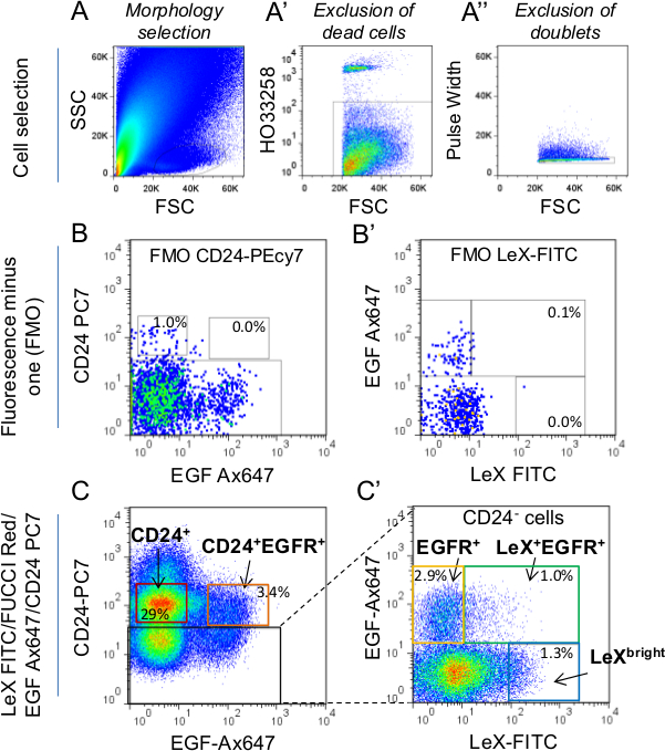
Figure 1: Strategy of cell selection by FACS. (A) Cells were first selected according to their morphology using Side scatter (SSC) vs Forward scatter (FSC) parameters. For further details on morphology gate selection, refer to Daynac et al. 9 (A’) Dead cells were labeled using HO marker and excluded from the selection. (A’’) Doublets were excluded using pulse width parameter. In order to set up the sorting gates, Fluorescence minus one (FMO) controls for CD24-PC7 (LeX-FITC/FUCCI-Red/EGF-Ax647 labeling; B) and LeX-FITC (FUCCI-Red/EGF-Ax647/CD24-PC7 labeling; B’) were used. (C,C’) Then, sorting gates were determined in LeX-FITC/FUCCI-Red/EGF-Ax647/CD24-PC7 labeled tubes along with FMO controls. The mean percentages of total cells are represented within the gates. If selecting CD24+ cells (neuroblasts), be careful to exclude CD24bright expressing cells from the gate. Indeed, CD24bright correspond to ependymal cells2,9. C’ represents the CD24 negative cells (black square in C). Only LeX+EGFR+ and EGFR+ (C’) sorting gates were used for this study.
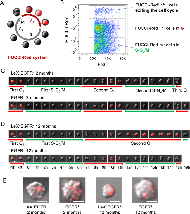
Figure 2: Live analyses of cell cycle using FUCCI-Red mice. (A) Schematic representation of FUCCI-Red cell cycle where cells are red-fluorescent during G1 phase and colorless during S-G2/M phases23. (B) FACS representation of FUCCI-Red fluorescence on SVZ cells. (C,D) Video microscopy with LeX+EGFR+ and EGFR+ cells sorted from young (C) and middle-aged (D) FUCCI-Red mice allows the tracking of the G1 phase with red fluorescence whereas the S-G2/M phases can be deduced when the red fluorescence switches off. Successive images are shown with a time scale presented in D. (E) Representative images of colonies (neurospheres) obtained 5 days after plating. Scale bar: 10 µm (C, D), 30µm (E). Please click here to view a larger version of this figure.

Figure 3: Strategy for live analyses of cell cycle using FUCCI-Green mice. (A) Schematic representation of FUCCI-Green cell cycle where cells are green during S-G2/M phase and colorless during G1 phase23. (B) FACS representation of FUCCI-Green fluorescence: FUCCI-Greenneg cells represent the cells in G1 phase of the cell cycle while FUCCI-Greenpos cells are cells in S-G2/M23. (C) In order to follow the green-fluorescent S-G2/M phases of LeX+EGFR+ and EGFR+ cells, SVZ cells had to be pre-sorted with separation columns. Cells were labeled with anti-mouse LeX antibody and LeX+ fraction was retained on the column using anti-mouse microbeads (see protocol 4.). Then, LeX+ and LeX- fractions were eluted and labeled with EGF-Ax647 and CD24-PC7 antibodies for FACS sorting. (D) Video microscopy of LeX+EGFR+ and EGFR+ cells sorted from young FUCCI-Green mice allows the tracking of the S-G2/M phases with green fluorescence. Successive images are shown with a time scale presented in D. Scale bar: 10 µm (D).
Discussion
The cell sorting technique described herein allows reliable discrimination between quiescent NSCs, activated NSCs and their progeny enabling studies of their properties and dynamics in the adult brain9. Coupled with the FUCCI technology which permits the visualization of cell cycle progression in living cells23, we developed a rapid and efficient technique to follow the G1 and S-G2/M phases of the cell cycle from young adult and aged mouse brain cells.
The cell sorting technique used in this protocol was the first validated combination of markers allowing the purification of the five main neurogenic populations from the SVZ9. It was also the first validated technique enabling the distinction of quiescent and activated NSCs. It is noteworthy that this technique does not require the use of transgenic mice per se, which is necessary when adapted to transgenic mouse models such as FUCCI. Since then, Codega et al.10 have used a GFAP-GFP/CD133/EGFR triple labeling combination to distinguish quiescent NSCs from their activated counterpart but it cannot be adapted to FUCCI technology as it requires the use of GFAP-GFP transgenic mice. Mich et al.11 have developed a Glast/EGFR/CD24 triple labeling strategy that shares common results with the technique used in this study9. Indeed, a high correlation between LeX and Glast NSCs markers was already observed in Daynac et al. study9. However, the Glast antibody used in the Mich et al. technique is coupled to phycoerythrin and thus cannot be adapted with the use of FUCCI-Red mice.
There are several important technical points that require attention before sorting SVZ cells. First, the dissociation step is very important as the brain tissue has to be dissociated into single cells while preserving the structural integrity of the proteins used for the antibody labeling strategy. 0.05% trypsin-EDTA has been shown to be very effective in dissociating SVZ tissue27 but the LeX antigen was found to be highly sensitive as almost all LeX-FITC immunofluorescence was lost (data not shown). Thus, papain was used as it was more efficient and less destructive than other proteases on brain tissue. Moreover, neither LeX nor EGFR nor CD24 were affected by papain treatment. It should be noted that the antibody labeling was altered if the papain treatment exceeded 15 min. We obtained optimal results with a 10 min papain treatment associated with a mechanical dissociation (pipette up and down 20 times).
The poly-D-Lysine coating allows culture of adherent cells and the formation of colonies after several days in culture. It is now widely accepted that in vitro assays comes with their limitations28,29. We recommend determining only the first cell cycle for the cells undergoing at least one subsequent division to stay as close as possible to the in vivo cell phenotype.
It is noteworthy to mention that the Geminin (green fluorescence) and Cdt1 (red fluorescence) proteins used to design the FUCCI system23 were previously shown to be abundantly expressed by neural progenitors during early neurogenesis in mice30 and in adult brain tissues9,25. Although the mKO2-hCdt1(30/120) construct was mainly used in the present study to follow the G1 phase with red fluorescence, the use of both constructs [mKO2-hCdt1(30/120) and mAG-hGem(1/110)] could be envisioned to allow the visualization of the major phases of the cell cycle (G1 and S-G2/M) as well as the G1/S transition23. The main drawback of the dual-color imaging is the limited compatible sets of fluorescence that can still be used for the cell labeling. One solution is to use separation columns. For example, we have successfully depleted the CD24-positive fraction from cells using separation columns before cell sorting and live-imaging as we used a CD24-PE antibody that shared the same emission wavelength than the FUCCI-Red fluorescence25.
Few studies have investigated the cell cycle length of adult mouse SVZ populations. The total cell cycle length obtained with our technique is close to the one estimated in vitro by Costa et al.21, but we were able for the first time to distinguish the different cell cycle phases. In an in vivo study, Ponti et al.22 used the incorporation of thymidine analogs to determine the proliferation dynamics of the different SVZ cell populations. However, they could neither perform a continuous monitoring of the cell cycle nor track the cells at the single-cell level. This could be an issue as it was shown that a strong heterogeneity exists within a given SVZ cell population22,25. We describe here an alternative in vitro technique, easy to set up, that allows the live imaging of the cell cycle phases of different adult neurogenic populations at a single-cell level.
Understanding the regulation of the cell cycle of neural stem cells and progenitors remains a challenge for the development of new therapeutic approaches in the context of aging or brain pathologies. Our protocol can thus have a wide range of applications. In the context of aging, this technique could also prove useful to understand the effects of aging on NSCs differentiation. Indeed, it is possible to identify neural stem/progenitor cells entering differentiation as their red fluorescent intensity is distinctively higher26. Finally, it could also be of interest to exploit the continuous live imaging at a single-cell level to study the cell intrinsic and extrinsic processes responsible for the lineage progression of adult NSCs.
Disclosures
The authors declare that they have no conflict of interest.
Acknowledgements
We are indebted to C Joubert, V Neuville, V Barroca, J Tilliet and all the staff of animal facilities; to J Baijer and N Dechamps for cell sorting; O. Etienne for technical assistance, and A. Gouret for her administrative assistance. Flow cytometry and cell sorting were carried out at the iRCM Flow Cytometry Shared Resource, established by equipment grants from DIM-Stem-Pôle, INSERM, Fondation ARC, and CEA. This work was supported by grants of Electricité de France (EDF). M.D. has a fellowship from La Ligue Contre le Cancer and L.M. from Région Ile-de-France (DIM Biothérapies). Marc-André Mouthon and François D. Boussin share co-senior authorship.
Materials
| Name | Company | Catalog Number | Comments |
|---|---|---|---|
| 96-well uncoated glass bottom culture plates | MatTek Corp. | P96G-1.5-5-F | |
| µ-Plates 96-well | Ibidi | 89626 | |
| Poly-D-Lysine | Millipore | A003E | |
| NeuroCult NSC Basal Medium | STEMCELL Technologies | 5700 | |
| NeuroCult NSC Proliferation Supplement | STEMCELL Technologies | 5701 | |
| Heparin | STEMCELL Technologies | 7980 | |
| EGF | Millipore | GF144 | |
| FGF-2 | Millipore | GF003 | |
| Papain | Worthington | LS003119 | |
| EBSS | Invitrogen | 24010-043 | |
| L-cysteine | Sigma | C-7352 | |
| 0.5M EDTA, pH 8.0 | Promega | V4231 | |
| DNase I | Sigma | D5025-15KU | |
| Trypsin inhibitor type II | Sigma | T9128 | Ovomucoid |
| DMEM:F12 medium | Life Technologies | 31330-038 | |
| 20 µm filter | BD Medimachine Filcon | 340622 | |
| Eppendorf microtubes 3810X | Sigma | Z606340-1000EA | |
| BSA | Sigma | A1595 | Bovine serum albumin solution |
| CD24 phycoerythrin-cyanine7 conjugate [PC7] | Life Technologies | A14776 | Mouse IgM; clone MMA. 1:50 |
| CD15/LeX fluorescein isothiocyanate [FITC] - conjugated | BD Biosciences | 332778 | Rat IgG2b; clone M1/69. 1:50 |
| Mouse anti-human LeX antibody | BD Pharmingen | 559045 | Mouse IgM; clone MAM. 1:50 |
| Alexa647 - conjugated EGF ligand | Life Technologies | E35351 | 1:200; Ax647-conjugated EGF ligand in the text |
| CompBeads | BD Biosciences | 644204 | |
| MACS LS separation columns | Miltenyi Biotec | 130-042-401 | separation columns (in the manuscript) |
| Anti-mouse IgM microbeads | Miltenyi Biotec | 130-047-301 | |
| Hoechst 33258 | Sigma | 861405 | HO (in the manuscript) |
| INFLUX cell sorter | BD Biosciences | FACS sorter (in the text) | |
| Nikon A1R confocal laser scanning microscope system attached to an inverted ECLIPSE Ti | Nikon Corp. | confocal laser scanning microscope (in the manuscript) | |
| NIS-Elements AR.4.13.01 64-bit software | Nikon Corp. | NIS-Elements software (in the manuscript) | |
| Plan Apo VC 20x DIC objective (NA: 0.75) | Nikon Corp. | ||
| ECLIPSE Ti thermostated chamber | Nikon Corp. | thermostated chamber (in the manuscript) | |
| ImageJ | RBS | ||
| FlowJo | Tree Star, Ashland, OR | ||
| FUCCI mice | RIKEN BioResource Center, JAPAN | Sakaue-Sawano et al. 2008 |
References
- Morshead, C. M., et al. Neural stem cells in the adult mammalian forebrain a relatively quiescent subpopulation of subependymal cells. Neuron. 13, 1071-1082 (1994).
- Pastrana, E., Cheng, L. C., Doetsch, F. Simultaneous prospective purification of adult subventricular zone neural stem cells and their progeny. Proc Natl Acad Sci USA. 106, 6387-6392 (2009).
- Doetsch, F., Caille, I., Lim, D. A., Garcia Verdugo, J. M., Alvarez Buylla, A. Subventricular zone astrocytes are neural stem cells in the adult mammalian brain. Cell. 97, 703-716 (1999).
- Kriegstein, A., Alvarez Buylla, A. The glial nature of embryonic and adult neural stem cells. Annu Rev Neurosci. 32, 149-184 (2009).
- Lois, C., Garcia Verdugo, J. M., Alvarez Buylla, A. Chain migration of neuronal precursors. Science. 271, 978-981 (1996).
- Fuentealba, L. C., Obernier, K., Alvarez Buylla, A. Adult neural stem cells bridge their niche. Cell stem cell. 10, 698-708 (2012).
- Rietze, R. L., et al. Purification of a pluripotent neural stem cell from the adult mouse brain. Nature. 412, 736-739 (2001).
- Fischer, J., et al. Prospective isolation of adult neural stem cells from the mouse subependymal zone. Nat Protoc. 6, 1981-1989 (2011).
- Daynac, M., et al. Quiescent neural stem cells exit dormancy upon alteration of GABAAR signaling following radiation damage. Stem Cell Res. 11, 516-528 (2013).
- Codega, P., et al. Prospective identification and purification of quiescent adult neural stem cells from their in vivo niche. Neuron. 82, 545-559 (2014).
- Mich, J. K., et al. Prospective identification of functionally distinct stem cells and neurosphere-initiating cells in adult mouse forebrain. Elife. 3, e02669(2014).
- Capela, A., Temple, S. LeX ssea 1 is expressed by adult mouse CNS stem cells identifying them as nonependymal. Neuron. 35, 865-875 (2002).
- Calaora, V., Chazal, G., Nielsen, P. J., Rougon, G., Moreau, H. mCD24 expression in the developing mouse brain and in zones of secondary neurogenesis in the adult. Neuroscience. 73, 581-594 (1996).
- Pineda, J. R., et al. Vascular derived TGF-beta increases in the stem cell niche and perturbs neurogenesis during aging and following irradiation in the adult mouse brain. EMBO Mol Med. 5, 548-562 (2013).
- Lugert, S., et al. Quiescent and active hippocampal neural stem cells with distinct morphologies respond selectively to physiological and pathological stimuli and aging. Cell stem cell. 6, 445-456 (2010).
- Blackmore, D. G., Golmohammadi, M. G., Large, B., Waters, M. J., Rietze, R. L. Exercise increases neural stem cell number in a growth hormone dependent manner augmenting the regenerative response in aged mice. Stem cells. 27, 2044-2052 (2009).
- Bouab, M., Paliouras, G. N., Aumont, A., Forest Berard, K., Fernandes, K. J. Aging of the subventricular zone neural stem cell niche evidence for quiescence associated changes between early and mid adulthood. Neuroscience. 173, 135-149 (2011).
- Enwere, E., et al. Aging results in reduced epidermal growth factor receptor signaling diminished olfactory neurogenesis and deficits in fine olfactory discrimination. The Journal of neuroscience the official journal of the Society for Neuroscience. 24, 8354-8365 (2004).
- Maslov, A. Y., Barone, T. A., Plunkett, R. J., Pruitt, S. C. Neural stem cell detection characterization and age related changes in the subventricular zone of mice. The Journal of neuroscience the official journal of the Society for Neuroscience. 24, 1726-1733 (2004).
- Tropepe, V., Craig, C. G., Morshead, C. M., van der Kooy, D. Transforming growth factor alpha null and senescent mice show decreased neural progenitor cell proliferation in the forebrain subependyma. The Journal of neuroscience the official journal of the Society for Neuroscience. 17, 7850-7859 (1997).
- Costa, M. R., et al. Continuous live imaging of adult neural stem cell division and lineage progression in vitro. Development. 138, 1057-1068 (2011).
- Ponti, G., et al. Cell cycle and lineage progression of neural progenitors in the ventricular-subventricular zones of adult mice. Proc Natl Acad Sci U S A. 110, 1045-1054 (2013).
- Sakaue Sawano, A., et al. Visualizing spatiotemporal dynamics of multicellular cell cycle progression. Cell. 132, 487-498 (2008).
- Azari, H., Rahman, M., Sharififar, S., Reynolds, B. A. Isolation and expansion of the adult mouse neural stem cells using the neurosphere assay. Journal of visualized experiments JoVE. , (2010).
- Daynac, M., et al. TGFbeta lengthens the G1 phase of stem cells in aged mouse brain. Stem cells. 32, 3257-3265 (2014).
- Roccio, M., et al. Predicting stem cell fate changes by differential cell cycle progression patterns. Development. 140, 459-470 (2013).
- Walker, T. L., Kempermann, G. One mouse two cultures isolation and culture of adult neural stem cells from the two neurogenic zones of individual mice. Journal of visualized experiments JoVE. , e51225(2014).
- Reynolds, B. A., Rietze, R. L. Neural stem cells and neurospheres re evaluating the relationship. Nat Methods. 2, 333-336 (2005).
- Pastrana, E., Silva Vargas, V., Doetsch, F. Eyes wide open a critical review of sphere formation as an assay for stem cells. Cell stem cell. 8, 486-498 (2011).
- Spella, M., et al. Licensing regulators Geminin and Cdt1 identify progenitor cells of the mouse CNS in a specific phase of the cell cycle. Neuroscience. 147, 373-387 (2007).
Reprints and Permissions
