We propose a protocol to identify fatty acids without the need to purify them. It combines information on the retention times with the mass spectra of three types of fatty acid derivatives: fatty acid methyl esters (FAMEs), 4,4-dimethyl oxazoline derivatives (DMOX), and 3-pyridylcarbinyl esters (picolinyl).
A subscription to JoVE is required to view this content. Sign in or start your free trial.
Method Article
Identification of Fatty Acids in Bacillus cereus
In This Article
Summary
Abstract
The Bacillus species contain branched chain and unsaturated fatty acids (FAs) with diverse positions of the methyl branch (iso or anteiso) and of the double bond. Changes in FA composition play a crucial role in the adaptation of bacteria to their environment. These modifications entail a change in the ratio of iso versus anteiso branched FAs, and in the proportion of unsaturated FAs relative to saturated FAs, with double bonds created at specific positions. Precise identification of the FA profile is necessary to understand the adaptation mechanisms of Bacillus species.
Many of the FAs from Bacillus are not commercially available. The strategy proposed herein identifies FAs by combining information on the retention time (by calculation of the equivalent chain length (ECL)) with the mass spectra of three types of FA derivatives: fatty acid methyl esters (FAMEs), 4,4-dimethyl oxazoline derivatives (DMOX), and 3-pyridylcarbinyl ester (picolinyl). This method can identify the FAs without the need to purify the unknown FAs.
Comparing chromatographic profiles of FAME prepared from Bacillus cereus with a commercial mixture of standards allows for the identification of straight-chain saturated FAs, the calculation of the ECL, and hypotheses on the identity of the other FAs. FAMEs of branched saturated FAs, iso or anteiso, display a constant negative shift in the ECL, compared to linear saturated FAs with the same number of carbons. FAMEs of unsaturated FAs can be detected by the mass of their molecular ions, and result in a positive shift in the ECL compared to the corresponding saturated FAs.
The branching position of FAs and the double bond position of unsaturated FAs can be identified by the electron ionization mass spectra of picolinyl and DMOX derivatives, respectively. This approach identifies all the unknown saturated branched FAs, unsaturated straight-chain FAs and unsaturated branched FAs from the B. cereus extract.
Introduction
Fatty acid methyl ester (FAME) gas chromatography (GC) is an essential method for lipid characterization. It rapidly separates and quantifies the various fatty acids (FAs) of a sample after a short extraction step. Derivatives of methyl esters are highly volatile, stable and inert toward the chromatographic column, thereby avoiding tailing peaks. Their identification is rather straightforward when the sample consists of well-known FAs because the chromatographic profiles are either published or compared to standards. In addition, the repeated injection of calibration standards for quantification of various FAs is not required, given their almost constant response to f....
Access restricted. Please log in or start a trial to view this content.
Protocol
1. Bacterial Cultures
- Prepare a lawn of the bacteria (Bacillus cereus strain ATCC 14579) by spreading 100 µl of an overnight culture of the strain incubated at 30 °C in LB (Luria-Bertani medium), over the surface of a plate of LB agar medium. Incubate the plate overnight at 30 °C.
2. ECL: Equivalent Chain Length
- Calculate ECL as follows:
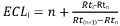 with:
with:
i, the solute of interest;
n, the....
Access restricted. Please log in or start a trial to view this content.
Results
The strategy of FA identification from bacterial cells is presented in Figure 1. Each step provides complementary spectral information or information about chromatographic retention. Step 1 consists of preliminary FA identification using a standard solution. Step 2 allows for the interpretation of FAME EI spectra and their ECL, in order to tentatively identify the products. Step 3 identifies the exact branching location in branched chain-FAs. Finally, step 4 identifies th.......
Access restricted. Please log in or start a trial to view this content.
Discussion
The FAs chromatogram profiles shown in Table 1 correspond to B. cereus ATCC 14579 grown on an agar plate surface. Similar profiles were obtained when the bacterium was grown in aerated liquid media at the same temperature8. In the case of bacteria grown in liquid media, the bacterial biomass is collected by centrifugation of the growth medium and can be washed according to previously described protocols depending on the growth conditions8,19. The identification of the vario.......
Access restricted. Please log in or start a trial to view this content.
Disclosures
The authors declare that they have no competing financial interests.
Acknowledgements
Authors are grateful to Thomas Mison for his technical support, and to Rachel Kopec for revising the manuscript.
....Access restricted. Please log in or start a trial to view this content.
Materials
| Name | Company | Catalog Number | Comments |
|---|---|---|---|
| GC/MS | Shimadzu | QP2010 | |
| capillary column ZB WAX | Phenomenex | 7HG-G007-11 | 30 m x 0.25 mm x 0.25 µm |
| Methanol Lichrosolv | VWR | 1.06018.2500 | |
| potassium hydroxide | Aldrich | P1767 | |
| THF | Hipersolv Chromanorm | 28559.320 | |
| Dichloromethane | Hipersolv Chromanorm | 23373.320 | |
| Hexane | Hipersolv Chromanorm | 24575.320 | |
| 3-pyridinemethanol | Aldrich | P6-680-7 | |
| potassium tertiobutoxide | Aldrich | 156671 | |
| 2-amino-2-methyl-1-propanol | A-9879 | ||
| MilliQ Academic | Millipore | ZMQS50001 | |
| Bacterial Acid Methyl Ester (BAME) Mix | Sigma-Aldrich | 47080-U Supelco |
References
- Christie, W. W., Han, X. Lipid Analysis 4th Edition. , Oily press. (2010).
- HÜbschmann, H. -J. Handbook of GC-MS: fundamental and application. Third edition. , Wiley-vch. (2015).
- Sasser, M.
Access restricted. Please log in or start a trial to view this content.
Reprints and Permissions
