Adipose-derived stromal cells may be useful for engineering new tissue from a patient's own cells. We present a protocol for the isolation of a subpopulation of human adipose-derived stromal cells (ASCs) with increased osteogenic potential, followed by application of the cells in an in vivo calvarial healing assay.
Method Article
Rapid Isolation of BMPR-IB+ Adipose-Derived Stromal Cells for Use in a Calvarial Defect Healing Model
In This Article
Summary
Abstract
Invasive cancers, major injuries, and infection can cause bone defects that are too large to be reconstructed with preexisting bone from the patient's own body. The ability to grow bone de novo using a patient's own cells would allow bony defects to be filled with adequate tissue without the morbidity of harvesting native bone. There is interest in the use of adipose-derived stromal cells (ASCs) as a source for tissue engineering because these are obtained from an abundant source: the patient's own adipose tissue. However, ASCs are a heterogeneous population and some subpopulations may be more effective in this application than others. Isolation of the most osteogenic population of ASCs could improve the efficiency and effectiveness of a bone engineering process. In this protocol, ASCs are obtained from subcutaneous fat tissue from a human donor. The subpopulation of ASCs expressing the marker BMPR-IB is isolated using FACS. These cells are then applied to an in vivo calvarial defect healing assay and are found to have improved osteogenic regenerative potential compared with unsorted cells.
Introduction
Major bone defects resulting from injury, infection, or invasive cancer have a significant impact on a patient's recovery and quality of life. Techniques exist to fill these defects with healthy bone from elsewhere in the patient's own body, but this transfer carries its own morbidity and risk of complications1,2,3. Furthermore, some defects are so large or complex that sufficient donor bone is not available to fill the defect. Prosthetic devices are a potential option for filling bony defects but these are associated with several disadvantages including infection risk, hardware failure, and foreign body reaction4.
For these reasons there is great interest in the possibility of engineering biological bone substitutes using a patient's own cells5. Adipose-derived stromal cells (ASCs) have potential for this application because they are abundantly available in the patient's own fat tissue and they have demonstrated the ability to heal bone defects by generating new bone tissue6,7. ASCs are a diverse population of cells and several studies have shown that selecting for specific cell surface markers can produce cell populations with enhanced osteogenic activity8,9. Selecting ASCs with the highest osteogenic potential would increase the likelihood that a scaffold seeded with these cells could regenerate a large bone defect.
Bone morphogenetic protein (BMP) signaling is critical for regulating bone differentiation and formation10 and the BMP Receptor type IB (BMPR-IB) is known to be important for osteogenesis in ASCs11. Recently, we have shown that expression of BMPR-IB can be used to select for ASCs with enhanced osteogenic activity12. Here we demonstrate a protocol for the isolation of BMPR-IB-expressing ASCs from human fat followed by an assay of their osteogenic activity using an in vivo calvarial defect model.
Protocol
NOTE: Samples were obtained from patients who gave informed consent. All protocols were reviewed and approved by the appropriate Stanford University Institutional Review Board. While handling human tissue and cells, always adhere to Biosafety Level 2 (BSL2) precautions, as specified by your institution.
1. Preparation of Reagents
- Prepare FACS buffer: Add 10 mL FBS, 5 mL Poloxamer 188 and 5 mL Pen-Strep to 500 mL sterile phosphate-buffered saline (PBS).
- Prepare digest mixture: Add 0.375 g C. hemolyticum collagenase type II powder and 5 mL Poloxamer 188 to 500 mL sterile 199/EBSS medium.
- Prepare standard medium: Add 50 mL FBS and 5 mL Pen-Strep to 500 mL Dulbecco's Modified Eagle Medium.
2. Harvesting and Isolation of ASCs
NOTE: Ensure that adequate institutional approvals are in place for using human tissue and for isolating human stem cells. Obtain human abdominal, flank, or thigh subcutaneous fat from a healthy donor undergoing elective liposuction. Keep the fat in a plastic suction canister.
- Add sterile PBS in a 1:1 ratio to the fat in the original plastic canister. (If there is 500 mL fat, add 500 mL PBS). Mix by gentle agitation for 30 s. Allow the aqueous layer to settle to the bottom (Figure 1) and then aspirate and discard the aqueous layer with a 10 mL plastic pipette attached to suction.
- Decant the fat into a large plastic container and add digest mixture in a 1:1 ratio. Close the container and clean the outside with 70% ethanol. Seal the cap with paraffin film.
- Agitate this container in an orbital shaker at 180 rpm at 37 °C for 30 min.
- Neutralize the digestion by adding standard medium in a 1:1 ratio. (If there is 1,000 mL fat mixture, add 1,000 mL standard medium).
- Distribute the mixture equally into an even number of 250 mL plastic conical centrifuge tubes and centrifuge the mixture at 300 x g for 20 min at 4° C.
- Aspirate the supernatant and be careful to leave the pellet intact. Resuspend pellets in 5 mL standard medium for each conical tube.
- Filter the suspension through a 100 micron filter into one 50 mL conical centrifuge tube. Then, centrifuge at 300 x g for 15 min at 4 °C.
- Aspirate the supernatant and resuspend the pellet in 5 mL room temperature RBC lysis buffer. Allow the mixture to sit for 5 min at room temperature then centrifuge at 300 x g for 15 min at room temperature (RT).
- Aspirate the supernatant and resuspend the pellet in 15 mL standard medium. Again filter through a 100 micron filter into a 50 mL conical centrifuge tube.
- In a new 50 mL conical, add 15 mL polysucrose solution designed for density-based separation (see list of reagents). Then, holding the tube at a 45° angle, carefully pipette the cells onto the side of the tube so that the cell suspension gently layers on top of the polysucrose solution (Figure 2).
NOTE: It is important to pipette cells onto the surface of the solution. Do not add polysucrose solution to a suspension of cells as this will create an irreversible mixture. Similarly, do not pipette the cells into the center of the polysucrose solution. - Centrifuge this mixture at 300 x g for 10 min at RT. Use low acceleration (2 - 3 out of 10) and set the brake function of the centrifuge to zero for this step.
NOTE: Three layers will be visible: clear on the bottom, cloudy in the middle, and salmon-colored on the top. The middle layer contains the cells of interest. - Using a 10 mL pipette carefully remove the middle layer and transfer to a new 50 mL conical tube.
NOTE: It is better to completely transfer the middle layer and in the process take some of the top and bottom layers than to leave behind some of the middle layer. - Count the cells using a hemocytometer and determine total live cell number.
NOTE: We typically use trypan blue added to the cell mixture to a final concentration of 0.8% to assist in counting cells. Viable live cells will not take up the blue dye and will appear white.
3. FACS Sorting for BMPR-IB Positive Cells and Preparation of Cell-containing Scaffolds
- Centrifuge the cell suspension at 300 x g for 5 min at 4 °C. Resuspend the cells in FACS buffer at a concentration of 1 million cells per 100 µL (based on the cell count done in step 2.13).
- Transfer at least 10 million cells to a plastic centrifuge tube labeled "BMPR-IB." Separately, transfer one million cells in 100 µL FACS buffer to a separate plastic centrifuge tube labeled "Unstained." Add 1 mL FACS buffer to the "Unstained" tube. Keep the remainder of the cells in FACS buffer on ice while the FACS sorting portion of the experiment is being done. Label these cells "Unsorted." Use these as controls later in the experiment.
- Add an appropriate amount of a fluorescent anti-human BMPR-IB antibody to the "BMPR-IB" tube according to the manufacturer's instructions. Pipette the mixture up and down gently to distribute the antibody.
NOTE: We use a Human BMPR-IB/ALK-6 APC-conjugated Antibody at a 1:10 dilution (see list of reagents for details). However, antibodies from different manufacturers will have different recommended concentrations. - Cover the ice bucket in a way that keeps out light. Allow the cell/antibody mixture to sit for 30 min (or according to the instructions of the antibody manufacturer).
NOTE: If the anti-BMPR-IB antibody comes as a primary unconjugated antibody that requires a secondary antibody, perform a separate staining step on ice with the secondary antibody according to the manufacturer's instructions. - Centrifuge both tubes at 300 x g for 5 min at 4 °C. Aspirate the supernatant, being careful not to aspirate the pellet. Resuspend the pelleted cells in 1 mL FACS buffer. Again centrifuge the tubes at 300 x g for 5 min. Carefully aspirate the supernatant and resuspend the cells in FACS buffer to a concentration of 1 million cells per 1 mL.
- Filter the "BMPR-IB," "Unstained," and "Unsorted" cells through 40 micron cell strainers into new labeled glass FACS tubes. Rinse the filters with 500 μL FACS buffer to ensure that cells are not left behind in the filter. Also add 2 mL standard medium to two separate plastic centrifuge tubes labeled "BMPR-IB-positive" and "BMPR-IB-negative." Use these two to collect the sorted cells in the FACS machine.
- On the FACS machine, use the unstained cells to define a negative gate for the fluorescent marker.
- Using a 100 micron nozzle, sort BMPR-IB positive and negative cells into the respective tubes containing standard medium. Keep these tubes chilled at 4 °C during the sort if the FACS machine has this ability.
NOTE: Please refer to the JOVE article13 by Sharon et al. for an excellent protocol for FACS sorting. - Using a hemocytometer, count the cells in each tube. Centrifuge the "BMPR-IB-positive," "BMPR-IB-negative," and "Unsorted" tubes at 300 x g for 5 min at 4 °C and aspirate the supernatant, being careful not to disturb the cell pellet. Then resuspend the cells in standard medium to a concentration of 1 million cells per 250 µL.
- Obtain pre-cut 4 mm PLGA (poly[lactic-co-glycolic acid])scaffolds coated with hydroxyapatite. Place each scaffold into a well of a 24-well plate. Cover each scaffold with 50 µL of cell suspension (i.e., 200,000 cells) from one of the three groups: Unsorted, BMPR-IB-positive, and BMPR-IB-negative (Figure 3).
NOTE: Please refer to the JOVE article14 by Lo et al. for details on the construction of PLGA scaffolds. - Partially fill the surrounding empty wells with standard medium (to prevent desiccation of the scaffolds), cover the plate with its lid, and incubate in a cell culture incubator at 37° C for 30 min.
4. Creation of Calvarial Defects and Application of ACS-containing Scaffold
NOTE: Ensure that adequate institutional approvals are in place for the creation of calvarial defects in live mice. This protocol has been approved by the Stanford University Institutional Animal Care and Use Committee.
- Anesthetize a CD-1 nude mouse using either inhaled isoflurane gas or an injected long-acting anesthetic cocktail.
- To use gas alone, place the mouse into an anesthesia chamber and start the flow of oxygen at 2 - 3 L/min with a 2% concentration of isoflurane. Once the mouse is fully unconscious, place it prone onto a warm water recirculating blanket with an absorbant pad and place its snout into an anesthesia nose cone with the same concentration of oxygen and isoflurane. Carefully monitor the respiratory rate and adjust the concentration of isoflurane as needed.
- To use an injected long-acting anesthetic cocktail, anesthetize the mouse in an anesthesia chamber as described above and then inject the anesthetic cocktail. Remove the mouse from the anesthesia chamber and observe its behavior.
NOTE: The mouse may briefly emerge from anesthesia but within several minutes should be again fully anesthetized under the effect of the injected anesthetic. Transfer the mouse to a warm water recirculating blanket covered with an absorbant pad.
NOTE: For an anesthetic cocktail, we recommend a mixture containing ketamine 10 mg/mL and xylazine 1 mg/mL in normal saline, delivered intraperitoneally. The standard dose of this mixture is 300 µL for a 30 g mouse, which equates to ketamine 100 mg/kg and xylazine 10 mg/kg. See the Discussion section for more details regarding the injectable anesthetic. - Use a toe pinch maneuver to determine the depth of anesthesia. An adequately anesthetized mouse will not retract its paw when it is lightly pinched by the surgeon.
- Apply eye ointment to prevent desiccation of the cornea.
- Administer 1 mg/kg buprenorphine SR subcutaneously.
NOTE: Providing analgesia prior to the surgery results in superior pain relief. - During the entire procedure, monitor the respiratory rate of the mouse.
NOTE: A mouse properly anesthetized under isoflurane should have a respiratory rate of 50 - 100 breaths/min. An increased respiratory rate indicates that anesthesia is too light, while a decreased respiratory rate may indicate that anesthesia is too deep.
- Prep the skin of the dorsal aspect of the skull with povidone-iodine solution followed by 70% ethanol three times. Drape with sterile drapes, leaving the surgical site exposed.
NOTE: Wear sterile surgical gloves and maintain sterile technique during the procedure. Only touch sterile surfaces and objects such as the sterile drape and the autoclaved instruments. Have an assistant adjust lighting, open suture packets, etc. - Using a 15-blade scalpel, make a sagittal midline incision that extends over the majority of the dorsal skull. Using a fine toothed forceps, retract the skin on the right side of the incision to expose the right parietal bone.
- Using a drill with an autoclaved 4 mm diamond-coated trephine drill bit, drill a defect through the parietal bone. Do not extend the defect past bone into the dura mater layer. (Figure 4)
- Place a scaffold into the defect, and then close the skin incision with running nylon suture.
- Monitor the mouse and provide standard postoperative care according to institutional guidelines.
- Keep the mouse on a clean paper towel inside a clean cage while it recovers. One half of the cage should be placed over a warm water recirculating blanket and the mouse should initially be placed in this half.
- Do not leave a mouse unattended until it has regained sufficient consciousness to ambulate. Do not return a mouse that has undergone surgery to a cage with other mice until it is fully recovered.
- Repeat the above steps with a new mouse for each scaffold that is to be tested. Sterilize surgical instruments in between surgeries using a hot bead sterilizer or other suitable method.
- Shortly after the procedure, and then at 2, 4, 6, and 8 weeks, use micro CT scanning to analyze the rate of calvarial defect closure.
NOTE: See the article by Levi et al.6 for a description of micro CT scan of calvarial defects. - When the experiment is complete, euthanize the mice by placing them into a clean euthanasia chamber and starting a flow of carbon dioxide gas into the chamber at a rate of 2 L/min. After respirations have ceased completely (after roughly 10 min) perform cervical dislocation to confirm euthanasia.
- Optionally, after the experiment is complete, use sectioning and histological staining to assess bone formation within the defect.
NOTE: See the article by McArdle et al.12 for details of preparing bone specimens for histology. For a review of histological and staining techniques, see a histological manual such as the Manual of Surgical Pathology15.
Results
Micro CT scan done on the day of surgery will clearly show the skull defect. At this time there will be no ingrowth into the 4 mm defect. Subsequent scans are obtained over time to quantify the size of the defect over time compared with the baseline. Defects seeded with BMPR-IB+ cells should demonstrate more rapid closure of the defect when compared with BMPR-IB- and Unsorted cells (Figure 5). In addition, the portion of the skull containing the defect can be decalcified and processed for histology using standard methods 12. Sections stained with Movat's pentachrome stain will reveal greater bone regeneration in the defect treated with BMPR-IB cells compared with the other cell populations (Figure 6).
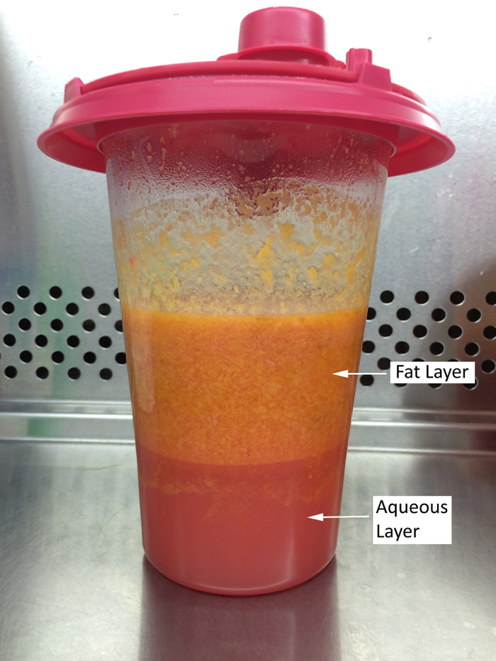
Figure 1: Lipoaspirate after PBS Wash. A canister of lipoaspirate after PBS is added and the mixture is allowed to settle. The top tissue layer is the adipose tissue which hosts ASCs, while the bottom layer is the aqueous layer, mainly comprised of saline and blood. Please click here to view a larger version of this figure.
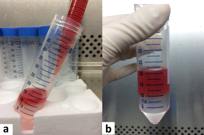
Figure 2: Placement of Cell Suspension onto Polysucrose Solution. (a) The cell suspension must be pipetted very gently onto the side of the tube, allowing it to layer on top of the polysucrose solution. (b) This is the correct appearance of a cell suspension layer on top of the polysucrose prior to centrifugation. Please click here to view a larger version of this figure.
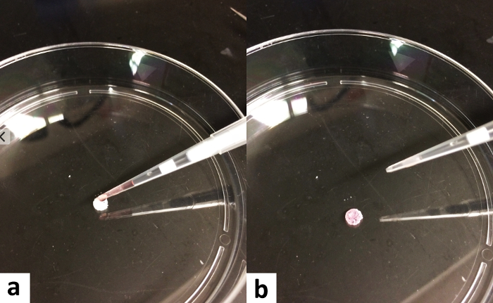
Figure 3: Seeding of Cells onto the Scaffold. (a) On the sterile surface of one well of a 24-well cell culture plate, load a dry scaffold with approximately 50 μL of cell suspension. (b) Incubate the scaffold in a cell culture incubator for 30 min to allow cell adhesion. Note: In the figure, a 10-cm plate is used for demonstration purposes. Please click here to view a larger version of this figure.
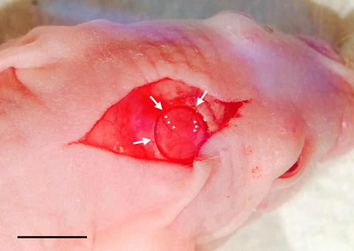
Figure 4: Creation of the Calvarial Defect. The calvarial defect (arrows) is visible within the open skin incision. Note the intact dural blood vessels (arrowheads), indicating that the drilling has not breached the dura. Scale bar, 5 mm. Please click here to view a larger version of this figure.
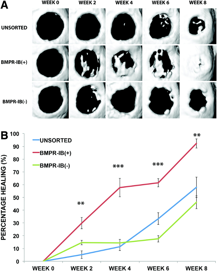
Figure 5: Healing of Critical-sized Calvarial Defects with Different ASC Subpopulations. (A) Three-dimensional micro- CT reconstructions were performed for calvarial defects re- paired with unsorted, BMPR-IB(+), or BMPR-IB(-) ASCs. (B) Quantification of healing at 8 weeks demonstrated significantly greater bone regeneration with BMPR-IB(+) ASCs (92%) compared with unsorted and BMPR-IB(-) ASCs (58% and 46%, respectively, **p < 0.01). Significant differences in healing were also seen at 2 weeks (**p < 0.01), 4 weeks (***p < 0.001), and 6 weeks (***p < 0.001). Micro-CT, micro-computed tomography. Reprinted with permission from McArdle et al.12. Error bars represent standard deviation. Please click here to view a larger version of this figure.
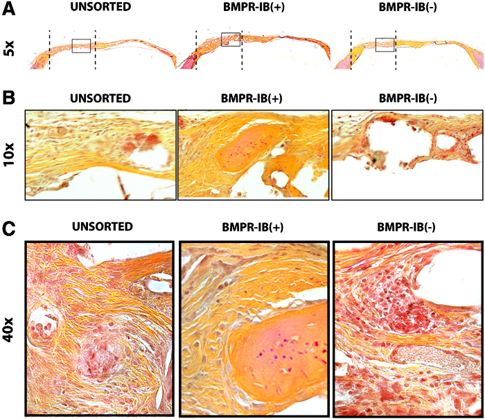
Figure 6: Histological Staining of Bone Regenerate. (A) Movat's pentachrome staining of bone regenerate in defects repaired with unsorted, BMPR-IB(+), or BMPR-IB(-) ASCs at 5X magnification using bright field microscopy. Note more robust bone formation in BMPR-IB(+) group compared with BMPR-IB(-) group. The dotted line represents the extent of the defect area. The area within the black rectangle is shown on higher magnification using (B) 10X and (C) 40X bright field microscopy of defect area. Reprinted with permission from McArdle et al.12. Please click here to view a larger version of this figure.
Discussion
Critical Steps within the Protocol
During the harvest of ASCs, the critical step is adequate digestion of fat with collagenase. Inadequate digestion will result in a low yield of ASCs. During FACS sorting of BMPR-IB+ cells, it is important to carefully define the gate for positivity. Defining gates too loosely may result in sorted populations that are not pure. During creation of the calvarial defect, it is critical to drill the defect through the bone of the skull but to not advance into the dura mater. This will cause profuse bleeding and exposure of the brain, which will necessitate euthanasia of the animal.
Modifications and Troubleshooting
Regarding anesthesia for the calvarial defect surgery, our lab prefers to use inhaled isoflurane, but another option is to intraperitoneally inject a mixture containing a final concentration of ketamine 10 mg/mL and xylazine 1 mg/mL in normal saline. The standard dose of this mixture is 300 µL for a 30 g mouse. This provides roughly 30 min of reliable anesthesia that does not require the mouse to be in the isoflurane nose cone during surgery. A disadvantage of using injected anesthetic is that it is not possible to decrease the dose after the injection has been given. In contrast, inhaled isoflurane can easily be titrated up or down in real time.
When adding cell suspension to the scaffold, researchers may find that the fluid volume is too small and will partially evaporate in the 30 min of incubation. Evaporation can cause cell death, which is a particularly harmful confounder in bone healing experiments. To combat this, load the scaffold inside a well of a 24 well plate. Fill the immediately surrounding wells with plain medium. This will create a humidified micro-environment which helps keep the loaded scaffold from drying out.
Histological staining of bone is predicated on correct and thorough decalcification of the calvarium prior to embedding and sectioning. It is accepted that skulls of differing ages require differing durations of decalcification, traditionally done in an EDTA solution
Limitations of the Technique
This protocol utilizes a 4 mm calvarial defect to assess the ability of a population of cells to enhance closure of the defect. This model may not simulate the healing that occurs in a more complex environment, such as a long bone fracture or a tumor resection. For this reason, alternate animal models may need to be used to understand the contribution of ASCs to bone healing in these settings.
Significance of the Technique with Respect to Existing/Alternative Methods
Using flow cytometry to isolate specific ASC populations is a promising technique for enhancing healing and regeneration in the context of multiple tissue and injury models, such as angiogenesis, adipogenesis, and osteogenesis. The utility of pro-osteogenic cells is explored in this study, showing a profound effect with cells positively selected for BMPR-IB in healing a calvarial defect.
Prior protocols for the isolation of ASC involve culturing the cells for several days on cell culture plates 16. However, cells may experience significant phenotypic drift while in culture. For this reason, we use an ASC isolation protocol that allows for rapid isolation of these cells, followed by further subpopulation purification using FACS. The entire procedure is accomplished in less than a day, minimizing the effects of cell surface marker or phenotypic drift.
Future Applications and Directions
We have shown that ASCs highly expressing BMPR-IB demonstrate a greater ability to enhance regenerative bone defect healing compared with other ASCs. The mechanisms underlying this phenomenon are not known. For example, it is not clear whether the cells themselves directly differentiate into bone-forming cells, or whether they produce factors that encourage other cells to do so. Future studies will need to be performed to answer these questions.
Disclosures
The authors have no disclosures to make.
Acknowledgements
C.D.M. was supported by the American College of Surgeons (ACS) Resident Research Scholarship. M.S.H. was supported by the California Institute for Regenerative Medicine (CIRM) Clinical Fellow training grant TG2-01159. M.S.H., H.P.L., and M.T.L. were supported by the American Society of Maxillofacial Surgeons (ASMS)/Maxillofacial Surgeons Foundation (MSF) Research Grant Award. H.P.L. was supported by NIH grant R01 GM087609 and a gift from Ingrid Lai and Bill Shu in honor of Anthony Shu. H.P.L. and M.T.L. were supported by the Hagey Laboratory for Pediatric Regenerative Medicine and The Oak Foundation. M.T.L. was supported by NIH grants U01 HL099776, R01 DE021683-01, and RC2 DE020771. D.C.W. was supported by NIH grant 1K08DE024269, the Hagey Laboratory for Pediatric Regenerative Medicine, and the Stanford University Child Health Research Institute Faculty Scholar Award.
Materials
| Name | Company | Catalog Number | Comments |
|---|---|---|---|
| 100 micron cell strainer | Falcon | 352360 | |
| 15 blade scalpel | Miltex | 4-515 | |
| 24 well plate | Corning | 3524 | |
| 40 micron cell strainer | Falcon | 352340 | |
| 50 mL conical centrifuge tubes | Falcon | 352098 | |
| 6-0 Ethilon nylon suture, 18", P-3 needle, | Ethicon | 1698G | |
| Anti-BMPR-IB primary antibody | R&D systems | FAB5051A | |
| BioGel PI surgical gloves | Mölnlycke Health Care | ALA42675Z | |
| Buprenorphine SR | ZooPharm | ||
| Castro-Viejo needle driver | Fine Science Tools | 12565-14 | |
| CD1 nude mouse | Charles River | 086 | |
| Collagenase Type II powder | Gibco | 17101-015 | |
| DMEM medium | Gibco | 10564-011 | |
| Drill: Circular knife 4.0 mm | Xemax Surgical | CK40 | |
| Drill: Z500 Brushless Micromotor | NSK | NSKZ500 | |
| FBS | Gicbo | 10437-077 | |
| Fisherbrand Absorbent Underpads, 20" x 24" | Fisher Scientific | 14-206-62 | |
| Fisherbrand Sterile cotton gauze pad, 4" x 4" | Fisher Scientific | 22-415-469 | |
| Heating pad | Kent Scientific | DCT-20 | |
| Hyclone 199/EBSS medium | GE Life Sciences | SH30253.01 | |
| Isothesia isoflurane | Henry Schein | 050033 | |
| Micro Forceps with teeth | Roboz | RS-5150 | |
| Micro Forceps with teeth | Roboz | RS-5150 | |
| Paraffin film (Parafilm) | Bemis | PM996 | |
| PBS | Gibco | 10010-023 | |
| Pen-Strep | Gibco | 15140-122 | |
| PLGA scaffolds | Proprietary Formulation | ||
| Poloxamer 188, 10% | Sigma | P5556-100ML | |
| Polylined Sterile Field, 18" x 24" | Busse Hospital Disposables | 696 | Cut a rectangular hole of the appropriate size |
| Polysucrose Solution: Histopaque 1119 | Sigma | 11191 | |
| Povidone Iodine Prep Solution | Medline | MDS093944H | |
| Puralube petrolatum ophthalmic ointment, 1/8 oz. tube | Dechra Veterinary Products | ||
| RBC lysis buffer | Sigma | 11814389001 | |
| Webcol alcohol prep swabs | Covidien | 6818 |
References
- Silber, J. S., et al. Donor site morbidity after anterior iliac crest bone harvest for single-level anterior cervical discectomy and fusion. Spine (Phila Pa 1976). 28 (2), 134-139 (2003).
- Giannoudis, P. V., Dinopoulos, H., Tsiridis, E. Bone substitutes: an update. Injury. 36, S20-S27 (2005).
- Laurencin, C., Khan, Y., El-Amin, S. F. Bone graft substitutes. Expert Rev Med Devices. 3 (1), 49-57 (2006).
- Walmsley, G. G., et al. Nanotechnology in bone tissue engineering. Nanomedicine. 11 (5), 1253-1263 (2015).
- Chapekar, M. S. Tissue engineering: challenges and opportunities. J Biomed Mater Res. 53 (6), 617-620 (2000).
- Levi, B., et al. Human adipose derived stromal cells heal critical size mouse calvarial defects. PLoS One. 5 (6), e11177(2010).
- Gimble, J. M., Katz, A. J., Bunnell, B. A. Adipose-derived stem cells for regenerative medicine. Circ Res. 100 (9), 1249-1260 (2007).
- Levi, B., et al. CD105 protein depletion enhances human adipose-derived stromal cell osteogenesis through reduction of transforming growth factor beta1 (TGF-beta1) signaling. J Biol Chem. 286 (45), 39497-39509 (2011).
- Chung, M. T., et al. CD90 (Thy-1)-positive selection enhances osteogenic capacity of human adipose-derived stromal cells. Tissue Eng Part A. 19 (7-8), 989-997 (2013).
- Wozney, J. M., et al. Novel regulators of bone formation: molecular clones and activities. Science. 242 (4885), 1528-1534 (1988).
- Wan, D. C., et al. Osteogenic differentiation of mouse adipose-derived adult stromal cells requires retinoic acid and bone morphogenetic protein receptor type IB signaling. Proc Natl Acad Sci U S A. 103 (33), 12335-12340 (2006).
- McArdle, A., et al. Positive selection for bone morphogenetic protein receptor type-IB promotes differentiation and specification of human adipose-derived stromal cells toward an osteogenic lineage. Tissue Eng Part A. 20 (21-22), 3031-3040 (2014).
- Sharon, Y., Alon, L., Glanz, S., Servais, C., Erez, N. Isolation of normal and cancer-associated fibroblasts from fresh tissues by Fluorescence Activated Cell Sorting (FACS). J Vis Exp. (71), e4425(2013).
- Lo, D. D., et al. Repair of a critical-sized calvarial defect model using adipose-derived stromal cells harvested from lipoaspirate. J Vis Exp. (68), (2012).
- Lester, S. C. Manual of surgical pathology. , Saunders/Elsevier. (2010).
- Bunnell, B. A., Flaat, M., Gagliardi, C., Patel, B., Ripoll, C. Adipose-derived stem cells: isolation, expansion and differentiation. Methods. 45 (2), 115-120 (2008).
Reprints and Permissions
