Whole-cell recordings from Drosophila melanogaster photoreceptors enable the measurement of spontaneous dark bumps, quantum bumps, macroscopic responses to light, and current-voltage relationships under various conditions. In combination with D. melanogaster genetic manipulation tools, this method enables the study of the ubiquitous inositol-lipid signaling pathway and its target, the TRP channel.
Method Article
Electrophysiological Method for Whole-cell Voltage Clamp Recordings from Drosophila Photoreceptors
* These authors contributed equally
In This Article
Summary
Abstract
Whole-cell voltage clamp recordings from Drosophila melanogaster photoreceptors have revolutionized the field of invertebrate visual transduction, enabling the use of D. melanogaster molecular genetics to study inositol-lipid signaling and Transient Receptor Potential (TRP) channels at the single-molecule level. A handful of labs have mastered this powerful technique, which enables the analysis of the physiological responses to light under highly controlled conditions. This technique allows control over the intracellular and extracellular media; the membrane voltage; and the fast application of pharmacological compounds, such as a variety of ionic or pH indicators, to the intra- and extracellular media. With an exceptionally high signal-to-noise ratio, this method enables the measurement of dark spontaneous and light-induced unitary currents (i.e. spontaneous and quantum bumps) and macroscopic Light-induced Currents (LIC) from single D. melanogaster photoreceptors. This protocol outlines, in great detail, all the key steps necessary to perform this technique, which includes both electrophysiological and optical recordings. The fly retina dissection procedure for the attainment of intact and viable ex vivo isolated ommatidia in the bath chamber is described. The equipment needed to perform whole-cell and fluorescence imaging measurements are also detailed. Finally, the pitfalls in using this delicate preparation during extended experiments are explained.
Introduction
Extensive genetic studies of the fruit fly, Drosophila melanogaster (D. melanogaster), initiated more than 100 years ago, have established the D. melanogaster fly as an extremely useful experimental model for the genetic dissection of complex biological processes. The methodology described below combines the accumulated power of D. melanogaster molecular genetics with the high signal-to-noise ratio of whole-cell patch clamp recordings. This combination allows for the study of D. melanogaster phototransduction as a model of inositol-lipid signaling and TRP channel regulation and activation, both in the native environment and at the highest resolution of single molecules.
Application of the whole-cell recording method to D. melanogaster photoreceptors has revolutionized the study of invertebrate phototransduction. This method was developed by Hardie1 and independently by Ranganathan and colleagues2 ~26 years ago and was designed to exploit the extensive genetic manipulation tools of D. melanogaster and use them to uncover mechanisms of phototransduction and inositol-lipid signaling. At first, this technique suffered from a rapid reduction in light sensitivity and a low yield of ommatidia during the dissection process, which prevented detailed quantitative studies. Later, the addition of ATP and NAD to the patch pipette dramatically increased the suitability of the preparation for prolonged quantitative recordings. Thereafter, extensive characterization of the signal-transduction mechanism at the molecular level was realized.
Currently, D. melanogaster phototransduction is one of the few systems in which phosphoinositide signaling and TRP channels can be studied ex vivo at single-molecule resolution. This makes D. melanogaster phototransduction and the methodology developed to study this mechanism a highly sensitive model system. This protocol describes how to dissect the D. melanogaster retina and mechanically strip the isolated ommatidia from the surrounding pigment (glia) cells. This enables the formation of a giga-seal and a whole-cell patch clamp on the photoreceptor cell bodies. Fortunately, most signaling proteins are confined to the rhabdomere and do not diffuse. In addition, there is an immobile Ca2+ buffer called calphotin, located between the signaling compartment and the cell body3,4, and a high expression level of the Na+/Ca2+ exchanger (CalX) in the microvilli5. Together, the protein confinement to the rhabdomere, the calphotin buffer, and the high expression of the CalX allow for relatively prolonged (i.e. up to ~20 min) whole-cell recordings, without the loss of essential components of the phototransduction process and while maintaining high sensitivity to light. The following protocol describes how to obtain isolated ommatidia and perform whole-cell recordings that appear to preserve the native properties of the phototransduction cascade. Whole-cell patch clamp experiments on dissociated cockroach (Periplaneta americana)6 and cricket (Gryllus bimaculatus)7 ommatidia were performed similarly to that described for D. melanogaster. In addition, patch clamp experiments on dissociated photoreceptors of the file clam, (Lima scabra) and scallop (Pecten irradians) were performed in a slightly different manner from that conducted on D. melanogaster, allowing both whole-cell8 and single-channel measurements9. Here, the major achievements obtained in D. melanogaster using this technique are described. The Discussion includes the description of some pitfalls and limitations of this technique.
Protocol
1. Reagent Preparation
NOTE: Prepare all solutions according to the instructions in Tables 1-4.
- Fill a 10 mL syringe with Extracellular Solution (ES or ES-0Ca2+; as required; see Table 1) and keep it on ice.
- Prepare one vial of Trituration Solution (TS; see Table 2; i.e. ES or ES-0Ca2+ + FBS and sucrose) and keep this on ice .
- Prepare sufficient ES for the experiment and keep this on ice until needed.
NOTE: Continuous bath perfusion is not required, so the volume of ES need not be more than a few dozen mL. - Using a 22 µm PVDF filter, load the intracellular solution (see Table 3 or Table 4) into a 1 mL syringe with an electrode filling elongated tip. Keep this on ice.
2. General Setup of Dissecting Tools
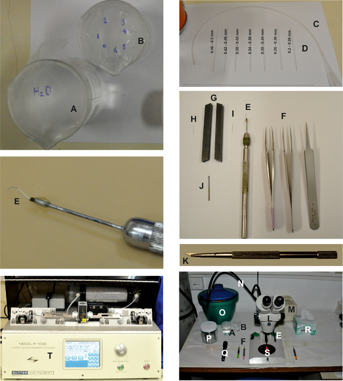
Figure 1: Tools and Devices Needed for Making the Isolated Ommatidia Preparation. The pictures show the various devices required for creating the isolated ommatidia preparation, as described in the detailed protocol above. Two beakers, one filled with water (A) and the other filled with ethanol (B) for cleaning the trituration pipettes (D), which are connected to tubing (C). The dissecting tools are: 2 pairs of fine and 1 pair of coarse tweezers (F) and a retina scooper (E). In order to prepare the retina scooper, a micro dissection needle (H) is pressed between two lathe tools acting as a vice (G) to flatten the top of the needle (I). Then it is connected to an elongated piece of metal using glue (J) and mounted onto a needle holder (E). Razor blade chip and holder: Break off and mount a small triangular chip of a razor blade using a razor blade holder (K). The dissection working area is composed of a binocular (L) and a cool red light source ((M),) with two light guides (N). The dissection tools, including tweezers (F), the retina scooper (E), beakers (A and B), a flashlight with a red filter (Q), and delicate wipers (R) are placed on both sides of the binocular. An ice bucket with the ES, the FBS-ES, the intracellular solution syringes, the 60 mm Petri dish (S), and the electrode holder (P) are also placed on the table. The recording electrodes are pulled using a horizontal puller (T). Please click here to view a larger version of this figure.
- Construct a retina scooper to isolate the retinae.
- Insert the point (1-4 mm) of a micro dissection needle (entomological needle, 12 mm length, 0.1 mm diameter) between the two vise jaws and flatten it by tapping with a small hammer.
- Mount the flattened needle on a micro dissecting needle holder (see the Table of Materials).
- Using a pair of tweezers, curve the flattened end of the needle to form a hook with a curvature of ~2.5 mm.
- Create trituration pipettes for ommatidia separation.
- Place a 1.2 x 0.68 mm (O.D. x I.D.) glass capillary over an open flame and fire polish it to reduce its opening.
- Measure the size of the capillary opening under a microscope. Sort the created ommatidial trituration pipettes into seven groups according to the size of their openings (i.e 0.2-0.5 mm). Store them in separate suitable containers (e.g. test tubes).
- Fill one small beaker with double-distilled water (DDW) and another small beaker with ethanol 70%. Cover each beaker with a paraffin film sheet.
- Punch one small hole in the paraffin film sheet covering the DDW beaker (see Figure 1A).
- Punch seven small holes in the paraffin film sheet covering the ethanol beaker and number the holes from 0 to 6 (see Figure 1B).
- Place one ommatidial trituration pipette from each size group in every paraffin film hole, such that the pipette with the largest opening is located in the 0th hole and the pipette with the smallest opening is located in the 6th hole.
- Connect a plastic 200 µL pipette tip to a 35 cm long, 1.57 x 1.14-mm (O.D. x I.D.) piece of polyethylene tubing. Connect the other end of the tubing to one of the ommatidial trituration pipettes with the largest opening (i.e. 0.46 - 0.5 mm), ensuring that the pointed end of the trituration pipette is not facing into the tubing.
- Prepare whole-cell recording pipettes by pulling patch clamp pipettes from a 1 x 0.58 mm (O.D. x I.D.) borosilicate filament containing glass capillaries.
NOTE: The resistance of the pipettes should be 8-15 MΩ when using potassium gluconate-based intracellular solution (IS1). Any suitable patch pipette puller can be used (for examples, see the Table of Materials). Fire-polishing is not necessary. - Prepare a recording chamber (see Figure 2) by fixing a coverslip (see the Table of Materials) to the bottom of the bath chamber using melted paraffin or high-vacuum silicone grease. Use any suitable homemade or commercial chamber that allows electrode access and perfusion.
- Mount the bath on the stage of an inverted microscope. Place the perfusion system tube, suction system, and ground (i.e. Ag-AgCl wire/pellet) in the bath (see the Table of Materials and Figure 2).
- Place two pairs of fine #5 tweezers (Figure 1) on the working surface for use during the retina dissection and ommatidia isolation steps.
3. D. melanogaster Rearing
- Raise D. Melanogaster flies at a low population density (i.e. ~20 flies in a 6 oz. bottle) in bottles containing standard cornmeal food at 19-24 °C.
NOTE: It is preferable to work on dark-adapted flies. To maintain high sensitivity to light, reduce the diversity and prevent retinal degeneration in mutant flies. - Rear the flies in the dark for at least 24 h prior to the experiment.
NOTE: The flies used for the experiments should be recently eclosed (<2 h) and still soft, pale, and display the meconium. Ommatidia can also readily be prepared from pupae, though their sensitivity to light is then steeply dependent on age10.
4. Retina Dissection and Ommatidia Isolation: Option 1
NOTE: Perform all of the following steps under the stereoscopic zoom microscope using an amplification suitable for properly viewing the preparation (See Figure 1).
- Place four drops of ES-0Ca2+ and one drop of TS solution on a 60-mm Petri dish that has been turned over.
- Using rough tweezers, catch a newly eclosed (<2 h after eclosion) fly by its wings or body. From this point on, perform all procedures rapidly and under dim red illumination at 20 ±1 °C.
- While still grasping the fly with the rough tweezers, use the first pair of fine tweezers to detach the fly head from the body. Submerge the head in the first ES-0Ca2+ drop.
- Dissect the head in half along the sagittal plane using the second pair of fine tweezers. Ensure that, at the end of this step, both eyes are still intact.
- Transfer one half of the head to the second ES-0Ca2+ drop and the other half to the third ES-0Ca2+ drop.
- Using the fine tweezers, remove as much of the tissue surrounding the eye as possible and ensure that no harm is caused to the retina.
- Firmly grasp the edge of one cornea with the fine tweezers and scoop out the retina using the scooper.
NOTE: Upon completion of this step, the cornea will be left empty and intact, separated from an intact retina. - Rinse the trituration pipette connected to the tubing with DDW and fill the pipette with a small amount of ES-0Ca2+ from the fourth drop.
NOTE: This step must be performed every time a new ommatidia-separating pipette is used and removed from the ethanol beaker (the solution filling the pipette should match the solution in which the retina is submerged). - Gently aspirate by mouth to draw the isolated retina into the pipette. Use extreme caution not to aspire air bubbles into the pipette.
- Transfer the isolated retina to the drop of TS. Perform steps 4.6-4.10 on the second eye as well.
- Wipe away the drops of ES-0Ca2+ using delicate wipes, leaving only the drop of TS containing both retinae on the Petri dish. Add six more drops of TS to the top of the Petri dish. Transfer both retinas to one of the other TS drops.
- Replace the trituration pipette with a pipette of a smaller-diameter opening. Rinse it as described in step 4.8, using TS as the solution to fill the pipette.
- Rapidly and repeatedly aspirate and expirate both retinae in the solution to begin the separation of isolated ommatidia stripped of pigment cells from the whole retina.
NOTE: The isolated ommatidia are visible in the TS drop, and as the isolation process progresses, the TS drop becomes less translucent. - Transfer the remaining retinae to the next TS drop. Fill the pipette with the entire former TS drop (containing the isolated ommatidia) and expirate the drop into the bath chamber.
- Repeat steps 4.12-4.14 to achieve maximal isolated ommatidia. Wait approximately 1 min to allow the isolated ommatidia to sink and bind to the bottom of the bath chamber.
- Using the perfusion system, start the flow of ES-0Ca2+ with 1.5 mM Ca2+ into the bath chamber. Ensure that the chamber is completely filled with the solution, from bottom to top, and that the ground is completely submerged in the solution. Continue to wash the bath 4-5x.
5. Retina Dissection and Ommatidia Isolation: Option 2
Note: Perform all of the following steps under the stereoscopic zoom microscope, using an amplification suitable for properly viewing the preparation (See Figure 1).
- Prepare a razor blade chip and holder. Break off and mount a small triangular chip of a razor blade using a razor blade holder (Figure 1).
- Prepare a silicone dissection dish/block according to the manufacturer's instructions (see the Table of Materials).
- For the dissection, create a large drop (<0.5 mL) of ES solution on the silicone dissection block. Add two "reservoir" drops (~50 µL each) of TS solution to a 60 mm Petri dish
- Immobilize a newly eclosed (<2 h post-eclosion) fly in a glass tube on ice and pick it up by its wings using tweezers. From this point on, perform all procedures rapidly and under dim red illumination at 20 ±1 °C.
- Holding the fly with tweezers, cut off the fly's head using a razor blade chip mounted in a holder. Pick up an insect pin (12 mm long, 0.1 mm in diameter) with the tweezers and pierce the head between the eyes.
- Briefly submerge the head in 70% ethanol; this prevents air bubbles from forming on the head/eye surface. Pin the head under the ES drop on the silicone dissection dish.
- Cut off both eyes using the razor blade chip by using a sawing motion along the line of the frontal margin of the eye.
- Firmly grasp the edge of one cornea with the fine tweezers.
- Scoop out the retina using the scooper.
NOTE: Upon completion of this step, the cornea will be left empty and intact, separated from an intact retina. - Without damaging the retina, use the tweezers and scooper to gently remove adhering air sacs and excess brain tissue.
NOTE: The preparation of isolated retinae is also useful for Western blot analyses of non-specific retinae proteins11, whole-mount histology, and whole-retina imaging. Patch clamp recordings can also be conducted on photoreceptors from the whole retina12. - Take the trituration pipette with the largest diameter, connect it to the tubing, and backfill the pipette with a small amount of TS from one of the reservoir drops in the Petri dish by gentle suction (i.e. by mouth). Perform this step every time a new ommatidia trituration pipette is used.
- Gently blow the TS over the two retinae by mouth and then draw the isolated retinae into the pipette using gentle suction. Use caution to ensure that no air bubbles enter the pipette.
- Transfer the isolated retinae to the Petri dish, forming a small drop (~20 µL), and wash them once or twice with TS from one of the reservoir drops.
- Incubate the retinae in the dark for 20-25 min.
- Replace the ommatidia trituration pipette with a pipette with a smaller-diameter opening (using fresh TS from one of the reservoir drops as in step 4.6 to backfill the pipette).
- Rapidly aspirate and expirate both retinae in a small drop (~20 µL) to begin the separation of the isolated ommatidia.
NOTE: At the first stage, the surrounding pigmented glia should disintegrate, leaving visible small debris in the solution. - After substantial small debris has accumulated, but before many ommatidia have separated, use fresh TS from one of the reservoir drops and transfer the retinae to a new small drop.
- Select a smaller-diameter trituration pipette, backfill, and continue to triturate.
NOTE: As ommatidia now begin to separate, their elongated forms should be clearly visible under the high power of the stereomicroscope. If necessary, keep changing the trituration pipettes to smaller diameters until a good yield of ommatidia is visible - Once a reasonable yield of ommatidia are visible and the drop is no longer translucent, fill the pipette with the entire drop containing the isolated ommatidia and expirate the drop gently into the bottom of the bath chamber pre-filled with ES.
- Wait approximately 1 min to allow the isolated ommatidia to sink and settle on the bottom of the bath chamber.
- Using the perfusion system, start the flow of ES into the bath chamber. Ensure that the chamber is completely full of the solution, from bottom to top, and that the ground is totally submerged in the solution. Continue to wash the bath 4-5x.
NOTE: Thereafter, continuous perfusion is not required, though the bath should be briefly flushed before introducing a new patch pipette.
6. Whole-Cell Recording
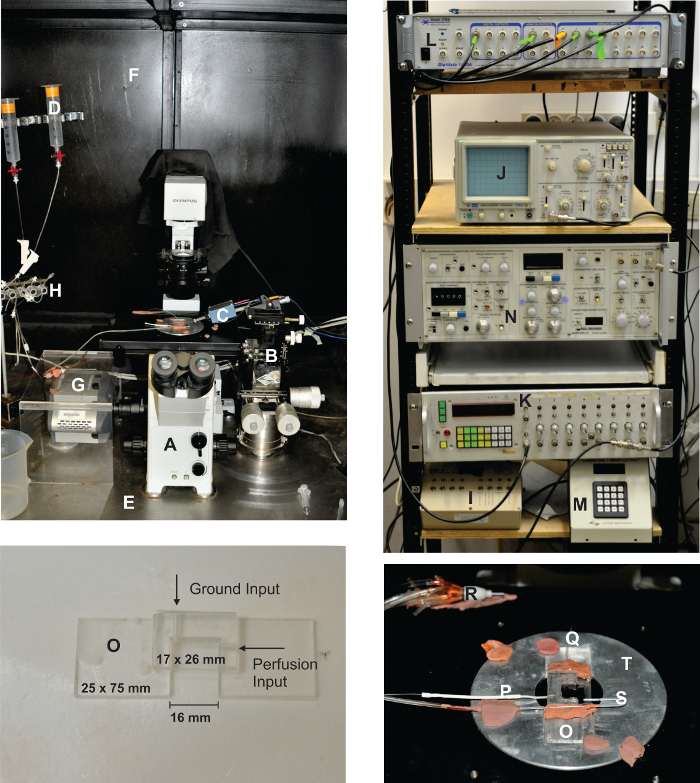
Figure 2: Overview of the Electrophysiological and Optical Setup. The setup contains an iron, black-painted Faraday cage (F). The front side is covered using a black curtain with copper mesh inside. This configuration enables the preparation to be completely sealed off from any stray light and is suitable for recording dark spontaneous bumps. The inverted fluorescence microscope (A) is fixed to an anti-vibration table (E). The photoreceptor recording apparatus consists of a homemade acrylic glass bath chamber (O) with a perfusion input (Q), a suction pipette (S), and a silver-silver chloride ground (P). The bath chamber is mounted on the microscope stage (T) with a homemade adaptor. The recording pipette is connected via silver-silver chloride wire to an acrylic glass holder, which is connected to the amplifier head stage (C). The head stage is then mounted on a coarse micromanipulator that is mounted on a fine XYZ mechanical micromanipulator (B). The perfusion system (D) is composed of a syringe set, while the flow of liquid is controlled by pinch valves (H). The rack is electrically connected to the same central ground to which all the equipment inside the Faraday cage is connected and consists of a patch clamp amplifier (N), an oscilloscope (J), a pulse/function generator (K), an A to D converter (L), a perfusion controller (I), and a filter wheel and shutter controller (M). For imaging experiments, a cooled (-110 °C, see the Table of Materials) CCD camera is connected via a side port (G). Please click here to view a larger version of this figure.
NOTE: During all of the following steps, use only dim red light illumination and ensure that the exposure of the ommatidia to light is minimal (i.e. work rapidly and turn off the dissecting light source and chamber red illumination used for viewing the ommatidia when performing tasks that do not demand viewing the ommatidium). In addition, perform all of the following steps according to standard electrophysiological protocol.
- Under an inverted microscope (40X objective), carefully inspect all of the ommatidia in the bath and choose a suitable ommatidium for the experiment.
- Ensure that the outer membrane of the ommatidium is smooth and intact, that the long axis is approximately at a right angle relative to the electrode approach direction (as seen in Figure 3A), and that the distal section of the ommatidium is not surrounded by any excess tissue. Place the chosen ommatidium at the optical axis of the objective lens (in the center of the field of vision) to ensure uniform illumination.
- Fill a patch pipette with intracellular solution (IS1 or IS2).
NOTE: To measure the intensity response and bump analysis, use IS1. To measure the reversal potential of the light-sensitive channels, use IS2. - Mount the patch pipette onto the electrode holder.
- Blow into the pipette by mouth, through the tube connected to the electrode holder, causing it to fill with positive pressure. Close the tube valve to maintain the pressure.
- Using the micromanipulator, insert the electrode into the bath chamber.
- Guide the electrode close to the distal section of the ommatidium, such that there is no contact between the electrode and the ommatidium, until a small dimple (due to the positive pressure in the patch pipette) can be observed in the ommatidium.
- Open the recording software (see the Table of Materials). Open the "membrane test module" to apply continuous square voltage pulses of 2 mV at a rate of 100 Hz.
- Set the junction potential to "zero," by adjusting the appropriate knob in the patch clamp amplifier to set the base of the square pulse to "zero" current
NOTE: The electrophysiological setup includes a head stage (i.e. first-stage amplification) connected to an amplifier (i.e. second-stage amplification). The amplified analogue signal is converted to a digital signal using the A/D converter, which is controlled by software installed on a PC computer. - Release the positive pressure in the pipette by opening the valve of the tube connected to the electrode holder. Gently create negative pressure in the pipette by sucking out of the tube, leading to the association of the pipette to the cell membrane. Close the tube valve to maintain the pressure.
- Ensure that the electrode resistance viewed on the computer screen is elevated to 100 - 150 MΩ. Release the negative pressure in the pipette by manually opening the valve of the tube connected to the electrode holder.
- Ensure that the electrode resistance is elevated to at least 1-2 GΩ.
NOTE: At this point, a seal has been formed between the electrode and the photoreceptor. - Offset the capacitive currents of the pipette by adjusting the appropriate knob in the patch clamp amplifier.
- Create rapid, short, and forceful bouts of negative pressure in the electrode, sucking by mouth out of the tube connected to the electrode holder to "break" into the photoreceptor membrane and create a whole-cell configuration. Alternatively, use the "Zap button" to apply short, rectangular electrical pulses, starting with a duration of "0.1 ms," or apply a combination of both methods.
NOTE: The generation of the whole-cell configuration is revealed by a sudden increase in pipette capacitance (typically ~60 pF for a wildtype R1-6 photoreceptor; a capacitance of only ~20 pF indicates a recording from an R7 photoreceptor; a capacitance above ~90 pF indicates a recording from two photoreceptors). - Set the holding potential of the photoreceptor to the required voltage (usually, -70 mV), manually using the appropriate knob in the patch clamp amplifier.
NOTE: It is possible to perform this step after a seal has been obtained (step 6.11) and before the whole-cell configuration has been achieved. - Offset the capacitive currents and series resistance (a measured series resistance value greater than 25 MΩ indicates that the electrode pipette has been clogged) and, if required (i.e. for larger currents), apply series resistance compensation using the appropriate knobs in the patch clamp amplifier.
- Close the black front curtain of the Faraday cage to obtain maximal darkness and electrical isolation.
- Begin the recording process using the software and administer light stimuli and/or pharmacological substances according to the desired experimental procedure.
7. Simultaneous Whole-Cell Recordings and Ca2+ Imaging
- For genetically encoded Ca2+ indicators, isolate the ommatidia as described above using D. melanogaster flies expressing GCaMP6f13. Use a CCD camera (see the Table of Materials) for the fluorescence measurement and ensure that the microscope is equipped with proper excitation and emission filters and a dichroic mirror (see the Table of Materials)4.
- For the use of exogenous Ca2+ indicator (Figure 4; see the Table of Materials), isolate the ommatidia as described above. In addition, ensure that the pipette solution includes a 20-100 µM calcium indicator.
- Use the imaging software (see the Table of Materials) to acquire images at a rate of 40 Hz. Perform image acquisition during a 10 s dark period followed by an intense 2 s light stimulation.
- Use the imaging software and define a Region of Interest (ROI). Measure the florescence intensity in the ROI. Average the dark fluorescence (FD) and subtract it from the fluorescence recordings during the light (FL) stimulation (FL-FD). Normalize these measurements according to the fluorescence intensity at the beginning of the light stimulation (FL0).
Results
The described method has enabled the accurate recording of the fundamental unitary currents that generate spontaneous and light-evoked quantum bumps, which sum to produce the macroscopic response to light, under defined conditions. It also allowed the comparison between wildtype and mutant flies that have defects in critical signaling molecules (Figures 3 and 5)14,15,16,17,18. In addition, the ability to measure reversal potential under bi-ionic conditions revealed fundamental biophysical properties of the TRP and TRP-like (TRPL) channels18,19. It also enabled the measurement of the effects of amino acid substitutions in the pore region of TRP that modified its Ca2+ permeability20.
The light response obtained by patch clamp whole-cell recordings depends linearly on light intensity for at least 4 orders of magnitude. This could not be resolved by using ERG and intracellular recording methods. Accordingly, a series of responses to brief flashes of increasing intensity and a plot of the intensity response function revealed a strict linearity of the flash response with increasing light intensity. The strict linearity holds up to at least several hundred pA, but it is debatable whether thereafter it is linearity or clamp control that breaks down (Figure 6). These results suggest that the macroscopic responses to light are a linear summation of the unitary responses to light (i.e. quantum bumps).
It has been well established using voltage recordings that dim light stimulation induces discrete voltage fluctuations (i.e. quantum bumps) in most invertebrate species. The D. melanogaster quantum bumps result from the concerted opening of ~15 TRP channels and ~2 TRPL channels at the peak of the bump18. Each bump is generated by the absorption of a single photon, while the macroscopic response to more intense lights is the summation of these elementary responses14,21. The bumps vary significantly in latency, time course, and amplitude, even when the stimulus conditions are identical. Bump generation is a stochastic process described by Poisson statistics, whereby each effectively absorbed photon elicits only one bump. The single-photon-single-bump relationship requires that each step in the cascade includes not only an efficient "turn-on" mechanism, but also an equally effective "turn-off" mechanism. The functional advantage is the production of a very sensitive photon counter with a fast transient response very well suited to both the sensitivity and the temporal resolution required by the visual system. The requirement for an efficient turn-off mechanism is revealed when either the active photopigment (i.e. metarhodopsin, M) or its target, the Gqα, fails to inactivate and leads to the continuous production of bumps long after light is turned off (Figure 3)15,22,23,24 .
The bump represents the cooperative activity of the TRP/TRPL channels in a microvillus. As such, any hypothesis of channel activation should also explain cooperative channel activation. Recently, Hardie and colleagues have demonstrated that light evokes rapid contractions of the photoreceptors, suggesting that the light-sensitive channels (TRP/TRPL) may be mechanically gated25. This mechanical activation, together with the observed protons released by PLC-mediated PIP2 hydrolysis, promote the opening of the TRP/TRPL channels and explain the cooperative nature of bump production26. Currently, D. melanogaster photoreceptors are one of the few systems in which phosphoinositide signaling and TRP channels can be studied in vivo, thus making D. melanogaster phototransduction and the methodology developed to study this mechanism a highly valuable model system.
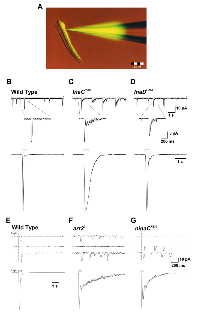
Figure 3: The inaCP209 and inaDP215 Mutants Reveal Slow Response Termination of the Macroscopic Response to Light and of the Single Quantum Bumps. (A)The isolated ommatidium preparation with a patch pipette filled with fluorescent Lucifer Yellow CH dye (excitation: 430 nm; emission: 540 nm) is presented during a whole-cell recording. Note that the fluorescent dye diffused and labeled a single photoreceptor cell body and that the photoreceptor cell bodies are detached from their elongated axons but still maintain viability. This preparation is suitable for simultaneous whole-cell recordings and imaging experiments. (B-D) Upper panels: Whole-cell voltage clamped quantum bump responses to continuous dim light (open bar) in WT, inaCP209, and inaDP215 mutant flies. A slow termination of the bumps is observed in inaCP209 and inaDP215 mutants relative to WT flies. The inset below displays the magnified shape of single bumps. Bottom panels: Normalized whole-cell recorded macroscopic responses to a 500-ms light pulse (1.5 x 10⁵ photons per s) of the above wildtype and mutant flies. (E-G) Upper panels: Whole-cell voltage clamped quantum bump responses to a brief (1 ms), dim light eliciting single-photon responses in wildtype, arr23, and ninaCP235 mutant flies. Note the train of bumps observed in arr23 and ninaCP235 mutant flies in response to a single photon absorption. Bottom panels: Whole-cell voltage clamped normalized responses to a 500 ms light pulse (1.5 x 10⁴ photons/s) in the corresponding mutants. Note the slow termination of the macroscopic responses observed in arr23 and ninaCP235 mutant flies relative to WT. Please click here to view a larger version of this figure.
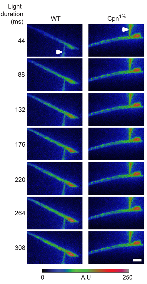
Figure 4: Cellular Ca2+ Dynamics Following Signal-induced Ca2+ Influx is Affected by Calphotin. A time series of photoreceptor images of wildtype and Cpn1% flies showing the fluorescence of the Ca2+ indicator during light stimulation. Raw intensity images are plotted using false-color coding (bar = 10 µm; arrowheads indicate the pipette). Figure reprinted with permission from Weiss et al.4. Please click here to view a larger version of this figure.
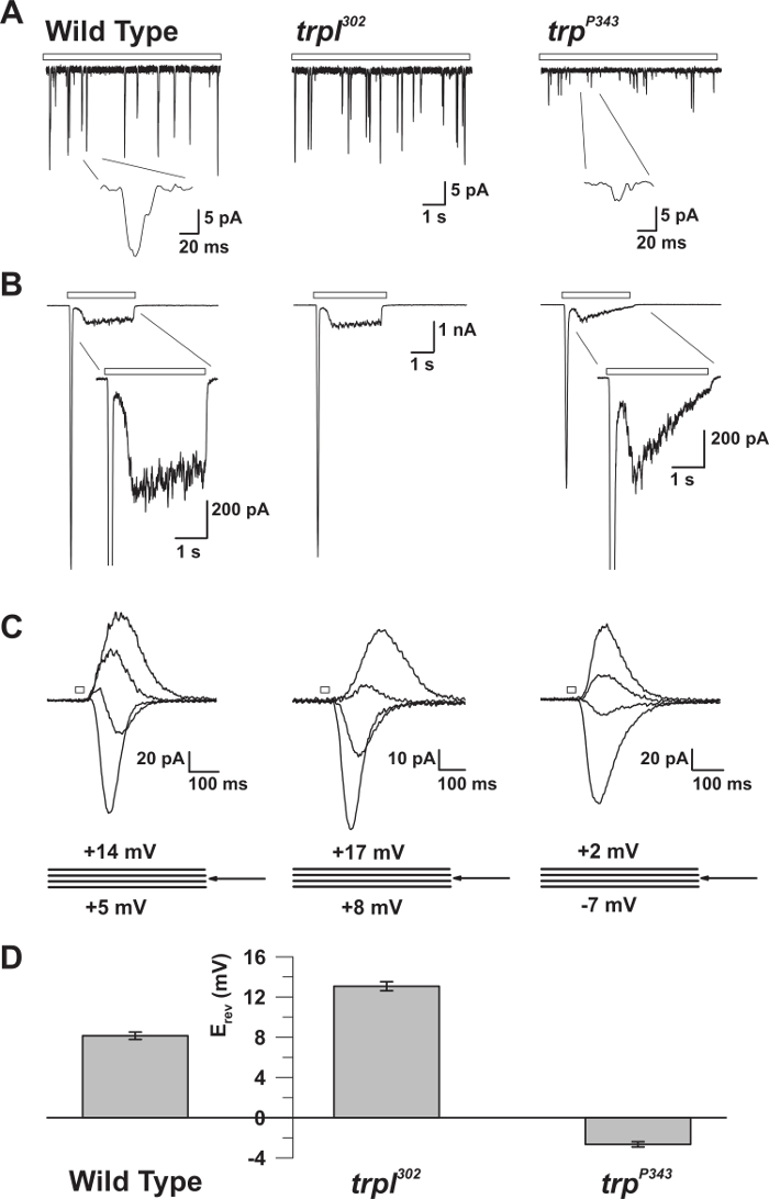
Figure 5: The Electrophysiological Properties of WT, trp, and trpl Mutants. (A) Whole-cell voltage clamp recordings of quantum bumps in response to continuous dim light (open bar) in WT, trpl302, and trpP343 null mutant flies. Highly reduced amplitudes of trpP343 bumps are observed. Inset: Magnified single quantum bumps of wildtype and trpP343 null mutant flies are shown. (B) Whole-cell voltage clamp recordings in response to a 3 s light pulse of wildtype and the corresponding mutants. The transient steady-state response of the trpP343 mutant is observed. Inset: Magnified light responses of WT and trpP343mutant are shown. (C) A family of superimposed light-induced currents of the above fly strains, elicited in response to a 20 ms light pulse, at voltage steps of 3 mV, measured around the reversal potential (Erev). (D) A histogram plotting the mean Erev of wildtype and the various mutants. The error bars are the S.E.M. The reversal potential (Erev) of WT is between the positive Erev of trpl302, which expresses only TRP, and the Erev of the trpP343 null mutant, which expresses only TRPL. Please click here to view a larger version of this figure.
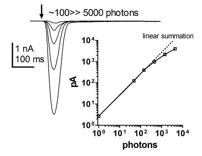
Figure 6: The Flash Response is Strictly Linear with Increasing Light Intensity.
A series of current responses to brief flashes of increasing light intensity and a plot of the dependence of the peak amplitude of the light response on the increasing intensity of brief light flashes. This relationship reveals a strict linearity between the flash response and increasing light intensity. This strict linearity holds up to at least several hundred pA, with light intensity spanning over 4 orders of magnitude, while it is debatable whether it is linearity or the clamp control that breaks down thereafter. Please click here to view a larger version of this figure.
| pH | 7.15 (adjust with NaOH) |
| Reagent | Concentration (mM) |
| NaCl | 120 |
| KCl | 5 |
| MgCl2 | 4 |
| TES | 10 |
| Proline | 25 |
| Alanine | 5 |
| Store at -20 °C. | |
| Note: This solution is nominally Ca2+ free but has no Ca2+ buffers added and hence will have approximately 5 - 10 µM trace Ca2+. Extracellular solution (ES) = ES-0Ca2+ with 1.5 mM CaCl2, made by adding CaCl2 from a 0.5 or 1 M stock solution to ES-0Ca2+. | |
Table 1: Ca+2-free Extracellular Solution (ES). Chemical description and the specific quantities required to produce Ca+2-free ES.
| Reagent | Amount |
| FBS | 15 mL |
| sucrose | 1.5 g |
| Divide into 150 µL aliquots in 1.5 mL vials and store at -20 °C. | |
| Trituration solution (TS) | Fill 1 vial of 150 mL of the stock solution with 1,350 mL ES or ES-0Ca2+, to match the solution used during the dissection. |
Table 2: Fetal Bovine Serum (FBS) + Sucrose - Stock Solution. Chemical description and the specific quantities required to producing fetal bovine serum (FBS) + sucrose - stock solution.
| pH | 7.15 (adjust with KOH) |
| Reagent | Concentration (mM) |
| Potassium gluconate (Kglu) | 140 |
| MgCl2 | 2 |
| TES | 10 |
| ATP magnesium salt (MgATP) | 4 |
| GTP sodium salt (Na2GTP) | 0.4 |
| β-Nicotinamide adenine dinucleotide hydrate (NAD) | 1 |
| Store at -20 °C. |
Table 3: Intracellular Solution (IS1). Chemical description and the specific quantities required to produce IS1, which is mostly used for intensity response and quantum bump measurements.
| pH | 7.15 (adjust with CsOH) |
| Reagent | Concentration (mM) |
| CsCl | 120 |
| MgCl2 | 2 |
| TES | 10 |
| ATP magnesium salt (MgATP) | 4 |
| GTP sodium salt (Na2GTP) | 0.4 |
| β-Nicotinamide adenine dinucleotide hydrate (NAD) | 1 |
| Tetra-ethyl-ammonium chloride (TAE) | 15 |
| Store at -20 °C. |
Table 4: Intracellular Solution (IS2). Chemical description and the specific quantities required to produce Intracellular Solution IS2, which is mostly used for reversal potential measurements of the light-induced current.
Discussion
The application of whole-cell recordings to D. melanogaster photoreceptors allowed for the discovery and the functional elucidation of novel signaling proteins, such as TRP channels27,28,29 and INAD30,31,32 scaffold protein. Ever since the initial introduction of this technique, it enabled the resolution of long-term basic questions regarding the ionic mechanism and voltage dependence of the light response. This occurred because of the conferred ability to accurately control the membrane voltage and extracellular and intracellular ionic composition19,28.
A major obstacle of the patch clamping technique in D. melanogaster has been the fragility of the isolated ommatidia preparation. Detailed studies have revealed that the integrity of the phototransduction machinery critically depends on the continuous supply of ATP, especially during light exposure, which leads to a large consumption of ATP. Unfortunately, the mechanical striping of the pigment (i.e. glia) cells, which is required to reach the photoreceptor membrane with the patch pipette, eliminates the main source of metabolites necessary for ATP production33. Application of exogenous ATP into the recording pipette only partially fulfills the requirement for large quantities of ATP. A short supply of ATP leads to spontaneous activation of the TRP channels and to the dissociation of the phototransduction machinery from the light-activated channels, causing a large increase in cellular Ca2+ and the abolishment of the normal response to light34,35. This sequence of events is not due to damage of the photoreceptors by the dissection procedure, but rather to the cellular depletion of ATP. To prevent this sequence of events from occurring and to maintain normal light responses, the photoreceptors should not be exposed to intense lights, which consume large quantities of ATP. Also, NAD must be included in the recording pipette, presumably to facilitate ATP production in the mitochondria18,36. For measurements of spontaneous and quantum bumps, the above difficulty is minimal because only dim lights are used. In practice, a stable whole-cell recording can be maintained for ~20-25 min, although there is a tendency for response kinetics to slow down over this period. A single preparation of dissociated ommatidia may remain viable for up to 2 h.
An additional shortcoming of the isolated ommatidia preparation is the inaccessibility of the microvilli, which translates to the inaccessibility of the TRP and TRPL channels to the recording pipette, preventing single-channel recordings. Using a method they developed, Bacigalupo and colleagues succeeded at directly recording single-channel activity from the rhabdomere37. However, this channel activity differs from that of TRPL channels heterologously expressed in tissue culture cells38 and from TRP channel activity derived from shot noise analysis obtained from isolated ommatidia34. Presumably, the dissection procedure greatly damaged the photoreceptor cells when using this method.
Disclosures
The authors have nothing to disclose.
Acknowledgements
The experimental part of this research was supported by grants from the US-Israel Bi National Science Foundation (to B.M. and I.L.), the Israel Science Foundation (ISF), the Deutsch-Israelische Projektkooperation (DIP) (to B.M.), and the Biotechnology and Biological Sciences Research Council (BBSRC Grant numbers: BB/M007006/1 and BB/D007585/1) to R.C.H.
Materials
| Name | Company | Catalog Number | Comments |
|---|---|---|---|
| 10 mL syringe | |||
| 5 mL syringe | |||
| 1 mL syringe with elongated tip | |||
| Petri dish | 60 mm | ||
| Syringe filters | Millex | 22 µm PVDF filter | |
| Capillaries (for omatidia separation) | Glass, 1.2 x 0.68 mm (~7.5 cm each) | ||
| Polyethylene Tubing | Becton Dickinson | 1.57 x 1.14 mm (35 cm) | |
| 2 small beakers | 50 mL or less | ||
| 2 paraffin film sheets | Parafilm M | ~5 x 5 cm | |
| Bath chamber | home-made | ||
| Cover slips | 22 x 22 mm No. 0 | ||
| Paraplast Plus | Sigma | Paraffin – polyisobutylene mixture | To glue the coverslip onto the bottom of the bath chamber |
| Ground | Warner Instruments | 64-1288 | Hybrid Assembly Ag-AgCl Wire Assembly |
| Headless micro dissection needle | Entomology, 12 mm | ||
| Micro dissecting needle holder | |||
| Vise | |||
| 2 fine tweezers + 1 rough tweezers | Dumont #5, Biology | 0.05 x 0.02 mm, length 110 mm, Inox | |
| Stereoscopic zoom Microscope | Nikon | SMZ-2B | |
| Cold light source | Schott | KL1500 LCD | |
| Filter (Color) for cold light source | Schott | RG620 | |
| Delicate wipers | Kimtech | Kimwipes | |
| Electrode holder | Warner Instruments | QSW-T10P | Q series Holders compatible with Axon amplifiers, straight body style |
| Silver Wire | Warner Instruments | 0.25 mm diameter, needs to be chloridized | |
| Micromanipulator | Sutter Instruments | MP 85 | Huxley-Wall Style Micromanipulator |
| Faraday cage | home made | Electromagnetic noise shielding and black front curtain | |
| Anti-vibration Table | Newport | VW-3036-OPT-01 | |
| Osilloscope | GW | GOS-622G | |
| Perfusion system | Warner Instruments | VC-8P | Pinch valve control system |
| Perfusion valve controller | Scientific instruments | BPS-8 | |
| Suction system | |||
| Amplifier | Molecular Device | Axopatch-1D | |
| Head-stage | Molecular Device | CV - 4 | Gain: x 1/100 |
| A/D converter | Molecular Device | Digidata 1440A | |
| Clampex | Molecular Device | 10 | software |
| pCLAMP | Molecular Device | 10 | software |
| Light source (Xenon Arc lamp) | Sutter Instruments | Lambda LS | |
| Light detector | home made | phototransistor | |
| Filter wheel and shutter controller | Sutter Instruments | Lambda 10-2 with a Uniblitz shutter | |
| Filters (Natural density filter) | Chroma | 6,5,4,3,2,1,0.5,0.3 | |
| Filter (Color) | Schott | OG590, Edge filter | |
| Xenon Flash Lamp system | Dr. Rapp OptoElecftronic | JML-C2 | |
| Light guide | Quartz | ||
| Pulse generator | AMPI | Master 8 | |
| Microscope | Olympus | IX71, Inverted | |
| Red illumination filter (Microscope) | RG630 / RG645 ø45mm | ||
| Microscope objective | Olympus | X60/0.9 UplanFL N air or X60/1.25 UplanFI oil | |
| CCD Camera | Andor | iXon DU885K | |
| NIS Element | Nikon | AR | software |
| Ca+2 indicator | Invitrogen | Calcium green 5N | |
| Excitation & emission filters and dichroic mirror | Chroma | 19002 - AT - GFP/FITC Longpass set | |
| Vertical pipette puller | Narishige | Model PP83 | Use either vertical or horizontal puller, as preferred. |
| Horizontal pipette puller | Sutter Instrument | Model P-1000 Flaming/Brown Micropipette Puller | |
| Filament | Sutter Instrument | 3 mm trough or square box | |
| Capillaries | Harvard Apparatus | borosilicate glass capillaries | 1 x 0.58 mm |
References
- Hardie, R. C. Whole-cell recordings of the light induced current in dissociated Drosophila photoreceptors: evidence for feedback by calcium permeating the light-sensitive channels. Proc. R. Soc. Lond. B. 245, 203-210 (1991).
- Ranganathan, R., Harris, G. L., Stevens, C. F., Zuker, C. S. A Drosophila mutant defective in extracellular calcium- dependent photoreceptor deactivation and rapid desensitization. Nature. 354, 230-232 (1991).
- Martin, J. H., Benzer, S., Rudnicka, M., Miller, C. A. Calphotin: a Drosophila photoreceptor cell calcium-binding protein. Proc. Natl. Acad. Sci. U.S.A. 90 (4), 1531-1535 (1993).
- Weiss, S., et al. Compartmentalization and Ca2+ buffering are essential for prevention of light-induced retinal degeneration. J Neurosci. 32 (42), 14696-14708 (2012).
- Wang, T., et al. Light activation, adaptation, and cell survival functions of the Na + /Ca 2+ exchanger CalX. Neuron. 45 (3), 367-378 (2005).
- Weckstrom, M., Hardie, R. C., Laughlin, S. B. Voltage-activated potassium channels in blowfly photoreceptors and their role in light adaptation. J. Physiol. Lond. 440, (1991).
- Frolov, R. V., Immonen, E. V., Weckström, M. Performance of blue- and green-sensitive photoreceptors of the cricket Gryllus bimaculatus. J Comp Physiol A Neuroethol Sens Neural Behav Physiol. 200 (3), 209-219 (2014).
- Nasi, E. Whole-cell clamp of dissociated photoreceptors from the eye of Lima scabra. J Gen Physiol. 97 (1), 35-54 (1991).
- Nasi, E., Gomez, M. P. Light-activated ion channels in solitary photoreceptors of the scallop Pecten irradians. J. Gen. Physiol. 99, 747-769 (1992).
- Hardie, R. C., Peretz, A., Pollock, J. A., Minke, B. Ca 2+ limits the development of the light response in Drosophila photoreceptors. Proc. R. Soc. Lond. B. Biol. Sci. 252, 223-229 (1993).
- Kohn, E., et al. Functional Cooperation between the IP3 Receptor and Phospholipase C Secures the High Sensitivity to Light of Drosophila Photoreceptors In Vivo. J Neurosci. 35 (6), 2530-2546 (2015).
- Hevers, W., Hardie, R. C. Serotonin modulates the voltage dependence of delayed rectifier and Shaker potassium channels in Drosophila photoreceptors. Neuron. 14 (4), 845-856 (1995).
- Chen, T. W., et al. Ultrasensitive fluorescent proteins for imaging neuronal activity. Nature. 499 (7458), 295-300 (2013).
- Henderson, S. R., Reuss, H., Hardie, R. C. Single photon responses in Drosophila photoreceptors and their regulation by Ca 2. J. Physiol. Lond. 524 (Pt 1), 179-194 (2000).
- Scott, K., Zuker, C. S. Assembly of the Drosophila phototransduction cascade into a signalling complex shapes elementary responses. Nature. 395 (6704), 805-808 (1998).
- Elia, N., Frechter, S., Gedi, Y., Minke, B., Selinger, Z. Excess of G betae over G qalphae in vivo prevents dark, spontaneous activity of Drosophila photoreceptors. J. Cell Biol. 171 (3), 517-526 (2005).
- Katz, B., Minke, B. Phospholipase C-Mediated Suppression of Dark Noise Enables Single-Photon Detection in Drosophila Photoreceptors. J. Neurosci. 32 (8), 2722-2733 (2012).
- Hardie, R. C., et al. Molecular basis of amplification in Drosophila phototransduction. Roles for G protein, phospholipase C, and diacylglycerol kinase. Neuron. 36 (4), 689-701 (2002).
- Reuss, H., Mojet, M. H., Chyb, S., Hardie, R. C. In vivo analysis of the Drosophila light-sensitive channels, TRP and TRPL. Neuron. 19, 1249-1259 (1997).
- Liu, C. H., et al. In vivo identification and manipulation of the Ca 2+ selectivity filter in the Drosophila transient receptor potential channel. J. Neurosci. 27 (3), 604-615 (2007).
- Ahmad, S. T., Natochin, M., Barren, B., Artemyev, N. O., O'Tousa, J. E. Heterologous expression of bovine rhodopsin in Drosophila photoreceptor cells. Invest Ophthalmol Vis Sci. 47 (9), 3722-3728 (2006).
- Scott, K., Sun, Y., Beckingham, K., Zuker, C. S. Calmodulin regulation of Drosophila light-activated channels and receptor function mediates termination of the light response in vivo. Cell. 91 (3), 375-383 (1997).
- Liu, C. H., et al. Ca 2+ -dependent metarhodopsin inactivation mediated by calmodulin and NINAC myosin III. Neuron. 59 (5), 778-789 (2008).
- Cook, B., et al. Phospholipase C and termination of G-protein-mediated signalling in vivo. Nat. Cell Biol. 2 (5), 296-301 (2000).
- Hardie, R. C., Franze, K. Photomechanical responses in Drosophila photoreceptors. Science. 338 (6104), 260-263 (2012).
- Huang, J., et al. Activation of TRP channels by protons and phosphoinositide depletion in Drosophila photoreceptors. Curr. Biol. 20 (3), 189-197 (2010).
- Minke, B., Wu, C. F., Pak, W. L. Isolation of light-induce response of the central retinular cells from the electroretinogram of Drosophila. J. Comp. Physiol. 98, 345-355 (1975).
- Hardie, R. C., Minke, B. The trp gene is essential for a light-activated Ca2+ channel in Drosophila photoreceptors. Neuron. 8, 643-651 (1992).
- Niemeyer, B. A., Suzuki, E., Scott, K., Jalink, K., Zuker, C. S. The Drosophila light-activated conductance is composed of the two channels TRP and TRPL. Cell. 85 (5), 651-659 (1996).
- Huber, A., et al. The transient receptor potential protein (Trp), a putative store- operated Ca 2+ channel essential for phosphoinositide-mediated photoreception, forms a signaling complex with NorpA, InaC and InaD. EMBO J. 15 (24), 7036-7045 (1996).
- Shieh, B. H., Niemeyer, B. A novel protein encoded by the InaD gene regulates recovery of visual transduction in Drosophila. Neuron. 14 (1), 201-210 (1995).
- Tsunoda, S., et al. A multivalent PDZ-domain protein assembles signalling complexes in a G-protein-coupled cascade. Nature. 388 (6639), 243-249 (1997).
- Tsacopoulos, M., Veuthey, A. L., Saravelos, S. G., Perrottet, P., Tsoupras, G. Glial cells transform glucose to alanine, which fuels the neurons in the honeybee retina. J. Neurosci. 14 (3 Pt 1), 1339-1351 (1994).
- Hardie, R. C., Minke, B. Spontaneous activation of light-sensitive channels in Drosophila photoreceptors. J. Gen. Physiol. 103, 389-407 (1994).
- Agam, K., et al. Metabolic stress reversibly activates the Drosophila light-sensitive channels TRP and TRPL in vivo. J Neurosci. 20 (15), 5748-5755 (2000).
- Agam, K., Frechter, S., Minke, B. Activation of the Drosophila TRP and TRPL channels requires both Ca2+ and protein dephosphorylation. Cell Calcium. 35 (2), 87-105 (2004).
- Delgado, R., Muñoz, Y., Peña-Cortés, H., Giavalisco, P., Bacigalupo, J. Diacylglycerol activates the light-dependent channel TRP in the photosensitive microvilli of Drosophila melanogaster photoreceptors. J Neurosci. 34 (19), 6679-6686 (2014).
- Parnas, M., Katz, B., Minke, B. Open channel block by Ca2+ underlies the voltage dependence of Drosophila TRPL channel. J. Gen. Physiol. 129 (1), 17-28 (2007).
Reprints and Permissions
