This protocol describes how to use an in vitro isolated turtle head preparation to measure the kinematics of their eye movements. After removal of the brain from the cranium, cranial nerves can be stimulated with currents to quantify rotations of the eye and changes in pupil sizes.
A subscription to JoVE is required to view this content. Sign in or start your free trial.
Method Article
Ocular Kinematics Measured by In Vitro Stimulation of the Cranial Nerves in the Turtle
In This Article
Summary
Abstract
After animals are euthanized, their tissues begin to die. Turtles offer an advantage because of a longer survival time of their tissues, especially when compared to warm-blooded vertebrates. Because of this, in vitro experiments in turtles can be performed for extended periods of time to investigate the neural signals and control of their target actions. Using an isolated head preparation, we measured the kinematics of eye movements in turtles, and their modulation by electrical signals carried by cranial nerves. After the brain was removed from the skull, leaving the cranial nerves intact, the dissected head was placed in a gimbal to calibrate eye movements. Glass electrodes were attached to cranial nerves (oculomotor, trochlear, and abducens) and stimulated with currents to evoke eye movements. We monitored eye movements with an infrared video tracking system and quantified rotations of the eyes. Current pulses with a range of amplitudes, frequencies, and train durations were used to observe effects on responses. Because the preparation is separated from the brain, the efferent pathway going to muscle targets can be examined in isolation to investigate neural signaling in the absence of centrally processed sensory information.
Introduction
Rationale for Using Red-eared Slider Turtles in Electrophysiological Experiments:
Red-eared slider turtles (Trachemys scripta elegans), are considered one of world's worst invasive species1 and can indicate that an ecosystem is in trouble. The reason why red-eared slider turtles are so successful is poorly understood but it may in part be due to their tolerant physiology and possession of nervous tissues that can survive under hypoxic conditions2,3,4. Using them for experimentation does not threaten their numbers a....
Access restricted. Please log in or start a trial to view this content.
Protocol
NOTE: Red-eared slider turtles, both male and female, were purchased from a vendor. Turtles were housed in a warm animal suite containing two 60-gallon tubs equipped with brick islands for sunning under 250-W infrared lights. The environment was maintained on a 14/10-h light/dark cycle with the water temperature at 22 °C. Lights were turned on at 6:00 am and turned off at 8:00 pm. The tanks equipped with filtering systems were cleaned weekly, and turtles were fed ad libitum every other day. The care of red-eared slider turtles and all of the following experimental procedures described here32,33 w....
Access restricted. Please log in or start a trial to view this content.
Results
Figure 1 shows stills of images taken from a video describing the dissection. Images provide typical locations of the nerves prior to cutting from the brain.
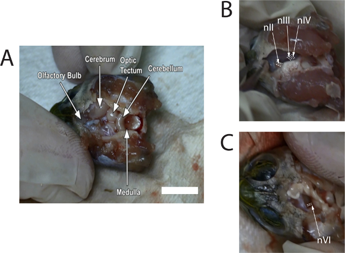
Figure 1: S.......
Access restricted. Please log in or start a trial to view this content.
Discussion
Critical Steps:
The critical steps within this protocol are the following: 1) the dissection and the care taken to maintain the viability of the transected nerves; 2) the matching of sizes by the suction electrodes to cranial nerves to provide consistent responses; and 3) the placement of the head in the gimbal to provide adequate calibration of the rotations of the eye.
Troubleshooting:
The dissection can be challenging, but after completing i.......
Access restricted. Please log in or start a trial to view this content.
Disclosures
The authors have nothing to disclose.
Acknowledgements
The authors thank Mrs. Paulette McKenna and Lisa Pezzino in this study for secretarial support, and Mr. Phil Auerbach for technical support. The authors also thank Drs. Michael Ariel and Michael S. Jones (Saint Louis University School of Medicine) for introducing us to the in vitro isolated head preparation. Funding for support of this collaboration was provided by the Department of Biology (Robert S. Chase Fund), the Academic Research Committee, and the Neuroscience Program at Lafayette College. Lastly, this work is dedicated to Mr. Phil Auerbach, who passed away 28 September 2016; he decommissioned a scanning electron microscope and recognized the usefulnes....
Access restricted. Please log in or start a trial to view this content.
Materials
| Name | Company | Catalog Number | Comments |
|---|---|---|---|
| Red-eared slider turtles | Kons Scientific | Trachemys scripta elegans | Large size (carapace length 15-20 cm) |
| Sodium chloride | Sigma-Aldrich Co. LLC. | S5886 | |
| Potassium chloride | Sigma-Aldrich Co. LLC. | P5405 | |
| Magnesium choride | Sigma-Aldrich Co. LLC. | M7304 | |
| Sodium bicarbonate | Sigma-Aldrich Co. LLC. | S5761 | |
| Dextrose | Sigma-Aldrich Co. LLC. | C5767 | |
| Concentrated hydrochloric acid | Sigma-Aldrich Co. LLC. | H7020 | |
| Calcium chloride | Sigma-Aldrich Co. LLC. | C7902 | |
| pH meter | Oakton | pH 6+ | |
| Suction stimulation electrode | A-M Systems | 573000 | Bipolar suction electrode. Note that 573000 has been replaced with 573050. |
| Capillary glass | A-M systems | 626000 | Single-barrel borosilicate capillary glass without microfilament, length 10 cm, outside diameter 1.0 mm, inner diameter 0.50 mm |
| Alternative suction stimulation electrode | A-M Systems | 573050 | Bipolar suction electrode. Requires larger diameter capillary glass: 627000, outside diameter 1.2 mm, inner diameter 0.68 mm |
| Stereoscope | Lieca | GZ7 | Magnification range, 10x – 70x |
| Fiber optic light source | Amscope | HL250-A | 150W Fiber optical microscope illuminator light box |
| Rongeurs | Carolina Biological Supply Company | 625654 | stainless steel, straight spring, 5.25" |
| Blunt dissection probe | Carolina Biological Supply Company | 627405 | Huber mall probe, double-ended probe and seeker, 6" |
| Microscissors | Carolina Biological Supply Company | 623555 | Iris microdissecting scissors, stainless steel, 0.5" blades, 4.75" long |
| Fine forceps | Sigma-Aldrich Co. LLC. | F6521 | Jewelers forceps, dumont No. 5, inox alloy, 4.25" |
| Curved forceps | Sigma-Aldrich Co. LLC. | Z168696 | Medium tip, curved forceps, stainless steel, 4" |
| Scalpel handle | Sigma-Aldrich Co. LLC. | S2896 | Scalpel handles, No. 3, stainless steel |
| Scalpel blade | Sigma-Aldrich Co. LLC. | S2771 | Scalpel blades, No. 11, steel |
| Guillotine | Harvard Apparatus | 73-1918 | Kleine guillotine type 7575 |
| Spatula | Sigma | Z648299 | Micro spoon and spatula weighing set. Use small spatula: 5.9” long x 0.07” diameter handle with square end: 0.17” x 1.3” long, other end round: 0.17” x 1.27” long |
| Hook | Autozone | 98069 | SureBilt hook and pick set. Use grinder to dull sharp points of hook to prevent injury to animals mouth. |
| 95/5% O2/CO2 | Airgas, Inc. | X02OX95C2003102 | 5% Carbon dioxide balance oxygen certified standard gas mixture, size 200 Cylinder, CGA-296 |
| Regulator | Airgas, Inc. | Y11244D296-AG | Single stage brass 0-100 psi analytical cylinder regulator CGA-296 with needle outlet. Use brass adjustable airline pipe valve to go from 3/8", inner diameter, vinyl airline tubing connected to regulator to a 3/16", inner diameter, airline connection going to airstone or glass pasteur pipette. |
| Adjustable airline pipe valve | Doctors Foster and Smith | CD-12061 | Brass valve |
| Rigid table | Unknown | Unknown | Auto-clave door laid on top of a sturdy table. Nine 5" diameter tennis balls isolate vibrations from the top surface of the table. |
| 5" tennis ball | Petco Animal Supplies, Inc. | 712868 | Petco Jumbo Pet Tennis Ball: balls are unsliced and held within an integrated frame on the underside part of the autoclave door. |
| Alternative vibration isolation table | Newport Corporation | INT1-36-6-N | Rigid vibration control system, integrity 1: Surface dimensions, 3' x 6' |
| Gimbal | ISI, International Scientific Instruments, Inc. | Stage from SUPER III-A Scanning EM | 5-axis eucentric stage: X, Y, and Z linear movements, ±20 mm, 0.1 mm precision; Rotations, vertical, ±10°, and horizontal, ±12.5°, with 1.25° precision. Note: from decommission instrument. |
| Chuck for gimbal | Unknown | Unknown | Chuck from an old microtome of unknown manufacture was machined to fit the shaft of the specimen holder of the Scanning EM stage |
| Alternative gimbal | ThorLabs, Inc. | GN2/M with MBT602/M | Dual-axis goniometer (GN2/M) mounted on 3-axis microblock stage with thumbscrew adjusters (MBT602/M): design a chuck to hold turtle head with eye at 12.7 mm above top surface of goniometer (distance to point of rotation) |
| Video-based eye tracking system | Arrington Research, Inc. | ViewPoint EyeTracker, PC-60 | Tracking method: Infrared video by dark pupil; Black and white camera (Item BC02): 30 Hz, 640 x 480; System requirements: Windows 2000, XP, 7, 8, 8.1, 10; Visual range: Horizontal +/- 44°; vertical +/- 20°; Accuracy ~0.5°; Spatial resolution ~0.15°; Pupil size resolution ~0.03 mm; Eye data: X, Y position of gaze, pupil height and width, torsion, delta time, total time, and regions of interest (ROI); Real-time communication (Item 0022): 4-Channel AnalogOut with eight TTL input channels to mark codes into the data file |
| Multi-position magnetic base | Harbor Freight Tools | Pittsburg, item #5645 | Magnetic holder reaches up to 12" and produces 45 lbs. of magnetic pull. Use to position camera. Machine thread holes onto the end of the rod to mount cameras. |
| Micromanipulator | Kopf | 900 | 5 axis manipulation for mount of suction electrode: X, Y, Z linear travel, 2 axis of rotation |
| Dissection scope on boom | Lieca | GZ6 | Magnification range, 6.7x – 40x |
| Nerve/muscle stimulator | Astro-Med Grass Telefactor | Grass S88 | Dual pulse voltage stimulator: two output channels that can be operated independently or synchronized to generate non-isolated constant voltage pulses (10 mv to 150 V). Pulses can be single (10 μsec to 10 sec), repetitive (0.01 Hz to 1 KHz), and trains (1 ms to 10 s) and synchronized with TTL inputs and output. Send TTL outputs via the output channels of a DB25 connector to the TTL input channels of the ViewPoint EyeTracker. Note: Astro-Med Grass Telefactor is no longer in business. |
| Current isolation device | Astro-Med Grass Telefactor | PSIU6 | Current stimulus isolation unit: enables safe delivery of constant currents by the S88 to the preparation. The PSIU6 connects by a BNC cable to one of the output channels of the S88. Multiplier switches on the PSIU6 allow the S88 to generate a wide array of current amplitudes ranging from 0.1 µA to 15 mA. |
| Alternative nerve/muscle stimulator with isolation | A-M Systems | 2100 | Isolated Pulse Stimulator: Unit has built-in isolator to produce constant currents. |
References
- Kikillus, K. H., Hare, K. M., Hartley, S. Minimizing false-negatives when predicting the potential distribution of an invasive species: A bioclimatic envelope for the red-eared slider at global and regional scales. Anim Conserv. 13, 5-15 (2010).
- Lutz, P. L., Rosenthal, M., Sick, T. J. Living without oxygen: turtle brain as a model of anaerobic metabolism. Mol Physiol. 8, 411-42....
Access restricted. Please log in or start a trial to view this content.
Reprints and Permissions
