Here, we present a protocol for the rapid virus nucleic acid extraction from the virus-inactivated whole blood. The extraction is performed directly in the blood collection tubes and requires no equipment or electricity. The method is not dependent on laboratory facilities and can be used anywhere (e.g., in field hospitals).
Method Article
Rapid, Safe, and Simple Manual Bedside Nucleic Acid Extraction for the Detection of Virus in Whole Blood Samples
In This Article
Summary
Abstract
The rapid diagnosis of an infection is essential for the outbreak management, risk containment, and patient care. We have previously shown a method for the rapid bedside inactivation of the Ebola virus during blood sampling for safe nucleic acid (NA) tests by adding a commercial lysis/binding buffer directly into the vacuum blood collection tubes. Using this bedside inactivation approach, we have developed a safe, rapid, and simplified bedside NA extraction method for the subsequent detection of a virus in lysis/binding buffer-inactivated whole blood. The NA extraction is directly performed in the blood collection tubes and requires no equipment or electricity.
After the blood is collected into the lysis/binding buffer, the contents are mixed by flipping the tube by hand, and the mixture is incubated for 20 min at room temperature. Magnetic glass particles (MGPs) are added to the tube, and the contents are mixed by flipping the collection tube by hand. The MGPs are then collected on the side of the blood collection tube using a magnetic holder or a magnet and a rubber band. The MGPs are washed three times, and after the addition of elution buffer directly into the collection tube, the NAs are ready for NA tests, such as qPCR or isothermal loop amplification (LAMP), without the removal of the MGPs from the reaction. The NA extraction method is not dependent on any laboratory facilities and can easily be used anywhere (e.g., in field hospitals and hospital isolation wards). When this NA extraction method is combined with LAMP and a portable instrument, a diagnosis can be obtained within 40 min of the blood collection.
Introduction
In virus outbreak situations, when patients are confined to the hospital isolation wards or when a fast diagnosis is needed, a safe, simple, and accurate point-of-care molecular diagnosis is imperative for the patient care and risk containment. The two recent viral outbreaks, of the Ebola virus (EBOV) in West Africa (2013) and the Zika virus in South America (2015), have increased the interest in improved point-of-care molecular diagnostic tests, such as reverse transcription loop-mediated isothermal amplification (RT-LAMP)1,2 and recombinase polymerase amplification (RPA)3,4. Both RT-LAMP and RPA are rapid, sensitive, and specific molecular tests that can be performed on simplified sample preparations. For the Zika virus, RT-LAMP has been combined with a lateral flow assay (LFA), which can detect Zika virus in non-purified whole blood samples within 30 min1; however, for EBOV, which is classified as a risk group 4 pathogen and is highly contagious, the samples need to be handled under biosafety level 4 (BSL-4) conditions and inactivated before any safe diagnostic procedures can be performed.
Simplified inactivation methods for EBOV, such as the addition of lysis buffers to the sample2,3,4,5, were used during the outbreak; however, these methods require handling under BSL-4 conditions with laboratory equipment, such as BSL-3 biosafety cabinets, centrifuges, heating blocks, and pipettes, at a minimum. This equipment is normally not present in isolation wards or out in field hospitals. To overcome this challenge, attempts have been made to perform diagnostics in suitcases3, and several portable devices and machines have been developed [e.g., a portable device for nucleic acid (NA) extraction]6. However, EBOV-positive samples still need to be inactivated before these devices can be used.
We have previously reported a rapid bedside virus inactivation method for the EBOV7, Vaccinia virus, and Cowpox virus8 by addition of a commercial lysis/binding buffer to ordinary vacuum blood collection tubes, allowing for the direct transfer of blood from the patient into the inactivation buffer7. This direct and immediate inactivation in a closed system eliminates the need for handling the samples using any rigorous containment, such as BSL-4 conditions7, and the samples can be handled under normal BSL-2 conditions. This inactivation method is compatible with several NA extraction systems, such as robots and hand purification kits7; however, these methods require laboratory equipment, such as robots, centrifuges, and electricity, which are not always present in field settings or inside hospital isolation wards.
In this report, we describe a safe, rapid, and simplified manual NA extraction method for the subsequent molecular detection of a virus in lysis/binding buffer-inactivated whole blood. The NA extraction method does not require any equipment other than a magnet/magnetic holder. No centrifuges, heating blocks, or electricity are needed for the NA extraction. Hence, this method is not dependent on laboratory facilities and can easily be used anywhere (e.g., in field hospitals, in hospital isolation wards, or with low-resource settings). The NA extraction method is rapid and simple and can be used directly in any downstream NA tests, such as qPCR, RT-qPCR, LAMP, or RT-LAMP. When this NA extraction method is combined with LAMP and a portable battery-driven isothermal instrument, a bedside diagnosis can be obtained within 40 min of the blood collection.
Protocol
The Committee on Biomedical Research Ethics, Capital region has given informed consent, and all methods described here have been exempted from a review by the ethical committee system, in accordance with the Danish law on assay development projects.
1. Preparation of Blood Collection Vacuum Tubes for Virus Inactivation and Rapid NA Extraction
CAUTION: The buffer used for this protocol contains guanidinium thiocyanate (GITC) and a non-ionic surfactant, which are irritants. Take appropriate laboratory safety measures, use a flow hood, and wear gloves when handling it. Avoid any skin and eye contact. If the buffer is spilled, the contaminated surface must never be disinfected directly with chloramine or sodium hypochlorite (the active ingredients in "bleach") because this mixture may lead to the formation of toxic cyanide. First, wipe up the spilled buffer with the absorbent tissue. Next, clean the surface with 70% ethanol and then with water, and finally, use chloramine or sodium hypochlorite.
- To prepare the blood collection vacuum tubes, inject 1.6 mL of the specific commercial lysis buffer into a 4 mL EDTA vacuum tube by puncturing the lid of the tube using a 25 G x 1 needle and a 3 mL syringe.
NOTE: Do not remove the lid of the vacuum tube. The vacuum must be maintained. - Store the vacuum tubes containing the buffer at room temperature until use.
NOTE: The tubes are stable for at least 1 year after their preparation.
2. Preparation of Buffers for NA Extraction
- To prepare the buffers for an NA extraction, place two 1.8 mL, two 4.5 mL, and one 3.6 mL tubes in a rack.
- Add, by pipetting, 960 µL of magnetic glass particles (MGPs) to a clean 1.8 mL tube and label it with MGPs. Resuspend the MGP suspension completely before pipetting it.
NOTE: The MGPs tend to quickly collect at the bottom of the tube. - Add, by pipetting, 4 mL of wash buffer I to a clean 4.5 mL tube and label it as WB-1.
CAUTION: Wash buffer I contains guanidinium chloride, which is irritant. Take appropriate laboratory safety measures, use a flow hood, and wear gloves when handling it. Avoid any skin and eye contact. - Add, by pipetting, 1.5 mL of wash buffer II to a clean 3.6 mL tube and label it as WB-2.
- Add, by pipetting, 3 mL of wash buffer III to a clean 4.5 mL tube and label it as WB-3.
- Add, by pipetting, 100 µL of elution buffer to a clean 1.8 mL tube and label it as EB.
- Store the aliquoted buffers at room temperature until use.
NOTE: The tubes are stable for at least 1 month after their preparation.
3. Blood Collection from Patients with Signs and Symptoms of a Virus Infection
CAUTION: Take appropriate laboratory safety measures when collecting whole blood from the patient. Wear gloves and glasses. If the patient is in isolation, please follow biosafety level 4 procedures.
- To collect intravenous whole blood from the patient, use a butterfly needle with small-bore extension tubing and a blood collection vacuum tube containing a lysis/binding buffer. Rest the patient's arm in a downward position and position the collection tube lower than the butterfly needle. Insert the butterfly needle into the vein of the patient and attach the blood collection vacuum tube to the small-bore extension.
NOTE: This will prevent back-flow. - After the blood collection, mix the contents of the tube by flipping the tube 5 - 10 times.
NOTE: Due to the remaining vacuum in the blood collection tube containing 1.6 mL of lysis/binding buffer, the volume of the sample collected will automatically be 1.6 mL. - Disinfect the outside of the tube using 70% ethanol.
- Incubate the tubes for 20 min at room temperature.
NOTE: The protocol can be paused here and the full blood collection tubes can be stored at -20 °C, 5 °C, 25 °C or 37 °C for at least 1 month7. - Continue directly to the simplified NA extraction method.
4. Simplified NA Extraction of the Whole Blood
- To purify NA from the lysis/binding buffer-inactivated collected blood, mix the contents of the tube by flipping the vacuum tube by hand 5 - 10x.
- Remove the lid of the tube carefully and discharge the lid.
- Pour the prepared aliquot of MGPs (1 mL) directly into the blood collection tube.
- Place a new lid from an unused blood collection tube on the tube containing the sample.
- Place a finger on the lid to ensure that the tube is tightly closed and mix the contents of the tube by flipping the blood collection tube by hand 5 - 10 times.
- Place the tube in the magnetic holder and keep a finger on the lid to ensure the tube is tightly closed.
- Flip the magnetic holder with the tube a few times by hand to make sure that all the MGPs are collected at the side of the tube with the magnet.
NOTE: The magnetic holder can be replaced with an elongated magnet and a rubber band. - Remove the lid of the tube and discard the contents of the tube either by using a disposable pipette or simply by pouring the contents into a 50 mL collection tube.
NOTE: Avoid aerosols from the 50 mL collection tube by closing the tube with a lid. - Pour the prepared aliquot of WB-1 (4 mL) directly into the blood collection tube.
- Place the lid on the tube and place a finger on the lid to ensure the tube is tightly closed.
- Remove the tube from the magnetic holder, keeping the lid securely tightened with a finger.
NOTE: If using a magnet and a rubber band, simply remove the rubber band and magnet from the tube. - Resuspend the MGPs by flipping the blood collection tube by hand 5 - 10 times.
- Repeat steps 4.6 - 4.8.
- Pour the prepared aliquot of WB-2 (1.5 mL) directly into the blood collection tube.
- Place the lid on the tube and remove the tube from the magnetic holder.
NOTE: If using a magnet and a rubber band, simply remove the rubber band and magnet from the tube. - Place a finger on the lid to ensure the tube is tightly closed.
- Resuspend the MGPs by flipping the blood collection tube for a few seconds by hand.
- Repeat step 4.6 - 4.8.
- Pour the prepared aliquot of WB-3 (3 mL) directly into the blood collection tube.
- Place the lid on the tube and remove the tube from the magnetic holder.
NOTE: If using a magnet and a rubber band, simply remove the rubber band and magnet from the tube. - Place a finger on the lid to ensure the tube is tightly closed.
- Resuspend the MGPs by flipping the blood collection tube by hand 5 - 10 times.
- Repeat steps 4.6 - 4.8.
- Pour the prepared aliquot of EB (100 µL) directly into the blood collection tube.
- Place the lid on the tube and remove the tube from the magnetic holder.
NOTE: If using a magnet and a rubber band, simply remove the rubber band and magnet from the tube. - Resuspend the MGPs in the EB by tapping the blood collection tube 5 - 10 times with a finger.
NOTE: The protocol can be paused here, and the tubes can be stored at -20 °C. - Transfer one droplet (5 - 8 µL) of the resuspended MGPs to the downstream NA amplification reaction mix using a 1.5 mL disposable pipette (any downstream diagnostic NA amplification assay such as LAMP/RT-LAMP or qPCR/RT-qPCR assays can be used).
NOTE: The NAs will stick to the MGPs, so be sure to use the MGPs in the downstream NA amplification reaction. Mix the MGP suspension before use. After mixing, the MGPs will collect at the bottom of the tube.
Results
The protocol presented here is simple and efficient and can be broadly applied to any molecular assay to be performed on infectious whole blood samples inactivated with the lysis/binding buffer. The workflow for the blood inactivation and NA extraction is shown in Figure 1, including the preparation of blood collection vacuum tubes7 (Figure 1A), the blood collection7 (Figure 1B), the NA extraction (Figures 1C - 1H), and the downstream NA analysis (Figure 1I).
Due to the simplified NA extraction protocol, the MGPs are not removed from the elution step, and, therefore, the NAs tend to stick to the MGPs. Any downstream molecular analysis, such as qPCR or LAMP analysis, must be performed directly on the MGPs (Figure 1I and Figure 2) to ensure a positive result.
The NA extraction method is efficient, and RNA- or DNA-virus-positive whole blood samples with viral loads as low as 130 - 3,000 copies/mL can be extracted and analyzed using either qPCR or RT-qPCR (Figure 3).
Using the NA extraction method, LAMP or RT-LAMP, and a portable battery-driven isothermal device for virus-positive whole blood samples, a diagnosis can be obtained within 40 min from the blood collection (Figure 4).
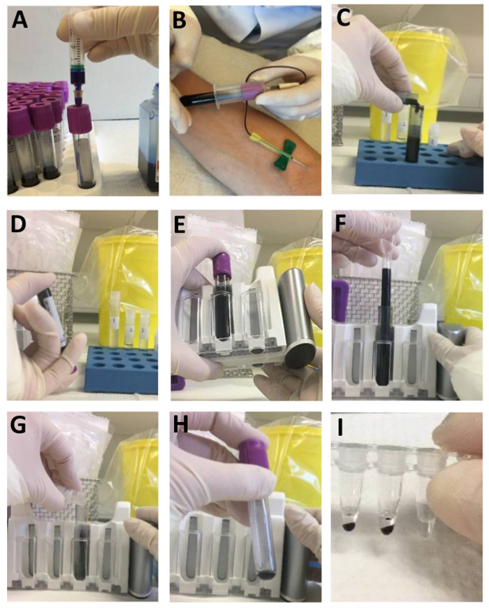
Figure 1: Workflow for simplified NA extraction of whole blood samples. (A) Prepare the lysis/binding buffer vacuum blood collection tubes using a needle and a syringe. (B) Collect the blood using a butterfly needle. (C) Place the blood collection tube in a holder, remove the lid, and pour the MGPs into the tube. (D) Close the tube with a new lid and mix the contents by flipping the tube by hand. (E) Collect the MGPs on the side of the blood collection tube by flipping the tube in the magnetic holder. (F) Remove the supernatant using a disposable pipette. (G) Pour the wash or elution buffers directly into the blood collection tube and repeat the actions shown in panels D - F. (H) This panel shows MGPs ready for direct use in a qPCR, RT-qPCR, LAMP, or RT-LAMP reaction. (I) Transfer one droplet (5 - 8 µL) of MGPs to a PCR or LAMP reaction tube using a 1.5 mL disposable pipette. Panel A and B: This figure has been modified from Rosenstierne et al.7. Please click here to view a larger version of this figure.
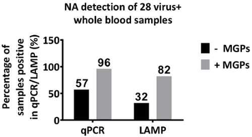
Figure 2: NA detection of 28 virus-positive whole blood samples extracted using the simplified NA extraction method. Whole blood samples (1.6 mL) tested negative for a virus were spiked with either a DNA-virus [Epstein-Barr virus (EBV) (2.0 x 10⁴ - 1.0 x 10³ copies/mL)] or an RNA-virus [Hepatitis C virus (HCV) (6.0 x 10⁵ - 3.0 x 10³ copies/mL)] or a Dengue virus (DENV) (1.7 x 10⁸ - 1.7 x 10⁶ copies/mL). NAs were extracted using the method described above, and the extracted NAs were analyzed in duplicate by specific in-house qPCR, RT-PCR, LAMP, or RT-LAMP, with or without MGPs. The x-axis = the NA detection method, the y-axis = the percentage of samples positive in qPCR/LAMP. Please click here to view a larger version of this figure.
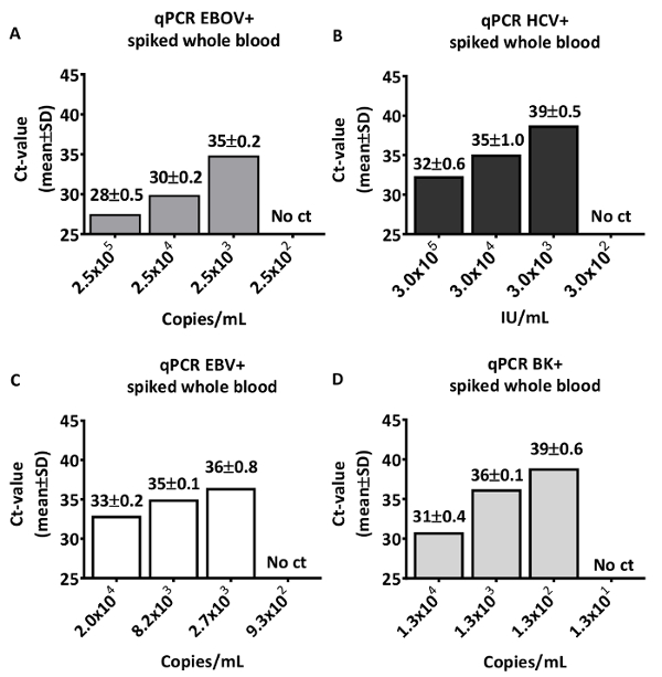
Figure 3: Sensitivity of the simplified NA extraction method. For a proof of concept of the method, virus negative whole blood was spiked with different RNA or DNA virus preparations containing a defined viral load, and NAs were extracted using the simplified NA extraction method. (A) Negative whole blood (1.4 mL) was spiked with 200 µL of 10-fold dilutions of the EBOV standard prepared for diagnostic purposes (ENIVD), which was prepared from the recent outbreak in Guéckédou/Guinea (2.0 x 10⁶ copies/mL). The extracted NAs were analyzed in duplicate by an in-house EBOV-specific RT-qPCR7 using an MX3005P thermal cycler. (B) Whole blood (1.2 mL) was spiked with 400 µL of 10-fold dilutions of an HCV WHO standardized serum sample (1.2 x 10⁶ IU/mL). The extracted NAs were analyzed in duplicate by an in-house HCV-specific RT-qPCR using an MX3005P thermal cycler. (C) Whole blood (1.2 mL) was spiked with 400 µL of different concentrations of EBV (2.0 x 10⁴ copies/mL, 8.0 x 10³ copies/mL, 2.7 x 10³ copies/mL, and 9.3 x 10² copies/mL). The extracted NAs were analyzed in duplicate by an in-house EBV-specific qPCR. (D) Whole blood (1.2 mL) was spiked with 400 µL of 10-fold dilutions of a BK virus (1.3 x 10⁴ copies/mL). The extracted NAs were analyzed in duplicate by an in-house BK-specific qPCR. The x-axis = the final concentration of a virus in the spiked whole blood sample (copies/mL or IU/mL), the y-axis = the Ct-value (mean ± SD). Please click here to view a larger version of this figure.
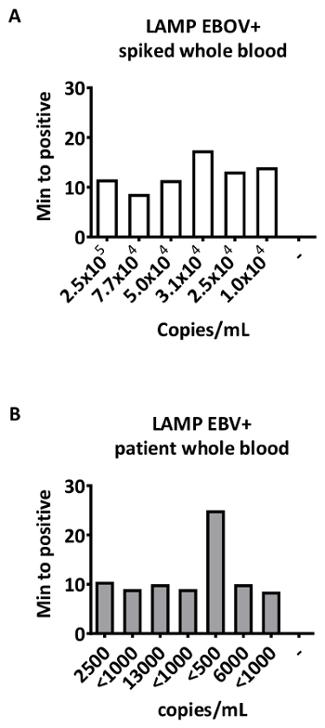
Figure 4: Simplified NA extraction and RT-LAMP or LAMP using a portable isothermal device. (A) Due to the lack of EBOV-positive whole blood samples at the Department of Virus & Microbiological Special Diagnostics, Statens Serum Institut, Denmark, 1.4 mL of negative whole blood was spiked with 200 µL of different dilutions of the EBOV standard prepared for diagnostic purposes (ENIVD). The spiked samples underwent an NA extraction using the simplified NA extraction method and were analyzed by an EBOV-specific RT-LAMP reaction. As a negative control, a negative whole blood sample was included. The x-axis = the final concentration of EBOV in the spiked whole blood sample (copies/mL), the y-axis = the min to positive. (B) For a proof of concept, whole blood (1.6 mL) aliquots from 7 patients, diagnosed positive for EBV (13,000 - 500 copies/mL) at the Department of Virus & Microbiological Special Diagnostics, Statens Serum Institut, Denmark, underwent an NA extraction using the simplified NA extraction method and were analyzed by an EBV-specific LAMP reaction using the portable battery-driven Genie II isothermal device. As a negative control, a Parvo B19-positive sample was included. The x-axis = the viral load of EBV in a patient sample (copies/mL), the y-axis = the min to positive. Please click here to view a larger version of this figure.
Discussion
In this report, we describe a safe, rapid, and simple manual bedside NA extraction method for the downstream molecular detection of a virus in lysis/binding buffer-inactivated whole blood. The described NA extraction method was developed to be performed directly on whole blood samples collected in vacuum blood collection tubes containing the lysis/binding buffer (Table of Materials)7. This specific buffer inactivates EBOV7 and is the critical component in the method. After the blood collection, we recommend incubating the tube for at least 20 min7 in order to ensure a complete inactivation of the sample. The bedside EBOV inactivation eliminates the need for handling any EBOV-positive samples under rigorous containment conditions, such as BSL-4 conditions, and allows the samples to be handled under normal BSL-2 conditions. It is likely that other risk group four pathogens, such as the Lassa virus, the Marburg virus, and the Crimean Congo hemorrhagic fever virus are also inactivated by this buffer, but this remains to be shown.
The described NA extraction method was developed to be performed next to a patient in a hospital isolation ward or in a field hospital and, therefore, requires no laboratory equipment, such as centrifuges, heating blocks, or electricity. All that the NA extraction method requires are tubes containing a fixed volume of MGPs or wash buffers, a magnetic holder, and disposable pipettes. The pre-preparation of the tubes containing buffers simplifies the protocol because it eliminates the need for pipettes, and instead, the aliquoted buffers are poured directly into the sample tubes. The magnetic holder catches the MGPs containing the NA and mediates an easy handling of the wash steps. The magnetic holder can be substituted with an ordinary magnet and a rubber band in case a magnetic holder is not available; however, the magnetic holder reduces the hands-on risk of dropping the tube and spilling the contents when applying the magnet and rubber band. Due to the viscosity of the lysis/binding buffer-inactivated whole blood sample, disposable pipettes are used to remove the supernatant/liquid/wash buffers from the tube. The disposable pipettes can be substituted by a direct pouring of the contents from the tube into a waste collection tube, but please be aware not to create any droplets outside the tube. If droplets are created, the contaminated surface must never be disinfected directly with chloramine or sodium hypochlorite (the active ingredients in "bleach") because this mixture may lead to the formation of toxic cyanide. Therefore, if spilled first, wipe up the spill with an absorbent tissue. Next, clean the surface with 70% ethanol and then with plenty of water, and finally, use chloramine or sodium hypochlorite. This includes all waste that has been in contact with the lysis/binding buffer (including the waste collection tube and the disposable pipettes). Instead, collect all waste in a separate waste container and only disinfect the outside of the container with 70% ethanol.
NAs are normally eluted from the MGPs during a heating step, but this heating step has been removed in the simplified NA extraction protocol to eliminate the use of electricity and equipment. Therefore, the NAs tend to stick to the MGPs, which, therefore, must be added to the downstream NA detection assay for a positive NA detection. The MGPs are transferred to the downstream reaction mix using one droplet from a 1.5 mL disposable pipette. This addition is not as precise as the normal addition using a fin tip pipette; however, the unprecise addition of NA/MGPs to the LAMP, RT-LAMP, qPCR, or RT-qPCR reaction mix does not inhibit the reaction or influence the fluorescence signal in our in-house diagnostic assay. Nevertheless, this may differ from assay to assay, and we recommend that every downstream NA test is validated, and that the sensitivity of the assay is tested using the simplified NA extraction method. The NA extraction method is relatively sensitive, and whole blood samples containing a DNA or RNA virus as low as 3,000 copies/mL can easily be extracted and detected in the downstream molecular tests, such as qPCR. The viral load in clinical samples is usually very high during the acute phase of an infection, and hemorrhagic fever viruses, such as the EBOV, Lassa virus, and Crimean Congo hemorrhagic fever virus, usually contain more than 105 copies/mL9,10,11. Therefore, viruses in these samples can easily be detected using this NA extraction method. However, the MPLB inactivation of viruses other than the EBOV7, Vaccinia, and Cowpox virus8 remains to be shown.
The simplified NA extraction method can be performed in hospital isolation wards or in field settings and is ideal for point-of-care molecular diagnostics. However, the method is not applicable for high-throughput NA extractions due to the hands-on handling of an open tube. If high-throughput NA extractions are needed, the inactivated whole blood samples can easily be purified using NA extraction robots7. Another disadvantage is the handling of harmful reagents such as the lysis/binding buffer [20 - 30% polyethylene glycol p-(1,1,3,3-tetramethylbutyl)-phenyl ether and 30 - 50% guanidinium thiocyanate (GITC)]17 and wash buffer I (30 - 50% guanidinium chloride)17 in an open tube outside a flow hood. The lysis/binding buffer in the concentrated form is contained within the vacuum tube and with the collection of blood 1:1 (v/v), the concentration is reduced before the tube is opened. The opening of the vacuum tube and the addition/removal of MGPs or wash buffer I is performed within a few seconds and, hence, the risk of inhalation of aerosols is minimal. After the removal of wash buffer-I, from the tube, no other harmful reagents are used in the protocol.
Many rapid point-of-care molecular tests and devices have been developed since the EBOV outbreak in 20133,6,12,13,14,15,16. However, many of these tests still require an up-stream handling of infectious material, BSL-3 biosafety cabinets, and at a minimum, laboratory equipment, such as pipettes, centrifuges, and heating blocks, or electricity. The use of biosafety cabinets complicates and prolongs a rapid diagnosis. The described bedside virus inactivation with the direct collection of blood into the virus inactivation tubes combined with the simplified NA extraction method eliminates the need for biosafety cabinets, electricity, and laboratory facilities. Additionally, because the simplified NA extraction method can be performed next to the patient and combined with a LAMP reaction in a portable battery-driven isothermal device, a safe bedside molecular diagnosis can be obtained within 40 min.
Disclosures
The authors have nothing to disclose.
Acknowledgements
We thank Susanne Lopes Rasmussen and Solvej Kolbjørn Jensen for their technical assistance and for handling the clinical samples. This project is part of the EbolaMoDRAD consortium, which has received funding from the Innovative Medicine Initiative 2 Joint Undertaking under grant agreement N°115843. This Joint Undertaking receives support from the European Union's Horizon 2020 research and innovation program and EFPIA.
Materials
| Name | Company | Catalog Number | Comments |
|---|---|---|---|
| Vacutainer K2 EDTA tubes (4 mL) | Becton Dickinson | 368861 | |
| Plus Blood Collection | Becton Dickinson | 362725 | |
| 23G x 1 needle | Becton Dickinson | 300800 | |
| LUER LOK syringe (3 mL) | Becton Dickinson | 309658 | |
| Butterfly needle with small-bore extension tubing | Jørgen Kruuse A/S | 121714 | |
| 1% Virkon | Nomeco A/S | 849265 | |
| MagNA Pure LC DNA Isolation Kit I - Lysis/Binding Buffer - Refill | Roche Diagnostics A/S | 03246752001 | |
| MagNA Pure LC Total Nucleic Acid Isolation Kit | Roche Diagnostics A/S | 3038505001 | |
| Nunc™ Biobanking and Cell Culture Cryogenic Tubes (1,8 mL) | Thermo Fisher Scientific | 375418 | |
| Nunc™ Biobanking and Cell Culture Cryogenic Tubes (3,6 mL) | Thermo Fisher Scientific | 379189 | |
| Nunc™ Biobanking and Cell Culture Cryogenic Tubes (4,5 mL) | Thermo Fisher Scientific | 379146 | |
| DynaMag™-5 Magnet | Thermo Fisher Scientific | 12303D | |
| Transfer pipette (3.5 mL) | Sarstedt | 86.1171.010 | |
| Thermo Scientific™ Samco™ Fine Tip Transfer Pipettes (1.5 mL) | Thermo Fisher Scientific | 231 | |
| Thermo Scientific™ Nunc™ 50 mL Conical Sterile Polypropylene Centrifuge Tubes | Thermo Fisher Scientific | 339652 |
References
- Lee, D., Shin, Y., Chung, S., Hwang, K. S., Yoon, D. S., Lee, J. H. Simple and Highly Sensitive Molecular Diagnosis of Zika Virus by Lateral Flow Assays. Analytical Chemistry. 88, 12272-12278 (2016).
- Benzine, J. W., Brown, K. M., et al. Molecular Diagnostic Field Test for Point-of-Care Detection of Ebola Virus Directly from Blood. Journal of Infectious Diseases. 214 (Suppl 3), S234-S242 (2016).
- Faye, O., Faye, O., et al. Development and deployment of a rapid recombinase polymerase amplification Ebola virus detection assay in Guinea in 2015. Euro Surveillance. 20 (44), 10-18 (2015).
- Yang, M., Ke, Y., et al. Development and Evaluation of a Rapid and Sensitive EBOV-RPA Test for Rapid Diagnosis of Ebola Virus Disease. Scientific Reports. 6, (2016).
- Smither, S. J., Weller, S. A., et al. Buffer AVL alone does not inactivate Ebola virus in a representative clinical sample type. Journal of Clinical Microbiology. 53 (10), 3148-3154 (2015).
- Byrnes, S., Fan, A., et al. A Portable, Pressure Driven, Room Temperature Nucleic Acid Extraction and Storage System for Point of Care Molecular Diagnostics. Analytical Methods. 5 (13), 3177-3184 (2013).
- Rosenstierne, M. W., Karlberg, H., et al. Rapid bedside inactivation of Ebola virus for safe nucleic acid tests. Journal of Clinical Microbiology. 54 (10), 2521-2529 (2016).
- Vinner, L., Fomsgaard, A. Inactivation of orthopoxvirus for diagnostic PCR analysis. Journal of Virological Methods. 146, 401-404 (2007).
- Schmitz, H., Köhler, B., et al. Monitoring of clinical and laboratory data in two cases of imported Lassa fever. Microbes and Infection. 4 (1), 43-50 (2002).
- De La Vega, M. A., Caleo, G., et al. Ebola viral load at diagnosis associates with patient outcome and outbreak evolution. Journal of Clinical Investigation. 125 (12), 4421-4428 (2015).
- Hasanoglu, I., Guner, R., et al. Dynamics of viral load in Crimean Congo hemorrhagic fever. Journal of Medical Virology. 90 (4), 639-643 (2017).
- Biava, M., Colavita, F., et al. Evaluation of a rapid and sensitive RT-qPCR assay for the detection of Ebola Virus. Journal of Virological Methods. 252, 70-74 (2018).
- Fernández-Carballo, B. L., McBeth, C., et al. Continuous-flow, microfluidic, qRT-PCR system for RNA virus detection. Analytical and Bioanalytical Chemistry. 410 (1), 33-43 (2017).
- Weller, S. A., Bailey, D., et al. Evaluation of the Biofire FilmArray Biothreat E-test (v2.5) for rapid identification of Ebola virus disease in heat-treated blood samples obtained in Sierra Leone and United Kingdom. Journal of Clinical Microbiology. 54 (1), 114-119 (2015).
- Semper, A. E., Broadhurst, M. J., et al. Performance of the GeneXpert Ebola Assay for Diagnosis of Ebola Virus Disease in Sierra Leone: A Field Evaluation Study. PLoS Medicine. 13 (3), e1001980(2016).
- Biocartis. Instructions for Use: Idylla TM Ebola Virus Triage Test. , https://www.fda.gov/downloads/MedicalDevices/Safety/EmergencySituations/UCM503944.pdf (2016).
- MPLC. Total Nucleic Acid Isolation Kit. Safety data sheet version 1.7. , https://pim-eservices.roche.com/DownloadDocument/SDS/DE/en/03038505001 (2015).
Reprints and Permissions
