This article presents a modular protocol for tissue lipidomics and transcriptomics, and plasma lipidomics in neurological disease mouse models targeting lipids underlying inflammation and neuronal activity, membrane lipids, downstream messengers, and mRNA-encoding enzymes/receptors underlying lipid function. Sampling, sample processing, extraction, and quantification procedures are outlined.
Method Article
Lipidomics and Transcriptomics in Neurological Diseases
Opens in a new tab
In This Article
Summary
Loading...
$$\rightleftharpoonup{xx}$$
$$\longleftharp{xx}$$,
$$\longrightharp{xx}$$,
Abstract
Loading...
$$\rightleftharpoonup{xx}$$
$$\longleftharp{xx}$$,
$$\longrightharp{xx}$$,
Lipids serve as the primary interface to brain insults or stimuli conducive to neurological diseases and are a reservoir for the synthesis of lipids with various signaling or ligand function that can underscore the onset and progression of diseases. Often changing at the presymptomatic level, lipids are an emerging source of drug targets and biomarkers. Many neurological diseases exhibit neuroinflammation, neurodegeneration, and neuronal excitability as common hallmarks, partly modulated by specific lipid signaling systems. The interdependence and interrelation of synthesis of various lipids prompts a multilipid, multienzyme, and multireceptor analysis in order to derive the commonalities and specificities of neurological contexts and to expedite the unravelling of mechanistic aspects of disease onset and progression. Ascribing lipid roles to distinct brain regions advances the determination of lipid molecular phenotype and morphology associated with a neurological disease.
Presented here is a modular protocol suitable for the analysis of membrane lipids and downstream lipid signals along with mRNA of enzymes and mediators underlying their functionality, extracted from discrete brain regions that are relevant for a particular neurological disease and/or condition. To ensure accurate comparative lipidomic profiling, the workflows and operating criteria were optimized and standardized for: i) brain sampling and dissection of regions of interest, ii) co-extraction of multiple lipid signals and membrane lipids, iii) dual lipid/mRNA extraction, iv) quantification by liquid chromatography multiple reaction monitoring (LC/MRM), and v) standard mRNA profiling. This workflow is amenable for the low tissue amounts obtained by sampling of the functionally discrete brain subregions (i.e. by brain punching), thus preventing bias in multimolecular analysis due to tissue heterogeneity and/or animal variability. To reveal peripheral consequences of neurological diseases and establish translational molecular readouts of neurological disease states, peripheral organ sampling, processing, and their subsequent lipidomic analysis, as well as plasma lipidomics, are also pursued and described. The protocol is demonstrated on an acute epilepsy mouse model.
Introduction
Loading...
$$\rightleftharpoonup{xx}$$
$$\longleftharp{xx}$$,
$$\longrightharp{xx}$$,
Recent advances in the function of lipids and their role in the onset and progression of neurological diseases open new research and development venues of new therapeutic targets and disease mechanism elucidation1. Documented differences in lipid composition in different brain regions, emphasized by modern molecular imaging techniques such as mass spectrometry imaging and advanced mass spectrometry profiling, shifts the paradigm of lipid investigation from whole brain toward functionally distinct and discrete brain regions. The fact that lipid composition varies in different brain regions prompts new conceptualization of both membrane lipid sensitivity and downstream lipid signaling in response to a brain insult or stimuli across the functionally distinct brain regions. Hence, lipid protocols require new developments to address the challenge of low tissue amounts for higher spatial resolution detection and quantification, and concurrently, analysis of multiple lipid components of cell membranes and signaling pathways. Also, determination of enzymes, lipid ligands, and receptors involved in the regulation of their levels and function is paramount to elucidate the signaling pathways affected in a neurological disease and guide new mechanistic investigations in a pathophysiological context.
In addition to the increased brain spatial resolution, there are two major difficulties challenging the development of new neurolipidomic approaches. First, the lipid signaling molecules are typically of very low abundance compared to membrane constitutive lipids. Second, the lipidome exhibits a high structural heterogeneity, difficult to dissect using a single analytical approach. Hence, extraction and analytical methods are tailored to different lipid categories and commonly performed in distinct tissue samples2. Shotgun lipidomic methods3 are excellent tools to rapidly reveal a broad profile of membrane lipids, while increased sensitivity and selectivity afforded by the targeted discovery and quantification mass spectrometric methods are capitalized upon for investigation of low abundant signaling lipids including: i) inflammatory lipids and ii) lipids involved in the modulation of neuronal activity, such as endocannabinoids (eCBs), amino acid-linked lipids, etc.4,5. To encompass lipid changes at both the cell membrane and signaling level occurring in brain regions of neurological disease models, typically the lipid extraction and analysis are carried out in distinct tissue samples, obtained from distinct animal batches or from different hemispheres, or by dissecting a larger tissue region into multiple pieces. When mRNA levels of enzyme receptors are also of interest, their investigation typically requires the procurement of a distinct tissue sample. For example, the investigation of membrane lipids, endogenous cannabinoids, and mRNA would require three different tissue samples, (e.g., two samples for the two lipid extraction methods-membrane lipids and signaling lipids- and subsequent two lipid analysis methods- and one sample for mRNA analysis). Investigation of inflammatory lipids and endogenous cannabinoids require two distinct tissue samples, extraction methods, and analysis methods, respectively. Another example is the investigation of mRNA and of any lipid category in a brain punch or laser microdissection sample which consequently requires two distinct animals to procure two samples per brain (sub)region. A substantial extent of variability and/or poor reproducibility of the results frequently occur in such cases, originating from biological variability and/or tissue heterogeneity. Guided by these practical limitations of multimolecular analysis, occurring particularly at high spatial resolution in the brain, a three-module neurolipidomics protocol was designed encompassing: 1) coextraction and co-analysis by LC/MRM of inflammatory lipids (e.g., eicosanoids (eiCs)) and lipids involved in modulation of neuronal activity, such as eCBs2; 2) co-extraction of phospholipids (PLs) and eCBs with subsequent multiscan LC/MRM and precursor/neutral loss scan analysis2; and 3) dual extraction of membrane (phospho)lipids and eCBs as well as mRNA, with subsequent LC/MRM and qPCR or RNA sequencing analysis6. Depending on the biological question to be addressed in a neurological disease and the brain region of interest, a combination of the first and the second protocol, or the first and the third protocol, can be applied on the same tissue specimen for tissues weighing around 4 mg. The first and third protocols can be independently applied for tissues around 2 mg. The second protocol can be applied for tissues weighing as little as 0.5 mg. Irrespective of the neurolipidomic protocol module selected, the tissue sampling and pre-analytical processing, the brain isolation and region dissection, as well as the procedure for sacrificing the animal model are standardized and identical for all three modules of the protocol. In our investigation of neurological diseases, peripheral organs that are relevant for the pathological consequences of the disease are always also collected and analyzed using these modular protocols. Additionally, blood is regularly sampled for plasma lipidomics to serve as a readout tool of neurological diseases with a view on prospective translational applications. The here presented modular lipidomics protocol is very versatile: scaleable to larger tissue amounts and readily applicable for virtually any tissue type and disease. For the application of the modular protocol (Figure 1) in neurological diseases, any standardized rodent model of onset and progression of neurological disorders, such as traumatic brain injury, Parkinson's disease, Alzheimer's disease, or epilepsy are amenable.
These protocols have been extensively applied to study changes in the tissue lipidome and/or transcriptome at the acute phase of epilepsy in the kainic acid (KA)-induced mouse model of epilepsy2,7, a model widely used in preclinical studies due to the resemblance to human temporal lobe epilepsy (TLE)8,9,10,11. Using these protocols, the therapeutic potential of drugs such as Palmitoylethanolamide (PEA)12,13 was assessed in the same mouse model of epilepsy. The study identified lipid and mRNA changes at high and low spatial resolution in the brain and periphery, at the time point of maximal acute seizure intensities (at 60 min postseizure induction), and upon subchronic and acute treatment with PEA at four different timepoints (20, 60, 120, and 180 min) post KA-seizure induction, a time-window covering the acute phase of epilepsy. Plasma, brains, and peripheral organs of untreated KA-injected mice, acute and subchronically PEA-treated mice, as well as vehicle and PEA-vehicle control mice, were collected at each time point12,13, and investigated with this molecular analysis. The molecular data were correlated with behavioral phenotypes obtained by seizure scoring, as well as with immunohistochemistry-derived data on neurodegenerative processes, in order to unravel the progression of the acute epilepsy phase and PEA's potential to alleviate it.
Protocol
Loading...
$$\rightleftharpoonup{xx}$$
$$\longleftharp{xx}$$,
$$\longrightharp{xx}$$,
All experimental procedures described here are in accordance with The European Community's Council Directive of 22 September 2010 (2010/63EU) and were approved by the local animal committee of the state Rhineland-Palatinate, Germany (file reference: 23 177-07/G16-1-075).
1. Animal model of acute and prophylactically treated KA-induced epilepsy
- Perform seizure induction, treatment, and behavioral scoring.
- Separate mice (minimum n = 6 mice per group) in single cages.
- Prepare seizure-induction injection solution and the corresponding vehicle (see Table 1), as well as treatment injection solution and the corresponding vehicle (see Table 2).
- Inject mice intraperitoneally (i.p.) (10 mL/kg mouse body mass) without anesthesia according to their group identity: disease, treated, or vehicle treated (i.e., KA, PEA-treated KA, and the two vehicle groups). See Figure 2, Table 1, and Table 2.
- Monitor and score the behavior according to the following standardized seizure intensity scale: 0 = no response; 1 = immobility and staring; 2 = forelimb and/or tail extension, rigid posture; 3 = repetitive movements, head bobbing; 4 = rearing and falling; 5 = continuous rearing and falling; 6 = severe clonic-tonic seizures; 7 = death14,15 (Figure 3).
- Perform sacrificing procedure for lipidomic and transcriptomic analysis.
- At four timepoints (i.e., 20, 60, 120, and 180 min post KA- or vehicle injection respectively), sacrifice six mice from each of the following groups: PEA treated, PEA-untreated epileptic vehicle 1, and vehicle 2, following IACUC approved protocols.
- Ten seconds after each time point is reached, anesthetize the mice using an isoflurane-soaked tissue in a glass chamber. Confirm anesthesia by the loss of the righting reflex, indicated by the inability to move while slowly overturning the glass chamber. Decapitate mice using surgical scissors and collect plasma, brain, and peripheral organs (see steps 2.2.2−2.2.4).
CAUTION: Ethical regulations for sacrificing procedures vary between local animal committees. Make sure to check and follow the regulations issued by the local animal committee.
- Follow sacrificing procedure for immunohistochemistry analysis.
- For immunohistochemical staining13, behaviorally score three mice from each of the following groups: PEA treated, PEA-untreated epilepsy, and vehicle groups, over the entire time course (180 min).
- Sacrifice and perfuse mice 5 days later. Deeply anesthetize mice (see step 1.3.1 for the mouse groups) by i.p. injection of pentobarbital (100 mg/kg) and buprenorphine (0.1 mg/kg) as narcotic drugs. Confirm deep anesthesia by the loss of reflex between the toes.
- Fix the mouse on a polystyrene plate and immediately open the thorax using fine scissors and standard forceps. Set the butterfly in the left heart ventricle and cut the right atrium using fine scissors.
- Adjust the speed and pressure of the pump to a constant peristaltic flow with a rate of 2−3 mL/min. Perfuse with ice-cold phosphate buffered saline (PBS) for ~5 min. If necessary, replace the butterfly.
NOTE: With proper perfusion, the inner organs (except the spleen) start to bleach out within 1−2 min. - Switch perfusion to ice-cold 4% paraformaldehyde (PFA) solution for ~5 min. Isolate whole brains as described in step 2.2.2 and fix for 24 h in 4% PFA solution at 4 °C.
- Incubate brains in 30% sucrose solution for 48 h at 4 °C. Remove the leftover sucrose with a dry tissue paper and freeze the brains on a metal plate (-80 °C). Store the brains at -80 °C for the staining procedures13.
2. Sampling procedures for lipidomic/transcriptomic analysis
- Prepare for sampling.
- Precool EDTA-tubes for plasma sampling to 4 °C. Precool the centrifuge and vortex apparatus to 4 °C. Precool the metal plate covered with aluminum foil (to snap frozen brain and/or other tissues of interest) on dry ice (-80 °C). Precool the labeled 2 mL and 5 mL tubes on dry ice (-80 °C) for plasma aliquots, frozen tissue, and brain collection, respectively. Use amber tubes to protect light sensitive molecules, such as eiCs.
- Clean the tissue sampling and isolation equipment (surgical scissors, straight sharp fine scissors, straight smooth standard forceps, fine forceps with mirror finish, curved forceps and spatula, polystyrene foam plate, and needles for fixation) with a disinfectant (e.g., 70% ethanol).
- Sampling protocols
- Determine the order of sampling.
- Collect blood immediately after decapitation in precooled 1 mL EDTA tubes (see step 2.2.3).
- Remove the whole brain and snap freeze immediately after removal. This step will take 1−2 min. Store frozen brains at -80 °C for further processing (see step 2.2.2).
- Remove peripheral organs of interest (e.g., lung, heart, liver), within 5 min post brain removal, and snap freeze and store at -80 °C for further processing (see step 2.2.4).
- Remove brain for dissection or punching procedures. This procedure takes 1−2 min/brain.
- After decapitation (see step 1.2), start making a midline incision in the skin using fine scissors. Free the skull by flipping the skin over the eyes. Reach to the top of the skull and make a small caudal incision starting at the level of the intraparietal bone. Avoid cutting through the brain.
- Cut firmly through the most anterior part of the skull between the eyes to ease brain removal. Tilt one side of the parietal bone using curved narrow pattern forceps and break off. Repeat the last step for the other side.
- Remove the brain meninges. Slide a spatula under the anterior part of the brain (i.e., the olfactory bulb) and tilt the brain gently upward. Slide the spatula further down to break the optic and other cranial nerves.
- Lift the brain out of the skull and snap freeze the whole brain immediately on a precooled (-80 °C) metal plate with the ventral side facing the metal plate (dorsal up).
- Allow the brains to completely freeze and transfer into precooled 5 mL tubes and store at -80 °C until the dissection of the brain regions or the punching procedure (see steps 3.1. and 3.2).
- Perform plasma sampling.
- Spike precooled EDTA-tubes with 10 µL of freshly prepared indomethacin-dilution to a target concentration of 10 µM.
- Collect the trunk blood immediately after decapitation by gentle squeezing of the body into precooled EDTA tubes to a maximum blood volume of 1 mL.
NOTE: In case of proper blood pressure, squeezing is not required. If blood volume is less than 1 mL, decrease the isoflurane incubation time to enable proper blood flow. - Immediately centrifuge blood tubes at 2,000 x g for 10 min at 4 °C.
- Remove the resulting upper plasma phase and for the purpose of multilipid analysis aliquot defined plasma volumes in precooled 2 mL tubes as follows: 50 µL for eCBs/eiCs analysis, 30 µL for PLs analysis, and the remaining plasma volume as the backup sample or for other types of analysis.
- Store the plasma samples at -80 °C for further extraction (see steps 4.1.2 and 4.1.4).
- Perform peripheral organ sampling.
NOTE: Use mouse anatomy references16 and documentation provided for the obligatory courses (FELASA) attended by animal investigators to identify the individual organs, their connective tissues, and/or blood vessels.- Immobilize the mouse torso to ease organ removal using needles. Make a ventral midline incision using a straight sharp scissor at the height of the pubis. Use blunt standard forceps to fixate the abdominal wall while cutting to open the abdominal cavity.
- Immobilize the skin to allow abdominal organ removal using blunt forceps. For heart or lung removal, continue medial cut in the direction of the breast cave. Remove the lung and/or heart using fine forceps.
- Open the breast cavity carefully to avoid bleeding. Cut the connective tissues and blood vessels anchoring the respective organ without cutting through the organ.
- Transfer tissue pieces immediately on precooled metal plates (-80 °C) and allow to completely freeze. Transfer tissue pieces to precooled tubes and store at -80 °C for further processing (see step 4.1).
- Determine the order of sampling.
3. Biological material processing
NOTE: For co-extraction of eCBs/eiCs use 2 mL amber tubes as extraction tubes and add in each tube seven precooled steel balls. For co-extraction of PLs/eCBs and for dual lipid and RNA co-extraction, use 2 mL of RNAse-free extraction tubes spiked with ceramic beads (Table of Materials).
- Perform brain dissection and peripheral organ processing.
NOTE: Use magnifying lamps to increase the visibility of the brain for dissection.- Clean the surgical tools, including the forceps with super fine tips, 2x with 70% ethanol.
- Transfer the frozen brains from -80 °C to a Petri dish containing precooled physiological buffer (pH 5.5) Ensure the Petri dish is at 4 °C. Allow brains to completely unfreeze to allow dissection. Test carefully using forceps.
CAUTION: Do not keep brains at 4 °C longer than required for thawing. - Precool a metal plate on ice (4 °C) and cover it with wet tissues soaked in ice-cold physiological buffer (pH 5.5) and transfer the brain with ventral side up carefully onto a precooled (4 °C) metal plate on ice. Cover it with wet tissues soaked in ice-cold physiological buffer (pH 5.5).
- Dissect brain regions within a maximum of 5 min using super fine tip forceps. Start with the hypothalamus (HYP) and turn the dorsal side up to proceed with the right side. Isolate the hippocampus (HCr), prefrontal cortex (PFCr), striatum (STRr), and cerebral cortex (cCTXr). Then dissect the left side. Isolate the hippocampus (HCl), prefrontal cortex (PFCl), striatum (STRl), and cerebral cortex (cCTXl). Dissect the cerebellum and the thalamic region. Use published anatomical references16,17 for brain region identification.
- Transfer each dissected piece directly on an aluminum foil-covered, precooled metal plate (-80 °C). Allow freezing and transfer the isolated brain regions in the labeled precooled 2 mL tubes.
- Pulverize the tissue pieces using a tissue pulverizer (at -80 °C), avoiding the thawing of the tissue. In the cold room, aliquot tissue powder in labeled, precooled extraction tubes containing ceramic beads or steel balls. For the dual lipidomics/transcriptomics analysis, weigh the tissue powder aliquots in the cold room. For lipidomics-only analysis, choose between normalization to the tissue weight or protein content. For the latter, proceed further without tissue weighing.
- Cut the peripheral organ tissues in pieces with a maximum weight of 20 mg. Proceed with tissue pulverization as in step 3.1.6.
- Proceed with the extraction of tissue samples (see steps 4.1.1, 4.1.3, or 4.1.5) or store tubes at -80 °C for later extraction.
NOTE: The brain matrix can also be used if the study design permits. However, the method is more suitable for a discrete and limited number of brain regions, and it is not practical to dissect and isolate all of the above-mentioned brain regions within the set time-frame.
- Perform brain punching.
- Mount the whole, frozen brains onto the mounting system (Table of Materials) in the cryostat. Set the thickness to 50 µm and slice in trim-mode close to the region of interest.
- Stain brain slice (18−20 µm) using toluidine blue (0.1%−1%) as a reference to localize the subregion of interest. Inspect the stained slices using a microscope to identify the regions of interest to be punched. Use a mouse atlas as reference to find the appropriate brain anatomical regions16. Take punches with a 0.8−1.0 mm diameter using sample corers in precooled tubes.
- Weigh frozen punches in the cold room in labeled, precooled 2 mL extraction tubes containing ceramic beads or steel balls. Use amber tubes for eCBs and eiCs co-extraction.
- Start the extraction (see steps 4.1.1, 4.1.3, or 4.1.5) or store tubes at -80 °C for further extraction.
- Perform plasma processing.
- Place the frozen plasma samples on ice (4 °C) and let them thaw (~20 min).
- Ensure that the plasma is entirely unfrozen before starting extraction.
- Proceed with the plasma extraction procedure (see steps 4.1.2 or 4.1.4).
NOTE: Plasma samples should remain at -80 °C until extraction. Avoid cycles of thawing and refreezing.
4. Extraction procedures
- Carry out Liquid-Liquid (LLE) lipid co-extraction protocols.
- Perform co-extraction of eCBs and eiCs from brain pieces, punches, or tissue powder samples.
NOTE: Accurate pipetting is required throughout the procedure.- Place the extraction tubes containing the tissue samples and seven steel balls on ice (4 °C). Add 600 µL of ice-cold MTBE and 50 µL of ACN/H2O (1:1; v/v) containing the internal standards. The target concentration of the internal standards in the final volume for analysis (50 µL) is as follows: 1 ng/mL AEA-d4, 125 ng/mL 2-AG-d5, 3,000 ng/mL AA-d8, 2 ng/mL OEA-d4; PEA-d4, 12.5 ng/mL 1-AG-d5, 2.5 ng/mL PGF2α-d4 and 5 ng/mL for PGD2-d4; PGE2-d9; 5(S)-HETE-d8; 12(S)-HETE-d8; 20-HETE-d6, and TXB2-d4, respectively.
- Add 400 µL of 0.1 M formic acid and homogenize with a tissue lyser (30 s-1 min). Centrifuge the homogenate for 15 min at 5,000 x g at 4 °C. Allow freezing of the aqueous lower phase for 10 min at -80 °C to ease the transfer of the upper organic phase.
- Transfer the organic phase in new tubes. Evaporate under a gentle stream of N2 at 37 °C and reconstitute in 50 µL of ACN/H2O (1:1; v/v) for further analysis.
- Store the aqueous phase at -20 °C or -80 °C for further protein content analysis.
- Perform co-extraction of eCBs and eiCs from plasma samples.
- Thaw plasma aliquots at 4 °C. Add 800 µL of MTBE and 50 µL of ACN/H2O (1:1; v/v) containing the internal standards (analogous to those used for tissue analysis, see step 5.1.1). Optimize the internal standard concentration for spiking using reference plasma samples.
- Add 600 µL of 0.1 M formic acid and vortex samples at 4 °C for 2 min. Centrifuge samples at 4,000 x g for 15 min at 4 °C.
- Transfer the organic phase into new tubes, evaporate under a gentle stream of N2 at 37 °C, and reconstitute in 50 µL of ACN/H2O (1:1; v/v) for LC/MRM analysis.
NOTE: If possible, avoid storage of dried extracts of eiCs and proceed immediately to LC/MRM analysis. If storage cannot be avoided, resort to short-time storage for only 2−3 days at 4° C with samples in LC injection solvent.
- Perform co-extraction of PLs and eCBs from brain regions, punches, or other tissue powder samples.
- Place the extraction tubes containing the tissue samples and ceramic beads on ice (4 °C). Add 800 µL of MTBE/MeOH (10:3; v/v) and 10 µL of MeOH containing the internal standards. The target concentration of the internal standards in the final volume for analysis (100 µL) is as follows: 150 ng/mL for PC 17:0/14:1, PE 17:0/14:1, PA 17:0/14:1, 100 ng/mL PG 17:0/14:1; PS 17:0/14:1; PI 17:0/14:1; LPC 17:0; LPA 17:0; SM d18:1/12: 0,1 ng/mL AEA-d4, 60 ng/mL 2-AG-d5, 4,000 ng/mL AA-d8, 2 ng/mL OEA-d2 and 3 ng/mL PEA- d4 respectively.
- Add 200 µL of 0.1% formic acid containing 25 µM tetrahydrolipstatin/URB597 and 50 µg/mL BHT. Homogenize with the tissue homogenizer (Table of Materials) and centrifuge at 5,000 x g and 4 °C for 15 min.
- Recover the upper organic phase in new tubes and evaporate under a gentle stream of N2 at 37 °C. Reconstitute in 90 µL of MeOH and either store at -20 °C or -80 °C or proceed with next step.
- Add 10% H2O to an aliquot of lipid extract (4.1.3.3) and inject 10 µL in the LC/MS for PLs analysis. For further eCB analysis proceed with step 4.3.5.
- Take an aliquot of the extract (4.1.3.3), evaporate to dryness and reconstitute in ACN/H2O (1:1; v/v). Use 20 µL for LC/MS injection for eCB analysis.
- Perform co-extraction of PLs and eCBs from plasma samples.
- Thaw plasma aliquots at 4 °C, add 1,000 µL of MTBE/methanol (10:3; v/v) and 10 µL MeOH containing internal standards (analogous to step 4.1.3) and vortex at 4 °C for 1 min.
- Add 250 µL of H2O and vortex for 45 min at 4 °C. Centrifuge samples for 15 min at 5,000 x g and 4 °C. Recover the upper organic phase, evaporate under a gentle stream of N2 at 37 °C, and reconstitute in 90 µL of methanol for further LC/MS analysis. Store at -20 °C or -80 °C or proceed to the next step.
- For PLs analysis, add 10% water to an aliquot of lipid extract (4.1.2.2).
- For eCB analysis, add 10% water to an aliquot of the lipid extract (see 4.1.4.3), evaporate to dryness, and reconstitute in 50% ACN/H2O (1:1; v/v).
- Perform dual extraction of RNA and lipids (co-extraction of PLs and eCBs) from tissue samples.
CAUTION: Ensure working under RNA-free conditions to avoid RNA degradation.- Thaw tissue powder aliquots or brain bunches (at 4 °C) and add 600 µL of RLT buffer containing 5 µM THL/URB597, 10 µg/mL BHT, and 1% β-mercaptoethanol (for percentages of the final volume of homogenization buffer, see step 4.1) together with 200 µL of chloroform to extraction tubes containing frozen brain punches and ceramic beads.
- Spike samples with 10 µL of internal standard mixture for PLs and eCB co-extraction (see step 4.1.3) and homogenize via tissue homogenizer (high speed, 20 s).
- Transfer lysates into new centrifuge tubes and centrifuge for 5 min at full speed and 4 °C to enable the phase separation.
- Recover and use the upper phase for RNA extraction using standard RNA extraction procedure kits (Table of Materials). Elute RNA in a total volume of 50 µL of RNase-free water and store at -80 °C.
NOTE: The sample at step 4.1.5.4 is amenable for both RNA sequencing and qPCR using the appropriate methods and instrumentation. - Use the lower chloroform-containing phase for lipid extraction. Add 800 µL of MTBE/methanol (10:3; v/v) and 200 µL of 0.1% formic acid and vortex for 45 min at 4 °C. Recover the upper organic phase and evaporate under a gentle stream of N2 at 37 °C.
- Reconstitute in 90 µL of methanol for further LC/MS analysis. Store at -20 °C or -80 °C or proceed to step 5.
- Add 10% H2O to an aliquot of lipid extract (see above step 4.1.5.6) and inject 10 µL in the LC/MS for PLs analysis. For further eCB analysis proceed with step 5.7.
- Take an aliquot of the extract (see above step 4.1.5.6) evaporate to dryness and reconstitute in ACN/H2O (1:1; v/v). Use 20 µL for LC/MS injection for eCB analysis (analogous to step 4.3.5).
- Perform co-extraction of eCBs and eiCs from brain pieces, punches, or tissue powder samples.
5. LC/MRM qualitative and quantitative profiling
- Prepare LC solvent systems, calibration solutions, and quality control samples.
- For PLs, prepare mobile phase A: methanol/water (1:1; v/v) containing 7.5 mM ammonium formate and 0.1% TEA. Prepare mobile phase B: methanol/isopropanol (2:8; v/v) containing 7.5 mM ammonium formate and 0.1% TEA. Store solvents in LC bottles.
- For eCB and eiC separation, prepare the following LC-solvents: 0.1% formic acid as mobile phase A, and 100% ACN containing 0.1% formic acid as mobile phase B. Store solvents in LC bottles.
- Prepare quality controls using the calibration standards and internal standards (Table 3) in an equimolar or user-defined concentration.
- Prepare calibration curves with seven concentration points. Use the same internal standard batch to spike in the calibration curve solution and in samples to be analyzed.
- Use the LC-MRM method for qualitative and quantitative lipid profiling.
- Open the Build Acquisition Method tab in the commercial software (e.g., Analyst) and select LC/MRM mode with polarity switching. Set in the method the ion transitions (as given in Table 3) for quantitative profiling. Set settling time to 50 ms.
- For PLs profiling, set the following ion source parameters: curtain gas = 40 psi; source heater temperature = 550 °C; ion spray voltage = -4,500 V in negative ion mode and = +5,200 V in positive ion mode.
- For co-analysis of eCBs and eiCs, set the following parameters: curtain gas = 40 psi; source heater temperature = 550 °C; ion spray voltage = -4,500 V in negative ion mode and = +4,500 V in positive ion mode.
- For PL analysis, set the column heating to 45 °C and flow rate to 200 µL/min and set the following gradient: min 0 = 40% B; min 3 = 40% B; min 42 = 90% B; min 43 = 99% B; min 50 = 99% B, and min 52 = 40% B. Set injection volume to 10 µL.
- For eCBs and eiCs analysis, set the column temperature at room temperature and set the following gradient: min 0 = 20% B; min 1 = 20% B; min 5 = 50% B, min 12 = 50% B; min 13 = 90% B, min 17 = 90% B; min 17.5 = 20% B; min 20 = 20% B. Set injection volume to 20 µL.
NOTE: MRM conditions may vary between instrumental platforms, hence fragmentation and MRM conditions have to be experimentally inferred and ionization parameters should be tested and adjusted if needed in order to get maximum sensitivity and selectivity.
- Perform batch analysis.
- Open Build Acquisition batch and fill in the required parameters for sample description, location in autosampler's sample rack, and acquisition method (set as step 5.2).
- Always include quality controls at the beginning and end of the analysis and within the batch. After every 25−30 samples, include at least three calibration curves within the batch and include a wash step with a solvent of choice after every quality control sample, calibration curve, and at the end of the sample batch.
- Transfer samples obtained at step 4 in LC/MS vials. Place the samples in the loading racks of the LC autosampler according to the position defined in the batch and load the sample rack in the LC autosampler.
- Submit batch and start queue analysis.
- Lipid quantification
- Use the commercial software (e.g., Analyst) and the embedded quantification module for eCBs and eiCs quantification and commercial software (e.g., Multiquant) for PLs quantification.
NOTE: The second software can also be used for eCBs and eiCs, particularly when one internal standard is used for the quantification of multiple analytes. - Open Build Quantification Method and fill in the parameters for analytes, internal standards, MRM transitions, and ascribe the internal standards to the analytes to be quantified (Table 3). Set the minimum peak height to 500 cps.
- Use the following criteria or combination thereof for lipid identity assignment and subsequent quantification6: retention time matching of lipid endogenous analytes with calibration standards, and/or deuterated internal standards, where available; fragment ion matching by positive and negative ion mode fragmentation for a given m/z of lipid analytes for which no standards are provided; literature-inferred elution behavior of lipids under similarly employed LC conditions to dissect isomeric and/or isobaric structures; and, where available, LC/MS and MS/MS analysis with high resolution mass spectrometry, using similar LC conditions as for targeted analysis (using LC/MRM), to accurately identify the identity and elution behavior of endogenous lipids of interest18,19.
- For the validation of batch analysis, use the following criteria: accuracy of quantification ≤ ±20%; regression coefficient ≥0.97 (ideally ≥0.99).
NOTE: Follow the guidelines for bioanalytical method development and validation to ensure a reliable, reproducible analysis of the target molecules.
- Use the commercial software (e.g., Analyst) and the embedded quantification module for eCBs and eiCs quantification and commercial software (e.g., Multiquant) for PLs quantification.
Results
Loading...
$$\rightleftharpoonup{xx}$$
$$\longleftharp{xx}$$,
$$\longrightharp{xx}$$,
The set of described protocols may be combined on different levels in an aim-specific fashion, such as choice of animal model, route of sampling, method of extraction and profiling (Figure 1).
In order to determine lipid level changes in the brain and periphery over a time course of an acute epileptic seizure state and to unravel the potential antiepileptic effect13 of PEA and its impact on lipidome changes in brain and periphery, three experimental animal groups were treated with vehicle, KA to induce acute epilepsy, and PEA as an antiepileptic drug candidate. PEA was administered via i.p. prior to the KA-injection (e.g., with a single PEA injection for acute treatment and two PEA injections for subchronic treatment). For the purpose of multiple molecular analysis and/or immunohistochemistry staining at 5 days after treatment, the animal experiments were repeated as needed (Figure 2).
KA-induction of acute epileptic seizures lead to a maximum seizure intensity 1 h post-injection15 (Figure 3). To unravel brain and peripheral lipid level changes at the state of maximal seizure intensities, mice were sacrificed at 1 h post KA-injection, followed by plasma, brain, and peripheral organ sampling. Frozen brains were dissected in six brain regions (see step 3.1). Brain regions and peripheral organ (heart and lung) tissues were pulverized to obtain homogenous tissue samples and subsequently aliquoted for the two lipidomic profiling of eCBs and eiCs (Figure 4A and step 4.1.1) and PLs and eCBs (Figure 4B and step 4.1.3).
The dual extraction protocol (see step 4.1.5) was applied to perform PLs/eCBs and RNA at a higher spatial resolution via profiling from brain punches in different regions: the hypothalamus (HYP), basolateral amygdala (BLA) and the ventral (vHC) and dorsal (dHC) hippocampus (Figure 5). Punches were sampled (see step 3.2) from KA-induced epileptic mice and control mice at the maximal seizure state at 1 h post-KA injection (Figure 2).
To assess the neurodegeneration extent and ascribe lipid changes to neurodegeneration extent with epilepsy and upon new epilepsy-treatment with PEA, immunohistochemical double staining was performed on brain sections (see step 1.3) sampled 5 days post KA-induced epileptic seizures (Figure 2) in mice without subchronic PEA treatment (middle), with subchronic PEA treatment (right), and in saline-injected mice (left) (Figure 6). KA-induced status epilepticus (SE) caused massive loss of NeuN signal, predominantly in the CA1, CA3, and hilus region of the hippocampus, accompanied by apoptotic events indicated by caspase-3 (CASP3) signal (Figure 6, middle image) compared to the control (Figure 6, left image). Subchronic PEA-treatment (right image) notably preserved neuronal nuclei protein (NeuN) signal, whereas (CASP3) signal is barely detectable.
| KA- and SAL injection solution. |
| KA injection solution (8 mL final volume): |
| 1. Weigh out 24 mg KA in 15 mL tube (caution: wear glothes and mask) |
| 2. Add 8 mL saline and vortex for 10 min |
| 3. Keep at 4°C for intermdediate storage |
| 4. Recondition to room temperature and vortex prior to injection |
| Vehicle 1 injection solution (8 mL final volume): |
| Skip step 1 and proceed with steps 2-4 |
Table 1: Preparation of seizure-inducing drug and vehicle 1 application. Steps to prepare the Kainic acid (KA) injection solution for 24 intraperitoneal injections (10 mL/kg) in a final concentration of 30 mg/kg and the corresponding vehicle injection solution (vehicle 1)1.
| PEA- and SAL/DMSO/Cremophor (18:2:1, (v/v/v)) injection solution. |
| PEA injection solution (8 mL final volume): |
| 1. Weigh out 32 mg PEA in 15 mL tube |
| 2. Add 0.4 mL DMSO and vortex for 10 min |
| 3. Add 3.4 mL SAL and vortex for 5 min |
| 4. Sonicate mixture for 5 min at 36°C |
| 5. Add 0.4 mL Cremophor and vortex for 5 min |
| 6. Sonicate for 1 min at 36°C and add 3.4 mL SAL |
| 7. Vortex for 30 sec and sonicate for 3 min at 36°C |
| 8. Keep solution at 36°C at low speed in shaker and vortex prior to injection |
| Vehicle 2 injection solution (8 mL final volume): |
| Skip step 1 and proceed with steps 2-8 |
Table 2: Preparation of antiepileptic drug and vehicle 2 application. Exemplified steps to prepare Palmitoylethanolamide (PEA) injection solution for 24 intraperitoneal injections (10 mL/kg) in a final concentration of 40 mg/kg and the corresponding vehicle injection solution (vehicle 2)1.
| Positive ion mode | ||||||
| Selected Calibration standards PLs | Corresponding internal standards | |||||
| Analyte | Precursor ion | Product ion m/z | Analyte | Precursor ion | Product ion m/z | |
| name | m/z | name | m/z | |||
| AEA | 348.3 | 62.1 | AEA-d4 | 352.3 | 66.1 | |
| 2-AG | 379.1 | 287.2 | 2-AG-d5 | 384.2 | 287.2 | |
| OEA | 326.2 | 62.1 | OEA-d2 | 328.2 | 62.1 | |
| PEA | 300.2 | 62.1 | PEA-d4 | 304.2 | 62.1 | |
| PC 16:0/18:1 | 760.59 | 184.07 | PC 17:0/14:1 | 718.54 | 184.07 | |
| PC 18:2_20:4 | 806.57 | 184.07 | ||||
| PC 18:0_20:4 | 810.6 | 184.07 | ||||
| LPC 18:0 | 524.37 | 184.07 | LPC 17:0 | 510.36 | 184.07 | |
| LPC 20:4 | 544.34 | 184.07 | ||||
| SM d18:1/18:0 | 731.61 | 184.07 | SM d18:1/12:0 | 647.51 | 184.07 | |
| Negative ion mode | ||||||
| Selected Calibration standards PLs | Corresponding internal standards | |||||
| Analyte | Precursor ion m/z | Product ion m/z | Analyte | Precursor ion | Product ion m/z | |
| name | name | m/z | ||||
| AA | 303.05 | 259.1 | AA-d8 | 311.04 | 267 | |
| 5(S)-HETE | 319.48 | 115 | 5(S)-HETE-d8 | 327.48 | 116 | |
| 8(S)-HETE | 319.48 | 155 | 12(S)-HETE-d8 | 327.48 | 184 | |
| 12(S)-HETE | 319.48 | 179 | ||||
| 15(S)-HETE | 319.48 | 219 | ||||
| 19(S)-HETE | 319.48 | 231 | ||||
| 20-HETE | 319.48 | 289 | 20-HETE-d6 | 325.48 | 295 | |
| LxA4 | 351.5 | 115.2 | LxA4-d5 | 356.5 | 115 | |
| PGF2α | 353.48 | 309.2 | PGF2α-d4 | 357.5 | 313.3 | |
| TxB2 | 369 | 169 | TxB2-d4 | 373 | 173 | |
| PGE2 | 351.47 | 315.3 | PGE2-d9 | 360.25 | 324.3 | |
| PGD2 | 351.47 | 315.3 | PGD2-d4 | 355.25 | 319.3 | |
| 11β-PGF2α | 353.24 | 193 | ||||
| RvD1 | 375.22 | 215.1 | RvD1-d5 | 380.22 | 180.2 | |
| PE 16:0/18:1 | 716.52 | 281.25 | PE 17:0/14:1 | 674.48 | 225.19 | |
| PE 38:4 | 766.54 | 303.23 | ||||
| PE 40:6 | 790.54 | 303.23 | ||||
| PE 40:4 | 794.57 | 303.23 | ||||
| PA 16:0/18:1 | 673.48 | 255.23 | PA 17:0/14:1 | 631.43 | 269.25 | |
| LPA 16:0 | 409.24 | 153 | LPA 17:0 | 423.25 | 153 | |
| LPA 20:4 | 457.24 | 153 | ||||
| LPI 20:4 | 619.29 | 303.23 | ||||
| PG 16:0/18:1 | 747.52 | 281.25 | PG 17:0/14:1 | 705.47 | 225.19 | |
| PG 16:1_20:4 | 767.49 | 303.23 | ||||
| PG 18:1_20:4 | 795.52 | 303.23 | ||||
| PI 16:0/18:1 | 835.53 | 281.25 | PI 17:0/14:1 | 793.49 | 269.25 | |
| PS 16:0/18:1 | 760.51 | 255.23 | PS 17:0/14:1 | 718.47 | 269.25 | |
| PS 36:4 | 782.49 | 303.23 | ||||
| PS 38:4 | 810.53 | 303.23 | ||||
| PI 16:0/18:1 | 835.53 | 281.25 | PI 17:0/14:1 | 793.49 | 269.25 | |
| PI 36:4 | 857.52 | 303.23 | ||||
| PI 38:4 | 885.55 | 303.23 | ||||
| C1P d18:1/16:0 | 616.47 | 78.9 | C1P d18:1/12:0 | 560.41 | 78.9 | |
| S1P d18:1 | 378.24 | 78.9 | S1P d17:1 | 364.23 | 78.9 | |
Table 3: Lipid standards and MRM transitions for targeted lipidomics analysis. Table content was originally published in Lerner et al.2
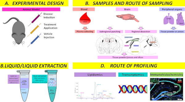
Figure 1: Overview of the workflow modules. Depending on the study aim and different routes of sampling, extraction and profiling can be combined to enable a significant outcome for the study. Please click here to view a larger version of this figure.
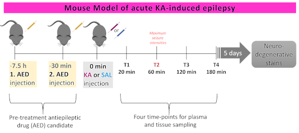
Figure 2: Experimental design of acute kainic acid (KA)-induced epileptic seizure model in mice. Mice are either treated with 1) Saline (10 mL/kg i.p.); 2) KA (30 mg/kg i.p.); and/or 3) (sub) chronically pretreated (1−2x 40 mg/kg i.p.) with the potential antiepileptic compound Palmitoylethanolamide (PEA). Twenty-four mice per group were treated as above and behavior was scored to evaluate seizure intensities. Six mice per group were sacrificed at each of the four different timepoints (T1−T4) to determine lipid level changes over a time course of acute epileptic seizure state in the brain, peripheral organs, and plasma. Please click here to view a larger version of this figure.
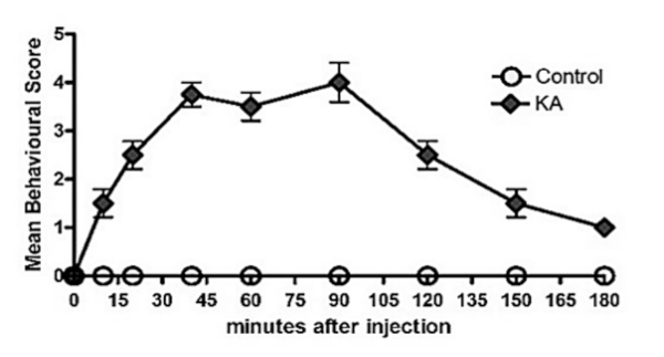
Figure 3: Behavioral scoring over a time course of acute kainic acid (KA)-induced epilepsy vs. controls. Assessment of seizure intensities over a time course of 180 min post KA-seizure induction (n = 24) or saline injection (n = 24) given as mean behavioral scores. No differences were found between the vehicle 1 and vehicle 2 injected groups. Error bars = SEM. ANOVA repeated measurement yielded significant interactions between the time points of the measurements and the test groups, indicating significant effects of KA treatment on behavioral scores. Seizure intensities of KA-treated mice were originally published in Post et al.13 Please click here to view a larger version of this figure.
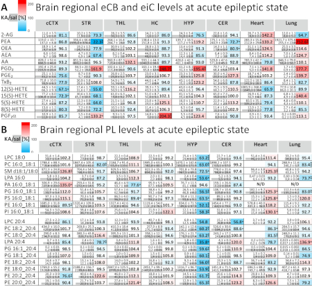
Figure 4: Brain and peripheral tissue lipid levels at the acute epileptic seizure state. Lipid level changes presented as mean value ± SEM at maximum seizure intensity (e.g., 1 h post-KA injection) across six brain regions (n = 9): cerebral cortex (cCTX), striatum (STR), thalamic region (THL), hippocampus (HC), hypothalamus (HYP), cerebellum (CER), as well as heart and lung tissue in KA-induced epileptic mice (upper value) and controls (lower value). The basal lipid levels in tissues are depicted in grey. The values of PLs, 2-AG, and AA are given in nmol/g. and the values for NAEs, eiCs, and AEA in pmol/g, respectively. All lipid levels are normalized to the tissue weight. To highlight the specific molecular changes, the lipid levels of the KA-treated mice are represented as percentage of the saline-injected (KA/sal) in a heat map displaying decreased values at acute seizure intensity compared to the control are depicted in light blue and the increased levels compared to control in red, respectively. They are considered significant at a p value <0.05. These data were originally published in Lerner et al.2 Please click here to view a larger version of this figure.
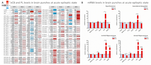
Figure 5: Dual extraction of eCBs/PLs and RNA for quantitative profiling from mouse brain punches. (A) Quantitative distribution of selected PLs and eCBs across: 1) a subregion of the hypothalamus (HYP); 2) the basolateral amygdala (BLA); and 3) the ventral (vHC) and dorsal (dHC) subregions of the hippocampus, from the KA-induced epileptic seizure mice (upper value) versus the controls (lower value) (n = 10). Levels are normalized to the tissue weight (punches correspond to approximately 0.5−1.5 mg) and expressed in nmol/g. Only AEA is expressed in pmol/g. The lipid levels are presented as mean value ± SEM. Mean variation per punched brain region (SEM as percentage of mean, averaged over all lipids) is: HYP = 7.83%; BLA = 7.80%; vHC = 6.28%; and dHC = 7.90%, respectively. (B) Relative expression levels of endogenous enzymes and receptors involved in lipid signaling, as well as markers for brain activity investigated at the mRNA level in different brain regions/subregions from mice subjected to KA-induced epileptic seizure (red) and controls (light grey). Statistical analyses of the difference between group means were carried out by using the two-tailed unpaired Student's t-test and considered significant at a p value <0.05 (n = 10). These data were originally published in Lerner et al.7 Please click here to view a larger version of this figure.
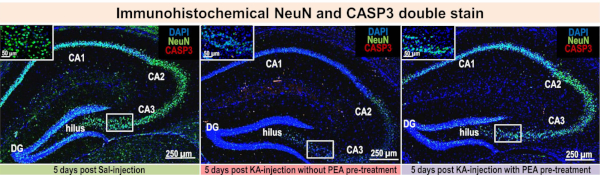
Figure 6: Immunohistochemical NeuN and CASP3 double stain. Five days post-KA injection immunohistochemistry was performed on brain sections from untreated saline-injected mice (left), subchronically PEA pretreated mice (middle), and epileptic mice without pretreatment (right). PEA pretreatment shows neuroprotective effects in comparison to untreated epileptic mice (n = 3). These data were originally published in Post et. al.13 Please click here to view a larger version of this figure.
Discussion
Loading...
$$\rightleftharpoonup{xx}$$
$$\longleftharp{xx}$$,
$$\longrightharp{xx}$$,
The neurolipidomic and transcriptomic methodology described here is a viable mean to investigate any disease or healthy development at high and low spatial resolution in the brain and peripheral organs. Due to the optimized plasma sampling and handling procedures, plasma lipidomic analysis can also be carried out from the same animals sacrificed for tissue lipidomics and transcriptomics, thus improving the reliability of tissue blood molecular correlates and biomarker discovery. The provision of a broad set of data by application of either of the three protocols or combinations thereof, is of value to investigate not only a neurological disease within a context (animal model experiment) but also across and between experimental model contexts. Moreover, a high level of standardization of sampling, processing, and molecular analysis facilitates high reproducibility of molecular data, hence reliably referencing molecular changes between and within studies and laboratories.
However, to attain this, the setup of an experimental design that offers the maximum readout potential for the defined study aim is critical. To attain a reliable comparison of the molecular changes between experimental groups, it is recommended to use a minimum of ten animals to compensate for animal variability and the biological range of lipid levels. When procurement of animals and/or logistics of animal handling are restrictive, use of a minimum of six animals per group is imperative to afford confident statistical analysis. Group size calculations need to compensate for model-related mortality rates (i.e., a minimum of six, ideally ten, animals per group are required for the study despite the possible mortality rate of the model). A critical requisite is to ensure age-, gender-, and strain-matched animals per experimental group. For the discovery phase, it is essential to use the same provider for experimental animals for all studies and the same animal batch if possible, in order to avoid bias of the findings due to possible behavioral and molecular phenotype differences between animal batches. To ensure reliability and reproducibility of the molecular and behavioral phenotype determined in the studies, it is critical to carry out a biological replicate analysis whenever possible.
Another crucial step is to set up a standardized, scheduled experimental work with animal groups. It is imperative to treat the animals within the same time-window of the day to circumvent circadian molecular variability. The time of the day should be set according to the known impact of circadian rhythm on synthesis and degradation of the target molecules or kept consistent for all experimental groups when no information on circadian rhythm effects is available. Similarly, the housing and feeding conditions prior to animal sacrifice must be maintained consistent and strictly controlled across experimental models. This is particularly relevant for lipidomic profiling due to influence of nutrition on lipid plasma and tissue metabolism. Administration of therapeutic or disease-inducing drugs should invariably be carried out in parallel with vehicle administration in control groups, whereby the vehicle must be the same as the one used for the drug administration. In order to choose the most suitable rodent strain and/or substrains for the purpose of the study, the drug-based treatment strategies should be carried out according to drug specificity in terms of time and frequency of administration, doses and route of administration, and the specific susceptibilities to drugs of different strains inferred from literature and/or prior experience. A time course investigation of a disease and response to therapy involving large cohort groups or multiple animal groups is impossible to carry out in one day in terms of treatment, sacrificing, and sampling. In such cases, animal group processing must be scheduled and carried out in consecutive days, but maintaining the same conditions in terms of time of the day, experimental design, processing time, researchers, etc. The preparation of chemical injections is another critical step. Drug- or disease-inducing compounds must be freshly prepared prior to administration and according to drug specifications. The use of the same production batch of the drug is recommended for all cohorts to be compared. This is especially important in the case of natural compound formulations such as kainic acid (KA) used in this study for epilepsy induction.
To enable reliable lipidomic and or transcriptomic profiling, the animal sacrificing procedure must be performed consistently across the animal groups within a timeframe of 5 minutes. If blood is collected after decapitation, it is important to maintain a constant amount of isoflurane in the glass chamber. For this purpose, soak frequently (after five uses) with isoflurane and do not exceed 10 s for the duration of anesthesia using isoflurane, in order to avoid onset of arrhythmia and palpitations and ensure proper blood pressure for plasma sampling.
Conditions for biological material sampling and handling (e.g., the time window and the order of biological material sampling and handling) must be strictly followed and maintained identical for all groups. To avoid variable degrees of tissue thawing and hence inconsistent ex vivo tissue changes of lipid and/or mRNA levels, it is essential to maintain strictly controlled time and temperature conditions for post-sampling
storage. tissue dissection or punching, and subsequent sample processing and analysis (see sections 3 and 4). Freshly sampled whole brains can be immediately dissected on a precooled metal plate (4 °C) without prior snapfreezing, if the size of the experimental animal groups allows for animal sacrifice, removal, and dissection without altering the timeframe indicated here for each of these procedures. If the size of experimental groups is not practical for these procedures, snap-freezing of the brains and subsequent dissection is recommended to allow comparable and controlled time for processing. When strictly following the protocols and timeline guidelines indicated here, no discrepancies were observed between molecular levels obtained by extraction of freshly dissected brain regions and brain regions dissected from frozen brains.
A critical aspect for attaining reproducible and minimal variability of the lipid levels within and between groups, apart from the sample processing under strictly controlled temperature conditions, is the provision of antioxidants (see sections 2 and 3). Avoiding any stress factors (e.g., the smell of blood) of the animals prior to sacrificing is of paramount importance, since many lipids involved in neuronal activity such as eCBs can rapidly change in response to stress.
For lipid extraction and analysis, it is essential to freshly prepare the internal standards, calibration solutions, and extraction solvents on the day of the extraction. The same source of internal standards must be used for both calibration curve preparation and for sample extractions. Also, following strictly controlled temperature conditions for sample extraction, storage, and analysis is paramount to minimize and control ex vivo enzymatic or chemical alterations of molecules. For LC/MRM analysis, the set of targeted lipids can be tailored to the study aim by adding or removing targets and correspondingly internal standards and calibrants, provided that the separation, detection, and MRM transitions for a new set of lipids are optimized. The presented extraction protocols allow the provision of two LC/MRM replicates for eCBs and eiCs, which is instrumental for cases of technical failure or when replicate analysis is of significance to the study. PL extraction protocols render sample/extract amounts suitable for at least 10 analyses per extract (e.g. multiple scan experiments based on precursor ion and neutral loss scanning2, respectively; additional LC/MRM analyses; or LC/MRM replicates to compensate for technical failure during a run). The lipid analysis is not restricted to LC/MRM; in fact the lipid extracts obtained with any of these protocols are amenable for untargteted, high-end mass spectrometriy analysis. Except for brain punches or minute amount of tissues obtained from discrete regions (less than 3 mg), frozen pulverized tissues of regions larger than 2−3 mg can be aliquoted and used for multiple extraction modules as described here for replicate analysis and/or for other investigations amenable in tissue powder.
A general advantage of the protocols described here compared to commonly used ones is the increased overall time-effectiveness and sensitivity for multicompound extraction and analysis at decreased expenditure of animal resources, consumables, and analysis costs. Importantly, the dual lipid/mRNA extraction protocol also affords a higher efficiency of mRNA extraction20,21 and integrity of the mRNA compared to corresponding available standard protocols, and simultaneously increased efficiency of the lipid extraction7. This is likely also due to the decreased matrix effect for each of the lipid and mRNA fractions when dually extracted. Due to this, the method is readily applicable for high spatial resolution profiling such as in brain punches.
However, a current limitation of the protocol is that the inflammatory lipids are not amenable for analysis and quantification using the dual lipid/mRNA extraction. Thus, the protocol is subject for further refinement. To this end, tissue and plasma inflammatory lipids and endocannabinoids can be co-extracted and co-analyzed, which is an optimized tool to concurrently investigate neuroinflammatory processes and endocannabinoids-modulated neuronal activity (see coextraction of eiCs and eCBs). Inclusion of phospholipids in this latter assay is expected to be feasible.
In view of prospective multi-omic approaches for neurological diseases, the proteomic analysis of protein fractions obtained after the lipid extraction protocol (i.e., co-extraction of eCBs and eiCs, as well as co-extraction of PLs and eCBs) is expected to be feasible. However, this is not yet possible when using the dual lipid/mRNA protocol. For the latter, the chemical environment of the extraction precludes even protein amount determination using standard protein assays such as the bicinchoninic acid assay (BCA). Further developments to overcome this limitation and expedite the inclusion of proteomic profiling in these protocols are planned.
Using the modular protocol described here, it was possible to attain a brain regional map of eiCs, eCBs, and PLs in an animal model of acute epileptic seizures (Figure 4). The protocol showed the hippocampal modulation of inflammatory processes by eiCs and of neuronal activity modulation by eCBs in treated and untreated mice with KA-induced acute seizures13. Subregional brain localization of phospholipid, endocannabinoid, and mRNA changes at acute epileptic seizure states, respectively, were also observed (Figure 5). These results highlight the value and applicability of the methods described here in advancing the knowledge on a broad spectrum of lipids involved in modulation of a complex neurological diseases such as epilepsy in brain regions and subregions. These protocols are of general applicability in neurological disease investigation and beyond while further development of the protocols and applications for cell populations continues.
Disclosures
Loading...
$$\rightleftharpoonup{xx}$$
$$\longleftharp{xx}$$,
$$\longrightharp{xx}$$,
The authors declare no conflict of interest.
Acknowledgements
Loading...
$$\rightleftharpoonup{xx}$$
$$\longleftharp{xx}$$,
$$\longrightharp{xx}$$,
We dedicate this article to Dr. Ermelinda Lomazzo. During the finalization of this manuscript, Dr. Ermelinda Lomazzo passed away. She is the embodiment of passion for science and selfless engagement in team work to fulfill a meaningful research purpose. She always dreamed of contributing meaningfully to the greater well-being of humans. Her goodhearted nature was never compromised by the strenuous roads of science and life. She will remain invaluable, and forever, in our hearts.
Julia M. Post was funded by Focus Program for Translational Neuroscience (FTN) at University Medical Center of the Johannes Gutenberg University Mainz and is currently funded by the SPP-2225 EXIT project to LB. Raissa Lerner was partially funded by DZHK project 81X2600250 to LB and Lipidomics Core Facility. Partial funding for these studies was provided by the Lipidomics Core Facility, Institute of Physiological Chemistry, and Intramural funds (to LB) from the University Medical Center of the Johannes Gutenberg University Mainz.
Materials
| Name | Company | Catalog Number | Comments |
|---|---|---|---|
| 12(S)-HETE | Biomol | Cay10007248-25 | Lipid Std |
| 12(S)-HETE-d8 | Biomol | Cay334570-25 | Lipid Std |
| 1200 series LC System | Agilent | Instrumentation/LCMS | |
| 2100 Bioanalyzer | Agilent | Instrumentation/qPCR | |
| 5(S)-HETE-d8 | Biomol | Cay 334230 | Lipid Std |
| ABI 7300 Real-Time PCR cycler | Applied Biosystems | Instrumentation/qPCR | |
| Acetonitrile LC-MS Chroma Solv | Honeywell | 9814920 | Solvent/LCMS |
| amber eppendorf tubes | Eppendorf | Sample Prep. | |
| Analyst 1.6.2 Software | AB SCIEX, Darmstadt | Software | |
| Analytical balance | Mettler Toledo | Instrumentation/Sample prep. | |
| Arachidonic Acid-d8 MS Standard | Biomol | Cay-10007277 | Lipid Std |
| Bessmann Tissue Pulverizer | Spectrum Laboratories, Inc. (Breda, Netherlands) | Instrumentation/Sample prep. | |
| Bino | Zeiss | Microscopy | |
| cleaved Caspase 3 antibody | Cellsignaling | 9661S | Microscopy |
| Cryostat, Leica CM3050 S | Leica Biosystems | Instrumentation/Sample prep. | |
| CTC HTC PAL autosampler | CTC Analytics AG | Instrumentation/LCMS | |
| Dumont Curved Forceps Dumoxel #7 | FST | 11271-30 | Surgical Tools |
| Dumont Forceps Super fine tip #5SF (x2) | FST | 11252-00 | Surgical Tools |
| EDTA 1000 A Röhrchen | Kabe Labortechnik | 078001 | Sample Prep. |
| EP-1 EconoPump | BioRAD | 700BR07757 | Instrumentation/Sample prep. |
| Fine Forceps Mirror Finish | FST | 11412-11 | Surgical Tools |
| Fine Iris Scissors straight sharp | FST | 14094-11 | Surgical Tools |
| Fine Scissor Tungsten Carbide straight | FST | 14568-09 | Surgical Tools |
| Iris Spatulae | FST | 10094-13 | Surgical Tools |
| Kainic acid | Abcam | ab120100 | Epileptic drug |
| Lipid View software | AB SCIEX, Darmstadt | Software | |
| LPC 17:0 | Avanis Polaris | 855676P | Lipid Std |
| LPC 18:0 | Avanis Polaris | 855775P | Lipid Std |
| Luna 2,5µm C18(2)- HAST 100A LC column | Phenomenex | 00D-4446-B0 | Instrumentation/LCMS |
| Magnifying lamp | Maul GmbH | Instrumentation/Sample prep. | |
| Methanol LC-MS Chroma Solv 99.9% | Honeywell | 9814920 | Solvent/LCMS |
| Motic Camara | Motic | Microscopy | |
| MTBE | Honeywell | 34875-1L | Solvent/LCMS |
| MultiQuant 3.0 quantitation software package | AB SCIEX, Darmstadt | Software | |
| NanoDrop 2000c Spectrophotometer | Thermo Scientific | Instrumentation/qPCR | |
| PA 16:0-18:1 | Avanis Polaris | 840857P | Lipid Std |
| PA 17:0-14:1 | Avanis Polaris | LM-1404 | Lipid Std |
| Palmitoyl Ethanolamide | Biomol | Cay90350-100 | Lipid Std |
| Palmitoyl Ethanolamide-d5 | Biomol | Cay9000573-5 | Lipid Std |
| PC 16:0-18:1 | Avanis Polaris | 850457P | Lipid Std |
| PC 16:0-18:1 | Avanis Polaris | 850457P | Lipid Std |
| PC 17:0-14:1 | Avanis Polaris | LM-1004 | Lipid Std |
| PE 16:0-18:1 | Avanis Polaris | 850757P | Lipid Std |
| PE 17:0-14:1 | Avanis Polaris | LM-1104 | Lipid Std |
| PG 16:0-18:1 | Avanis Polaris | 840457P | Lipid Std |
| PG 17:0-14:1 | Avanis Polaris | LM-1204 | Lipid Std |
| PI 17:0-14:1 | Avanis Polaris | LM-1504 | Lipid Std |
| Precelleys 24 | Peqlab | Instrumentation/Sample prep. | |
| Precellys Keramik-Kügelchen | Peqlab | 91-pcs-ck14p | Sample Prep. |
| Precellys Stahlkugeln 2,8mm | Peqlab | 91-PCS-MK28P | Sample Prep. |
| Precellys-keramik-kit 1,4 mm | VWR | 91-PCS-CK14 | Sample Prep. |
| Prostaglandin D2 | Biomol | Cay 12010 | Lipid Std |
| Prostaglandin D2-d4 | Biomol | Cay 312010 | Lipid Std |
| Prostaglandin E2 | Biomol | Cay10007211-1 | Lipid Std |
| Prostaglandin E2-d9 | Biomol | Cay10581-50 | Lipid Std |
| PS 17:0-14:1 | Avanis Polaris | LM-1304 | Lipid Std |
| Q Trap 5500 triple-quadrupole linear ion trap MS | AB SCIEX | AU111609004 | Instrumentation/LCMS |
| Real Time PCR System | Appliert Biosystem | Instrumentation/qPCR | |
| Resolvin D1 | Biomol | Cay10012554-11 | Lipid Std |
| Rneasy Mini Kit - RNAase-Free DNase Set (50) | Qiagen | 79254 | Sample Prep. |
| Security Guard precolumn | Phenomenex | Instrumentation/LCMS | |
| Shandon coverplates | Thermo Fisher | 72110017 | Microscopy |
| Shandon slide rack and lid | Thermo Fisher | 73310017 | Microscopy |
| SM 18:0 | Avanis Polaris | 860586P | Lipid Std |
| SM d18:1/12:0 | Avanis Polaris | LM-2312 | Lipid Std |
| Standard Forceps straight Smooth | FST | 11016-17 | Surgical Tools |
| Surgical Scissor ToughCut Standard Pattern | FST | 14130-17 | Surgical Tools |
| T3000 Thermocycler | Biometra | Instrumentation/qPCR | |
| Thromboxane B2 | Biomol | Cay19030-5 | Lipid Std |
| Thromboxane B2-d4 | Biomol | Cay319030-25 | Lipid Std |
| Tissue Lyser II | Qiagen/ Retsch | 12120240804 | Instrumentation/Sample prep. |
| Tissue Tek | Sakura Finetek | 4583 | Microscopy |
| Toluidinblau | Roth | 0300.2 | Microscopy |
| Vapotherm | Barkey | 4004734 | Instrumentation/Sample prep. |
| Wasser LC-MS Chroma Solv | VWR | 9814920 | Solvent/LCMS |
References
Loading...
$$\rightleftharpoonup{xx}$$
$$\longleftharp{xx}$$,
$$\longrightharp{xx}$$,
- Neuroinflammatory targets and treatments for epilepsy validated in experimental models. Epilepsia. 58, 27-38 (2017).">Aronica, E., et al. Neuroinflammatory targets and treatments for epilepsy validated in experimental models. Epilepsia. 58, 27-38 (2017).
- Targeting brain and peripheral plasticity of the lipidome in acute kainic acid-induced epileptic seizures in mice via quantitative mass spectrometry. Biochimica et Biophysica Acta - Molecular and Cell Biology of Lipids. 1862 (2), 255-267 (2017).">Lerner, R., Post, J., Loch, S., Lutz, B., Bindila, L. Targeting brain and peripheral plasticity of the lipidome in acute kainic acid-induced epileptic seizures in mice via quantitative mass spectrometry. Biochimica et Biophysica Acta - Molecular and Cell Biology of Lipids. 1862 (2), 255-267 (2017).
- Shotgun lipidomics on a LTQ Orbitrap mass spectrometer by successive switching between acquisition polarity modes. Journal of Mass Spectrometry. 47 (1), 96-104 (2012).">Schuhmann, K., Almeida, R., Baumert, M., Herzog, R., Bornstein, S. R., Shevchenko, A. Shotgun lipidomics on a LTQ Orbitrap mass spectrometer by successive switching between acquisition polarity modes. Journal of Mass Spectrometry. 47 (1), 96-104 (2012).
- A review of analytical methods for eicosanoids in brain tissue. Journal of Chromatography B: Analytical Technologies in the Biomedical and Life Sciences. 964, 50-64 (2014).">Puppolo, M., Varma, D., Jansen, S. A. A review of analytical methods for eicosanoids in brain tissue. Journal of Chromatography B: Analytical Technologies in the Biomedical and Life Sciences. 964, 50-64 (2014).
- Development and validation of a high-performance liquid chromatography-electrospray mass spectrometry method for the simultaneous determination of 23 eicosanoids. Journal of Pharmaceutical and Biomedical Analysis. 46 (4), 653-662 (2008).">Blewett, A. J., Varma, D., Gilles, T., Libonati, J. R., Jansen, S. A. Development and validation of a high-performance liquid chromatography-electrospray mass spectrometry method for the simultaneous determination of 23 eicosanoids. Journal of Pharmaceutical and Biomedical Analysis. 46 (4), 653-662 (2008).
- Simultaneous lipidomic and transcriptomic profiling in mouse brain punches of acute epileptic seizure model compared to controls. Journal of Lipid Research. 59, 283-297 (2017).">Lerner, R., et al. Simultaneous lipidomic and transcriptomic profiling in mouse brain punches of acute epileptic seizure model compared to controls. Journal of Lipid Research. 59, 283-297 (2017).
- Simultaneous lipidomic and transcriptomic profiling in mouse brain punches of acute epileptic seizure model compared to controls. Journal of Lipid Research. , 1-48 (2018).">Lerner, R., et al. Simultaneous lipidomic and transcriptomic profiling in mouse brain punches of acute epileptic seizure model compared to controls. Journal of Lipid Research. , 1-48 (2018).
- Animal models of temporal lobe epilepsy following systemic chemoconvulsant administration. Journal of Neuroscience Methods. 260, (2016).">Lévesque, M., Avoli, M., Bernard, C. Animal models of temporal lobe epilepsy following systemic chemoconvulsant administration. Journal of Neuroscience Methods. 260, (2016).
- Microglia-Neuron Communication in Epilepsy. Glia. 65 (1), 5-18 (2017).">Eyo, U. B., Murugan, M., Wu, L. J. Microglia-Neuron Communication in Epilepsy. Glia. 65 (1), 5-18 (2017).
- Kainic acid-induced neurodegenerative model: Potentials and limitations. Journal of Biomedicine and Biotechnology. 2011, 457079(2011).">Zhu, J., Zheng, X. Y., Zhang, H. L., Luo, Q. Kainic acid-induced neurodegenerative model: Potentials and limitations. Journal of Biomedicine and Biotechnology. 2011, 457079(2011).
- Role of α-CGRP in the regulation of neurotoxic responses induced by kainic acid in mice. Peptides. 44, 158-162 (2013).">Park, S. H., Sim, Y. B., Kim, C. H., Lee, J. K., Lee, J. H., Suh, H. W. Role of α-CGRP in the regulation of neurotoxic responses induced by kainic acid in mice. Peptides. 44, 158-162 (2013).
- Broad lipidomic and transcriptional changes of prophylactic PEA administration in control mice. Frontiers in Neuroscience. 13, 527(2019).">Lerner, R., Cuadrado, D. P., Post, J. M., Lutz, B., Bindila, L. Broad lipidomic and transcriptional changes of prophylactic PEA administration in control mice. Frontiers in Neuroscience. 13, 527(2019).
- Antiepileptogenic Effect of Subchronic Palmitoylethanolamide Treatment in a Mouse Model of Acute Epilepsy. Frontiers in Molecular Neuroscience. 11, (2018).">Post, J. M., et al. Antiepileptogenic Effect of Subchronic Palmitoylethanolamide Treatment in a Mouse Model of Acute Epilepsy. Frontiers in Molecular Neuroscience. 11, (2018).
- Genetic determinants of susceptibility to excitotoxic cell death: Implications for gene targeting approaches. Proceedings of the National Academy of Sciences. 94 (8), 4103-4108 (2002).">Schauwecker, P. E., Steward, O. Genetic determinants of susceptibility to excitotoxic cell death: Implications for gene targeting approaches. Proceedings of the National Academy of Sciences. 94 (8), 4103-4108 (2002).
- The Endocannabinoid System Controls Key Epileptogenic Circuits in the Hippocampus. Neuron. 51 (4), 455-466 (2006).">Monory, K., et al. The Endocannabinoid System Controls Key Epileptogenic Circuits in the Hippocampus. Neuron. 51 (4), 455-466 (2006).
- The mouse Brain in Stereotaxic Coordinates. Psychoneuroendocrinology. 28 (6), (2003).">Konsman, J. P. The mouse Brain in Stereotaxic Coordinates. Psychoneuroendocrinology. 28 (6), (2003).
- Neuroproteomics. Li, K. W. 57, 13-27 (2011).">Spijker, S. Dissection of Rondent Brain Regions. Neuroproteomics. Li, K. W. 57, 13-27 (2011).
- The evolution of lipidomics through space and time. Biochimica et Biophysica Acta - Molecular and Cell Biology of Lipids. 1862 (8), 731-739 (2017).">Gross, R. W. The evolution of lipidomics through space and time. Biochimica et Biophysica Acta - Molecular and Cell Biology of Lipids. 1862 (8), 731-739 (2017).
- Novel advances in shotgun lipidomics for biology and medicine. Progress in Lipid Research. 61, 83-108 (2016).">Wang, M., Wang, C., Han, R. H., Han, X. Novel advances in shotgun lipidomics for biology and medicine. Progress in Lipid Research. 61, 83-108 (2016).
- An improved high-throughput lipid extraction method for the analysis of human brain lipids. Lipids. 48 (3), 307-318 (2013).">Abbott, S. K., et al. An improved high-throughput lipid extraction method for the analysis of human brain lipids. Lipids. 48 (3), 307-318 (2013).
- Lipid extraction by methyl-tert-butyl ether for high-throughput lipidomics. Journal of Lipid Research. 49 (5), 1137-1146 (2008).">Matyash, V., Liebisch, G., Kurzchalia, T. V., Shevchenko, A., Schwudke, D. Lipid extraction by methyl-tert-butyl ether for high-throughput lipidomics. Journal of Lipid Research. 49 (5), 1137-1146 (2008).
Reprints and Permissions
Request permission to reuse the text or figures of this JoVE article
Request Permission