Presented here is a protocol for primary human blood monocyte isolation as well as their differentiation into macrophages and dendritic cells and assembly with epithelial cells into a multicellular human lung model. Biological responses of cocultures composed of immune cells differentiated from either freshly isolated or thawed monocytes, upon exposure to proinflammatory stimuli, are compared.
Method Article
Multicellular Human Alveolar Model Composed of Epithelial Cells and Primary Immune Cells for Hazard Assessment
* These authors contributed equally
In This Article
Summary
Abstract
A human alveolar cell coculture model is described here for simulation of the alveolar epithelial tissue barrier composed of alveolar epithelial type II cells and two types of immune cells (i.e., human monocyte-derived macrophages [MDMs] and dendritic cells [MDDCs]). A protocol for assembling the multicellular model is provided. Alveolar epithelial cells (A549 cell line) are grown and differentiated under submerged conditions on permeable inserts in two-chamber wells, then combined with differentiated MDMs and MDDCs. Finally, the cells are exposed to an air-liquid interface for several days. As human primary immune cells need to be isolated from human buffy coats, immune cells differentiated from either fresh or thawed monocytes are compared in order to tailor the method based on experimental needs. The three-dimensional models, composed of alveolar cells with either freshly isolated or thawed monocyte-derived immune cells, show a statistically significant increase in cytokine (interleukins 6 and 8) release upon exposure to proinflammatory stimuli (lipopolysaccharide and tumor necrosis factor α) compared to untreated cells. On the other hand, there is no statistically significant difference between the cytokine release observed in the cocultures. This shows that the presented model is responsive to proinflammatory stimuli in the presence of MDMs and MDDCs differentiated from fresh or thawed peripheral blood monocytes (PBMs). Thus, it is a powerful tool for investigations of acute biological response to different substances, including aerosolized drugs or nanomaterials.
Introduction
In vitro lung cell cultures offer cost-effective, robust, and well-controlled platforms to assess the hazards of aerosols1. As a model cell system for human alveolar pneumocytes, the epithelial A549 cell line isolated from a pulmonary adenocarcinoma is often used2. These cells represent squamous type II epithelial cells from the alveolar region3 and are a widely used lung cell line for hazard and toxicity assessment1,4,5,6,7,8,9,10. The A549 cell line possesses relevant features of alveolar epithelial type II cells, such as the presence of characteristic lamellar bodies containing densely packed phospholipids3.
It has been shown that when the cells are cultured at an air-liquid interface (ALI), surfactant is released on the apical side of air-exposed epithelial cells, reducing surface tension11,12,13. This feature is particularly important in nanomaterial respiratory hazard and toxicity investigations. Once inhaled nanomaterials/toxicants are deposited in the alveolar region, they first interact with pulmonary surfactant and are displaced by wetting forces into the aqueous hypophase, where the interaction with pulmonary cells takes place14,15. Even though A549 cells form a monolayer (which can overgrow into multilayers at later timepoints when cultivated at ALI) and produce surfactant, a drawback is their insufficient tight junction formation, resulting in low transepithelial electrical resistance values, but still presenting a functional barrier against intercellular (nano)particles translocation16,17,18.
In the lungs, there is a variety of immune cell populations, including phagocytic and professional antigen-presenting cells (i.e., macrophages and dendritic cells) that directly communicate through cell-cell contact or intercellular signaling to control and maintain homeostasis. Macrophages and dendritic cells are critical innate immune effectors and initiators of the adaptive immune response19. Dendritic cells residing within or beneath the epithelium can form protrusions across the epithelium to the lumen to catch antigens. Alveolar macrophages are located on the apical surface of the epithelium and act as sentinel cells, representing the first cellular defense against foreign material as well as bacterial, viral and fungal infections. Their phenotypic plasticity allows rapidly induction of proinflammatory reactions in response to such stimuli as well as shifting into triggering anti-inflammatory (i.e., inhibitory) reactions20.
To simulate the human alveolar epithelial tissue barrier, we established a triple coculture model with A549 cells supplemented with human blood monocyte-derived macrophages (MDMs) and dendritic cells (MDDCs) on the apical and basal sides, respectively17. Cultivation of this model at ALI has been previously reported16, even up to 72 h post-exposure21. Acute immune responses to carbon nanotubes exposures was significantly enhanced in cell culture exposed to the ALI as compared to submerged conditions22. The coculture model, cultured and exposed to different materials at ALI, has been previously used to investigate cytotoxicity, oxidative stress and inflammatory responses upon exposures to zinc oxide,23 graphene-related materials24, gold nanoparticles25,26, carbon nanotubes21, and volcanic ash and diesel exhaust particles27.
Furthermore, the important role of macrophages and dendritic cells as immune effector cells in an in vitro human lung model has been confirmed. In particular, an increased proinflammatory response in the model was observed only in the presence of immune cells in comparison to monoculture systems7. Potential drawbacks of using primary monocyte-derived immune cells are the limited accessibility of PBMs as well as donor-to-donor variation. As a solution to these potential drawbacks, presented here is a protocol introducing cryopreservation of freshly isolated PBMs28 for the cell culture model assembly. The aim of this study is to demonstrate the 3D human alveolar epithelial tissue model assembly, including the isolation of PBMs from human buffy coats. The responsiveness to proinflammatory stimuli is compared to the model composed of MDMs and MDDCs differentiated from fresh PBMs or differentiated from frozen/thawed PBMs.
Working with untested human blood samples involves specific care to prevent the potential transmission of infectious diseases, such as HIV (human immunodeficiency virus), hepatitis B, and hepatitis C. Therefore, the use of personal protective measures such as gloves, gowns, masks, and eye protection are crucial and must be in accordance with good laboratory practice principles. These protections reduce the risk of exposing the skin or mucous membranes to potentially infectious fluids. Additionally, for those involved in handling buffy coats and PBMs, vaccination against the hepatitis B virus is mandatory, and blood titer levels of antibodies anti-hepatitis B must be above 100 IU/L (country-specific legislative requirements need to be addressed). In addition, all work must be performed in biosafety level 2 laboratories (country-specific legislative requirements need to be addressed). Standard health and safety precautions associated with working in a laboratory environment and handling mammalian cell culture, including handling of waste, should be adopted when conducting the entire protocol.
Protocol
The work involving primary monocytes isolated from human blood was approved by the committee of the Federal Office for Public Health Switzerland (reference number: 611-1, Meldung A110635/2) for the Adolphe Merkle Institute.
1. Isolation of peripheral blood monocytes (PBMs) from human buffy coats
NOTE: The following section describes the isolation of immune cells from one 50 mL bag of a buffy coat, purchased from the Swiss Transfusion Centre in Bern, Switzerland.
- Preparation of reagents
- Prepare 100 mL of magnetic separation buffer per buffy coat: 0.5% [w/v] bovine serum albumin (BSA; in phosphate-buffered saline [PBS]) with 2 mM ethylenediaminetetraacetic acid (EDTA), and adjust to pH = 7.2, sterile filter with 0.22 µm pore size). Keep at 4 °C throughout the procedure.
- Prepare the cell culture medium (CCM): RPMI 1640 with 10% [v/v] fetal bovine serum (FBS), 1% [v/v] L-glutamine (here, 2 mM L-glutamine), and 1% [v/v] penicillin-streptomycin (here, 100 units/mL penicillin and 100 µg/mL streptomycin).
NOTE: The required amount of each reagent depends on the number of cells to be seeded in the following steps.
- Isolation of PBMs
NOTE: All glass and plasticware needs to be sterilized before use. For safety reasons, the use of plasticware is recommended when handling human blood samples to reduce the risk of injury with glassware.- Use scissors to cut open the hose end of the bag containing the buffy coat.
- Distribute the buffy coat by pouring the bag's contents through the bag's duct directly into two conical centrifuge 50 mL tubes (~25 mL each).
- Gently pour or pipette PBS into the tubes to reach 50 mL volumes. Mix the contents by turning the tube gently upside down 3x.
- Divide the buffy coat-PBS mixture into four new 50 mL conical centrifuge tubes by pipetting 25 mL of the mixture in each fresh tube.
- Slowly lay 13 mL of density gradient medium underneath the buffy coat-PBS mixture using a 10 mL serological pipette. Detach the filled pipette from the pipette holder and immediately plug the upper opening of the pipette with a thumb to prevent any additional leaking of the density gradient medium into the buffy coat-PBS mixture.
NOTE: Holding the upper opening with the thumb, place the filled pipette at the bottom of the conical centrifuge tube so that the density gradient medium slowly flows beneath the buffy coat-PBS mixture, leaving approximately 1 mL of the density gradient medium inside the pipette. - Repeat step 1.2.5 with the other three tubes containing the buffy coat-PBS mixture.
- Centrifuge all four tubes containing the mixtures for 20 min at 1,000 x g and 25 °C at a slow braking mode. Use holders with protective lids for centrifugation.
- Open the lid of each tube, remove the upper layer containing plasma and platelets using a serological pipette, and dispose in a biohazard liquid waste container.
- Use a serological pipette for collecting the peripheral blood mononuclear cell layer, which appears as a whitish turbid small fraction (~2–3 mm in thickness) between the plasma and the density gradient medium layers. The pellet contains red blood cells at the bottom. Avoid transferring erythrocytes that form the bottom-most layer. Repeat this for all four tubes.
NOTE: Peripheral blood mononuclear cells consist of PBMs and lymphocytes. PBMs will be separated from lymphocytes later during magnetic CD14+ separation. - Pool peripheral blood mononuclear cells from four tubes into two 50 mL tubes.
- Fill up the two tubes with PBS to 50 mL and cover with a lid.
- Discard the leftover erythrocytes and plasma from the original four tubes in a biohazard liquid waste container.
- Centrifuge the two tubes for 8 min at 500 x g and 18–20 °C at a regular centrifuge speed.
- After centrifugation, remove the supernatant with a serological pipette and discard it in a biohazard liquid waste container.
- Resuspend the cells with 5 mL of PBS using a serological pipette.
- Pool the cell suspensions into one 50 mL conical centrifuge tube and fill to 50 mL with PBS.
- Use 5 µL of the cell suspension to count the cells with a cell counter using the trypan blue (45 µL) exclusion method.
- Pipette 10 µL of the trypan blue-PBMs solution into a cell counter chamber and count the number of cells per the standard counting protocol. Use Equation 1 to calculate the total cell number, CT.

- After counting the cells, centrifuge the 50 mL tube as done in step 1.2.13.
- CD14 positive selection
- Gently open the lid of each tube, then remove and discard the supernatant using a serological pipette without disturbing the pellet.
- Add the calculated amount (Equation 2) of magnetic separation buffer (here, 80 µL of buffer per 1 x 10⁷ total cells) and resuspend the cell pellet by pipetting the solution up and down.

- Calculate using Equation 3 (here, 10 µL per 1 x 10⁷ total cells) the corresponding volume of CD14+ magnetic beads and pipette the appropriate volume.

- Mix well by pipetting up and down, close the lid, and incubate the solution at 4 °C for 15 min.
- Upon incubation, fill the tube up to 50 mL with magnetic separation buffer.
- Centrifuge as done in step 1.2.13.
- Aspirate and discard the supernatant using a serological pipette without disturbing the cell pellet.
- Pipette the corresponding amount of magnetic separation buffer (Equation 4; here, 500 µL of buffer per 1 x 10⁸ cells) and gently mix by pipetting up and down 3x.

- Disinfect the magnetic separation station by spraying and wiping it with a sterilizing agent. Place in the laminar flow hood along with the column for magnetic separation.
- Place the magnetic separation column in the magnetic field and place an empty 50 mL conical centrifuge tube directly under the column for collecting the wash and unlabeled cells (i.e., waste).
- Rinse the magnetic separation column by pipetting 3 mL of magnetic separation buffer into the column. Do not let the column dry out throughout the procedure.
- Prepare a 15 mL conical centrifuge tube and pipette 1 mL of magnetic separation buffer.
- Apply the cell suspension (prepared in step 1.3.8) to the magnetic separation column. In the 50 mL conical centrifuge tube under the filter, collect unlabeled cells that passed through.
NOTE: Do not exceed 2 x 10⁹ cells per column to avoid blocking of the column. - As soon as the column reservoir is empty (i.e., when the cells have passed through the column), apply 3 mL of magnetic separation buffer using a serological pipette and let it pass through the column. Repeat this 3x.
- Remove the magnetic separation column from the magnetic separator by gently pulling with hands, then place it into a 15 mL tube containing pre-pipetted 1 mL of magnetic separation buffer (prepared in step 1.3.12).
- Add 5 mL of magnetic separation buffer to the column and flush out the magnetically labeled cells by firmly pushing the plunger into the column.
- Reagent preparation for MDM and MDDC differentiation
- Count the cells with a cell counter using the trypan blue exclusion method as done in step 1.2.17.
- Calculate the required volumes of CCM or FBS for further steps as follows: either the volume of CCM corresponding to a cell density of 1 x 10⁶ cells/mL (step 1.5.1), or the volume of FBS corresponding to a cell density of 6 x 10⁶ cells per 0.9 mL of FBS (step 1.6.3).
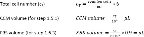
- Close the lid, place the tube in the centrifuge, and centrifuge as done in step 1.2.13. Remove and discard the supernatant without disturbing the cell pellet. Proceed to step 1.5 for cell seeding or step 1.6 for cell freezing.
- PBM seeding and differentiation into MDMs and MDDCs
- Resuspend the cell pellet in the calculated volume of CCM as calculated in step 1.4.2 (here, a final concentration of 1 x 10⁶ cells/mL) by pipetting up and down 3x.
- Pipette the number of cells intended to differentiate into MDMs and MDDCs in separate conical centrifuge tubes using a serological pipette.
- Pipette differentiating factors to the CCM with PBMs and mix well by pipetting up and down. Differentiating factors are applied as follows:
- For MDDCs: final concentration of 10 ng/mL interleukin-4 (IL-4) and 10 ng/mL granulocyte-macrophage colony-stimulating factor (GM-CSF).
- For MDMs: final concentration of 10 ng/mL macrophage colony-stimulating factor (M-CSF).
- Pipette the cell suspensions in CCM with the added differentiating factors into 6 well plates by distributing 3 mL of the suspension per well (corresponds to 3 x 10⁶ cells/well, i.e., 1 x 10⁶ cells/mL).
- Place the 6 well plates in a cell culture incubator (37 °C, 5% CO2) and let them differentiate for 6 days without refreshing the CCM.
NOTE: Differentiation ranges from 5–8 days depending on the local availability of buffy coats and experimental setup, provided that the differentiation efficiency is determined using the appropriate techniques (refer to the discussion section).
- PBM freezing
- Resuspend the cell pellet in a cryoprotective medium (here, FBS and dimethyl sulfoxide [DMSO; cytotoxic]) at a ratio of 9:1 (v/v) by pipetting a volume of prewarmed FBS. This corresponds to a final cell concentration of 6 x 10⁶ cells/mL, considering a further addition of 10% DMSO (v/v).
- Mark the desired number of cryovials in the laminar flow hood (i.e., record the date, isolation code, and number of cells).
- Pipette 0.9 mL of cell suspension in pure FBS (here, 6 x 10⁶ cells in 0.9 mL of FBS) to each cryovial. Subsequently, slowly pipette 0.1 mL of DMSO and mix the suspension well by turning the cryovials up and down 3x.
- Transfer the cryovials to a cell-freezing container and immediately set it to -80 °C for 24 h.
- After 24 h, remove the cryovials from the -80 °C freezer and container and place them into the liquid nitrogen tank suitable for cell storage.
- PBM thawing and differentiation into MDMs and MDDCs
- Warm all the required reagents to 37 °C in a water bath (~20–30 min).
- Prepare the appropriate number of 6 well plates corresponding to the number of thawed cells (here, one plate per 1.8 x 10⁷ cells, i.e. 3 cryovials). Pipette 2 mL of CCM to each well under aseptic conditions. Place plates in the incubator (5 % CO2, 37 °C) for 15 min to allow the pH to equilibrate.
- Take the required amount of cryovials with frozen cells from a liquid nitrogen tank and gently swirl them in a 37 °C water bath (1–2 min) to ensure uniform thawing of the cell suspension.
- Remove the cryovial from the water bath and decontaminate with a sterilizing agent, ensuring that the agent does not interact with the lid and O-ring.
NOTE: Hereon, all steps must be completed under aseptic conditions. - Prepare the appropriate number of 15 mL conical centrifuge tubes corresponding to the number of cryovials to be thawed (here, 6 x 10⁶ cells/tube). Pipette 9 mL of prewarmed CCM into each tube.
- Pipette slowly (drop-by-drop) the contents of the cryovial into a tube containing CCM. Close the lid, repeat for each tube, and centrifuge at 200 x g for 5 min at a regular centrifuge speed.
- Discard the supernatant without disturbing the pellet.
- Resuspend the pellet from each tube (which contains cells from one cryovial) in 2 mL of prewarmed CCM by pipetting up and down using a serological pipette (cell density corresponding to 3 x 10⁶ cells/mL).
- From each tube, pipette the resuspended cells into two wells (1 mL per well) of a 6 well plate containing 2 mL of previously prepared CCM to reach a cell density of 3 x 10⁶ cells/well (corresponding to a final concentration of 1 x 10⁶ cells/mL). Repeat this for all other tubes.
- Proceed with differentiation as described in section 1.5.3.
- Place the 6 well plates in a cell culture incubator (37 °C, 5% CO2) and let them differentiate for 6 days without refreshing the CCM.
2. Triple cell coculture model of human alveolar epithelial tissue
NOTE: This section provides instructions on volumes and cell numbers corresponding to 12 well plate inserts. Figure 1 summarizes a proposed timeline for model assembly.
- Epithelial cell (A549 cell line) seeding
- Culture the epithelial cells according to the recommendations provided by the supplier (ATTC). Briefly, subculture the cells in CCM at 80% cell confluency (approximately, 2x–3x per week).
NOTE: Subculture A549 for at least four passages before the coculture model composition, using A549 cells in a passage range of 5–25. - Pipette 1.5 mL of prewarmed CCM into 12 well plates (the number of wells corresponds to the desired number of models).
- Place individual 12 well cell culture inserts into wells of a 12 well plate using sterilized tweezers.
- Detach the cells from a flask according to the subcultivation protocol (i.e., using a detaching agent, remove the agent by centrifugation as done in step 1.2.13). Resuspend in the appropriate volume of CCM corresponding to the final cell concentration of A549 (here, 50 x 104 cells/mL; 0.5 mL of cell suspension per insert, i.e. 25 x 104 cells/insert, which corresponds to seeding density of 27.8 x 10⁴ cells/cm2).
- Pipette 0.5 mL of the cell suspension (i.e., 25 x 104 cells/insert) into the apical side of the insert using a 1 mL pipette.
- Cover the plates with lids and place them in a cell culture incubator (37 °C, 5 % CO2) for 4 days.
NOTE: Regularly check the confluency of A549 cells under a phase-contrast microscope.
- Culture the epithelial cells according to the recommendations provided by the supplier (ATTC). Briefly, subculture the cells in CCM at 80% cell confluency (approximately, 2x–3x per week).
- MDDC seeding
- Aspirate CCM with unattached cells in the 6 well plates containing MDDCs.
- Add 1 mL of fresh prewarmed CCM to each well.
- Use a cell scraper, detach (scrape) adherent MDDCs from each well, gently wash the wells with the existing 1 mL of CCM 3x, and combine them into one conical centrifuge tube.
- Count the cells with a cell counter using the trypan blue exclusion method using 10 μL of cell suspension and 10 μL of trypan blue solution.
- Centrifuge the cell suspension as done in step 1.2.13.
- Calculate the CCM volume required for resuspension (Equation 5):
Required MDDC density = 42 x 104 cells/mL; each insert requires 6.3 x 10⁴ cells, which corresponds to a seeded cell density of 7 x 10⁴ cell/cm2 (here, 150 µL added onto 0.9 cm2 in the step 2.2.10.).
CCM volume for cell resuspension (Vm; Equation 5):

- Gently aspirate and discard the CCM from the upper chamber of 12 well plates with growing A549 on the inserts.
- Place the inserts with A549 cells in an upside-down position in a sterile Petri dish using sterilized tweezers. Prepare a conical centrifuge tube (50 mL) with PBS and pre-moisten a cell scraper.
- Scrape off A549 cells from the basal surface of the insert (i.e., the upper part at the upside-down position), which should grow through the pores of the inserts.
NOTE: Rinse the scraper with PBS (prepared in a tube) between scraping individual samples and keep it wet throughout the procedure. - Upon centrifugation (step 2.2.5), aspirate and discard the supernatant, then redisperse the MDDC pellet in the calculated amount of CCM (step 2.2.6) and pipette up and down 3x.
- Pipette 150 µL of the cell suspension on top of each insert so that the entire basal surface of the insert is equally covered with the liquid and not containing bubbles.
- Cover the dish with a lid and place in a cell culture incubator for 70 min. Aspirate the CCM from the cell culture plates (where the inserts have initially been placed), discard it in a biohazard liquid waste, and pipette 1.5 mL of fresh CCM into each well. Cover the plate with a lid and place in the cell incubator (37 °C, 5% CO2).
NOTE: Do not exceed the time period mentioned above to avoid drying out the cells. - After the incubation, carefully hold each insert with sterilized tweezers and place them to the plates containing CCM in an ordinary position. Cover the plate with a lid and return it to the cell incubator (37°C, 5% CO2).
- Macrophage (MDM) seeding
- Take 6 well plates containing predifferentiated MDMs. Place them from the cell incubator to a laminar flow hood.
- Aspirate and discard CCM with unattached MDMs grown in 6 well plates, and pipette 1 mL of fresh prewarmed CCM in each well.
- Using a cell scraper, gently remove adherent MDMs from individual wells (as done in step 2.2.3 for the MDDCs).
- Pipette 10 µL of trypan blue into a well or tube and add 10 µL of the MDM suspension to achieve the final dilution of 1:1 (v/v). Count the number of MDMs using the appropriate counting protocol.
- Centrifuge the cell suspension as done in step 1.2.13.
- Calculate the required volume (Equation 6):
Required MDM density = 2.5 x 10⁴ cells/mL in CCM (here, each insert requires 1.25 x 10⁴ cells in 0.5 mL CCM, which corresponds to a seeded cell density of 1.4 x 10⁴ cell/cm2).
 (6)
(6) - Upon centrifugation, aspirate and discard the supernatant, redisperse the MDM pellet in the calculated amount of CCM (step 2.3.6), and pipette up and down 3x.
- Carefully pipette 0.5 mL of the MDM suspension (prepared in step 2.3.7) on the wall of the cell culture inserts with A549 and MDDCs (not directly on epithelial cells) using a 1 mL pipette. Cover plates with lids and place in a cell culture incubator (37 °C, 5 % CO2) for 24 h.
- Transfer of coculture model to air-liquid interface (ALI)
- Upon the end of the 24 h (±2 h) incubation period of the assembled model in a cell culture incubator, aspirate and discard CCM from both apical and basal parts of cell culture inserts and from the wells.
- Using sterilized tweezers, lift individual inserts from the wells and pipette 0.6 mL of fresh prewarmed CCM to each well using a 1 mL pipette. Do not add CCM to the apical side of the insert.
- Cover plates with lids and place in a cell culture incubator (37 °C, 5 % CO2) for 24 h prior further use.
3. Exposure to selected positive controls (known stimuli for inducing proinflammatory response)
NOTE: Exposure of the coculture models to a known proinflammatory stimulus endotoxin lipopolysaccharide (LPS)7 and proinflammatory cytokine tumor necrosis factor α (TNF-α)7 is used to illustrate responsiveness of the model. Furthermore, exposure to a detergent (Triton X-100) is used to confirm the sensitivity of a lactate dehydrogenase (LDH) assay.
- Prepare the positive control solutions: LPS stock (1 mg/mL in distilled water), TNF-α stock (100 μg/mL in distilled water) and Triton-X 100 (2% [v/v] in PBS).
- Upon 24 h incubation of the coculture model at ALI conditions, aspirate and discard the supernatant from the basal compartment. Using sterilized tweezers, lift individual inserts from the wells and pipette 0.6 mL of fresh prewarmed CCM into each well.
- Prepare individual positive controls working solutions by diluting the stocks in CCM in conical centrifuge tubes as follows: 1 μg/mL LPS, 1 μg/mL TNF-α, and 0.2% Triton-X 100. The volumes correspond to the number of tested inserts (here, 100 μL/insert). Mix the solutions well by pipetting up and down 3x.
- Apply 100 μL of each positive control solution by slowly pipetting onto the wall of the cell culture insert. Cover the well plate with a lid and place in the cell culture incubator (37 °C, 5% CO2) for 24 h. Upon incubation, aspirate and discard the liquid at the apical side of the insert by holding individual inserts using tweezers.
- Collect CCM in the basal compartments and store at 1) 4 °C for further LDH analysis, denoting cell membrane rupture-mediated cytotoxicity, and/or 2) store at -80 °C for further analysis of protein release via enzyme-linked immunosorbent assay (ELISA). Run the assays according to the kit supplier recommendations.
- Upon the removal of CCM, wash the inserts with PBS 3x and fix the cells on cell culture inserts in 4% [w/v] paraformaldehyde (in PBS, 15 min at room temperature) by ensuring both insert sides are well-covered with PFA solution. Subsequently wash 3x with PBS to remove PFA. Store the samples submerged in PBS at 4 °C for further immunostaining (an example of this method has been described previously17).
Results
Human lung coculture models, composed of alveolar epithelial cells and immune cells, were assembled either from fresh or frozen MDDCs and MDMs progenitors (here, human peripheral blood-derived monocytes). As presented in Figure 1, A549 cells were seeded 3 days after the first section involving monocyte isolation/thawing. After 6 days of differentiation, the differentiated MDMs appeared round-shaped, whereas MDDCs formed a more elongated shape with observable protrusions. They also appeared as agglomerates, especially when differentiated from fresh monocytes (Figure 2, Figure 3). Epithelial cells formed a dense cell layer of cells after 3 days of growth on membrane inserts (Figure 4), when the cocultures were assembled. After 24 h of assembling and an additional 24 h of being subjected to ALI conditions, the cocultures were prepared for exposures.
Responsiveness of the 3D cell culture models was investigated upon exposure to known proinflammatory stimuli using a pseudo-ALI approach, as described previously29. The proinflammatory stimuli, LPS and TNF-α, were added in low volumes (100 μL) onto the apical surface of the air-exposed cell model. In parallel, the absence of membrane rupture as a measure of cytotoxicity was assessed via LDH assay. A significant increase in LDH release in CCM of the basal compartment was observed upon exposure to the positive control for membrane rupture, a detergent Triton-X 100 (Figure 5). These results proved responsiveness of the model to a cytotoxic substance, whereas no increase in LDH release was observed upon apical stimulation with TNF-α or LPS.
A possible reason for the different measured values of LDH in the samples assembled with either fresh or previously frozen PBMs can be attributed to the sample storage. Samples from fresh PBMs were stored for a longer time at -80 °C; therefore, the activity of LDH enzyme may drop. Notably, LDH is stable for only up to 4 days in CCM; thus, it is recommended to perform the assay the latest 2 days after collecting the supernatants. Alternatively, it is possible to freeze down the supernatants directly after collection. However, it is important to consider that freezing can decrease the enzymatic activity of LDH.
Secretion of proinflammatory mediators (here, TNF-α and interleukins 6 [IL-6] and 8 [IL-8]) into the basal CCM was quantified via ELISA. Statistically significant (p < 0.05, one-way ANOVA) increases in the release of IL-6 and IL-8 were observed in both LPS- and TNF-α- treated samples compared to respective untreated cells, as well as in the cell culture models assembled from either PBMs source (Figure 6). Although concentrations (pg/mL) of all the tested cytokines in the basal CCM were higher in the cocultures composed of fresh PBMs, differences among the two cocultures and monocultures were not statistically significant (p > 0.05) (Figure 6). To confirm the added value of coculture models with respect to a 2D epithelial cell culture, A549 monocultures were also exposed to LPS or TNF-α. As expected, the release of all investigated mediators from A549 monocultures was lower compared to both coculture models; although, the difference between them was not statistically significant (p > 0.05, one-way ANOVA).
Cellular morphology of the 3D human alveolar epithelial tissue barrier was assessed via confocal laser scanning microscopy (LSM). To visualize the composition of each model, macrophages within the coculture models (MDMs) were stained with mature macrophage marker 25F9. MDDCs were stained with CD83, which is an important marker for activated dendritic cells30. Regarding cellular morphology, no difference was observed between the coculture models using MDMs and MDDCs from fresh PBMs compared to those using thawed PBMs. In LPS- and TNF-α-exposed cocultures, both composed of fresh and frozen immune cells, a disrupted epithelial layer in LSM images was observed, which was not the case in untreated cells (Figure 7, Figure 8).
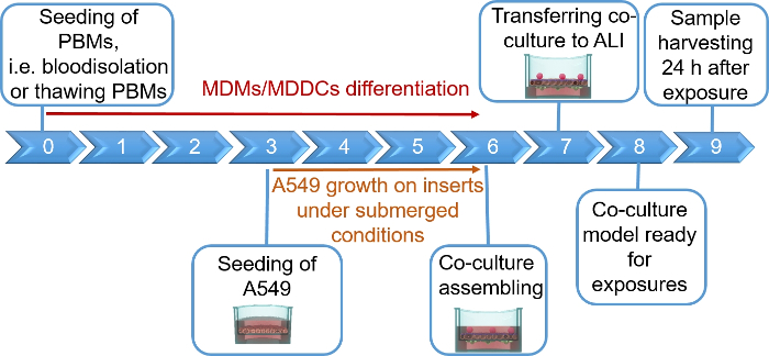
Figure 1: Schematic timeline of the protocol. Presentation of the 3D coculture model preparation, assembly, and application (exposure to a tested substance). ALI = air-liquid interface, MDDCs = monocyte-derived dendritic cells, MDMs = monocyte-derived macrophages, PBMs = peripheral blood monocytes. Please click here to view a larger version of this figure.
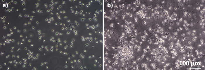
Figure 2: MDMs and MDDCs differentiated from fresh PBMs. Phase-contrast microscopy image of differentiated (A) MDMs and (B) MDDCs from fresh PBMs (6 days after cell isolation). MDMs are round-shaped, while MDDCs are often observed as agglomerates. Please click here to view a larger version of this figure.
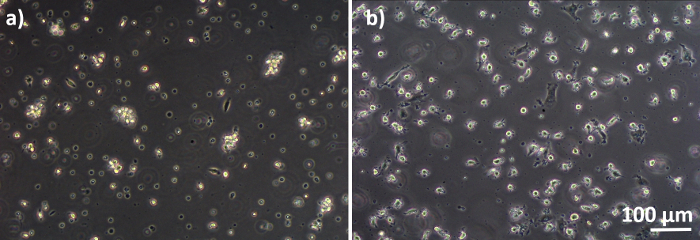
Figure 3: MDMs and MDDCs differentiated from frozen PBMs. Phase-contrast microscopy image of differentiated (A) MDMs and (B) MDDCs from thawed PBMs (6 days after thawing). MDMs are round-shaped, but some elongated cells can be observed. MDDCs also appear round-shaped with protrusions. Please click here to view a larger version of this figure.
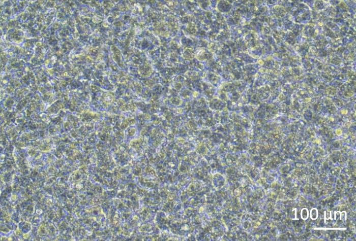
Figure 4: Epithelial cells growth on membrane inserts. Phase-contrast microscopy image of confluent A549 growing on a membrane insert 4 days after seeding, forming a dense layer of cells. Please click here to view a larger version of this figure.
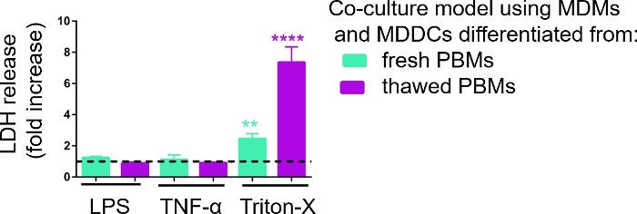
Figure 5: Cytotoxicity results investigated via membrane rupture-based (LDH) assay. The data is presented as a fold increase over untreated cells (mean ± SD, n = 3, asterisk denotes statistically significant increase compared to untreated cells, **p < 0.01, ****p < 0.0001). In green models MDMs and MDDCs from fresh PBMs are represented, and in purple models assembled from thawed PBMs are represented. Please click here to view a larger version of this figure.
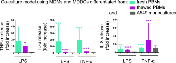
Figure 6: Proinflammatory reactions in the cocultures and monocultures. The proinflammatory mediators (TNF-α, IL-6, and IL-8) release in the cocultures upon 24 h challenge with LPS or TNF-α. The data is presented as relative to untreated cells (mean ± SD, n = 3, **p < 0.01, ***p < 0.001, ****p < 0.0001). In green models MDMs and MDDCs from fresh PBMs are represented, and in purple models assembled from thawed PBMs are represented. Grey represents A549 monocultures. Please click here to view a larger version of this figure.

Figure 7: Morphology of cocultures composed of fresh immune cells. LSM images of apical and basal sides of the coculture model with xz projections of apical sides of the model using MDMs and MDDCs from fresh PBMs. Cyan represents nuclei (DAPI), magenta represents cytoskeleton (rhodamine-phalloidin), white represents MDMs (25F9), and green represents MDDCs (CD 83). The white arrow denotes MDM, whereas the green arrow denotes MDDC. Please click here to view a larger version of this figure.
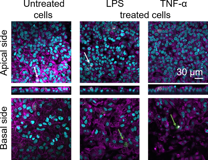
Figure 8: Morphology of cocultures composed of frozen immune cells. LSM images of the apical side of the coculture model with corresponding xz projections, and basal side of the model using MDMs and MDDCs from thawed PBMs. Cyan represents nuclei (DAPI), magenta represents cytoskeleton (rhodamine-phalloidin), white represents MDMs (25F9), and green represents MDDCs (CD 83). The white arrow denotes MDM, whereas the green arrow denotes MDDC. Please click here to view a larger version of this figure.
Discussion
Emerging production of novel materials, including chemicals and drugs, gradually increases the need for predictive in vitro models. To comply with the three principles of replacement, reduction, and refinement of animal testing32, in vitro cell models have become powerful tools regarding the replacement and reduction aspect for elucidating mechanisms of a drug’s or material’s action8,9,10,11. Presented here is a detailed protocol of assembling the multicellular model using immune cells that are either freshly isolated or thawed from previously frozen monocytes. Also described is the cultivation of the model at ALI. Finally, the protocol illustrates an example of exposure to proinflammatory stimuli and compares the response of the two models containing either fresh or frozen monocytes.
Various studies have been performed to confirm and justify the added value of the enhanced complexity of the models grown and exposed under ALI conditions compared to conventional submerged exposure7,22,31. Observation of the higher proinflammatory response in cocultures compared to monocultures of epithelial cells confirm a previous study. The study used the presented coculture model (stimulated with LPS) and showed a higher response at gene expression levels of TNF and IL1B compared to the A549 monoculture equivalent model7. On the other hand, both models showed higher variations within measured proinflammatory mediator release values compared to A549 monocultures. This can be explained by the use of immune cells from different donors (buffy coats) within biological repetitions (i.e., one repetition, one donor), as previously shown7. If desired, the variations among the replicates can be overcome by 1) using thawed PBMs from the same donor or 2) pooling PBMs from different donors prior to freezing the cells, then subsequent usage of the same pool in each repetition. Including more biological repetitions is also recommended.
The cell-freezing technique can be considered as a critical step; however, it is a common laboratory procedure for preserving cells for phenotypic and functional analysis. Various studies have demonstrated that the quality of frozen PBMs is vital to their survival, and an appropriate freezing technique is key to the success of subsequent assays with the same cells28,32. Modification of the protocol can be performed by freezing PBMs, which provides flexibility in the experimental setup, as the availability of buffy coats is usually limited. Another advantage of using frozen PBMs (in several vials) over freshly isolated ones is that they can be used in subsequent experiments even after 1 year. This decreases the potential issue of donor-to-donor variability if this is a desired or required parameter in an experimental.
Outcomes of an interlaboratory comparison performed after up to 13 months show that PBMs, when properly stored in a liquid nitrogen tank, can be used over a long period without any effect on cell viability or cell recovery33. Longer storage times (over 1 year) may be possible upon careful validation of cell viability and cell responsiveness before performing an experiment. Also, the temperature in the liquid nitrogen tank must remain stable at all times. The main factor affecting the viability of cryopreserved PBMs was found to be DMSO concentration, with an optimal concentration of 10%–20 % (v/v)28. To minimize potentially harmful effects of freezing, different sources of proteins, FBS or BSA (with a broad range of concentration from 40% up to 100 %34) are often added to the freezing medium as natural protective components that can increase cell survival.
Due to the high cytotoxic potential of DMSO, it is recommended first to disperse PBMs in FBS, then add DMSO to PBMs already dispersed in FBS. Notably, although higher FBS concentrations (>40%) did not show any improvement in cell viability, at the same time, they did not cause harm to the cells28. Nevertheless, freezing monocytes is a possible approach to overcoming issues of limited buffy coat availability. However, if the use of MDDCs and MDMs from fresh PBMs is desired, the immune cells can be differentiated and used 5–8 days after isolation7,16,17,35,36,37. If experimental planning allows, at least 6 days of differentiation in both MDDCs and MDMs is recommended. However, consistency among different repetitions in the same experiment, along with routine inspections of their specific surface marker expressions, are crucial. The responsiveness to a proinflammatory stimulus, such as LPS, after the differentiation time should also be regularly checked.
Many investigations using the A549 cell line have been performed at ALI, either as a monoculture or combined with other cell types (macrophages, dendritic cells, or fibroblasts) into 3D coculture model22,24,29,38. Using this 3D coculture model, the cytotoxicity, oxidative stress, or proinflammatory effects of (nano-)materials have been investigated for up to 72 h1,17,21,24,29. The model’s resemblance to in vivo tissue has previously been investigated based on confocal laser scanning imaging of the model16. When assembling the model, it is important to consider both cell proliferation (which can affect A549 in the model presented here) as well as performance of the primary (not proliferating) immune cells (here, MDDCs and MDMs). It is also important to consider that not all the CD14 positive monocytes differentiate into MDDCs and MDMs, and that the cells can be present in both attached and suspended forms. Based on the nature of the coculture assembly (here, both cell types need to attach to the existing epithelial layer), it is recommended to use only the adherent sub-populations of both immune cell types. Additionally, routine analyses of monocytes, MDDC and MDM monoculture responsiveness to LPS, and expression of specific surface markers (CD14, CD163, CD86, CD93, or CD206, data not shown) have suggested that 6 and 7 days of differentiation are the optimum timepoints.
Although a realistic number of alveolar epithelial cells in human lungs corresponds to ~160,000 cells/cm2, the number of A549 cells counted in the model is ~1,000,000 cells/cm2 after 9 days cultured on the insert16,18. Thus, this in vitro model’s limitations need to be considered. First, the density of epithelial cells was established based on their ability to form a confluent layer on the growing membrane. It is also important to mention that the A549 represents an epithelial type II cell with a cuboidal form, contrary to epithelial type I cells, which are flat and outspread. On the other hand, the required number of immune cells was established based on the literature and presented in this protocol as cell number/surface area39,40,41. The cell density of MDDCs in the range of 400 cells/mm2 (4 cells/cm2)16 is comparable to the steady-state cell density of 500–750 cells/mm2 (5- 7 cells/cm2) reported from in vivo studies39. The density of MDMs in this model is within the same range of in vivo situation in the human alveolar region40.
Mature macrophage marker staining (25F9) was observed both in the apical side (where MDMs are present) as well as basal side (i.e., at the site of dendritic cells). Translocation of immune cells through the membrane inserts pores is possible and has also been observed using this model16, which may explain the observed differences in staining intensities. However, another possible explanation is that the mature macrophage marker can also be expressed on dendritic cells, but the expression is highly donor-specific42. Also, the intensity of the 25F9 expression is much higher in MDMs (Figure 7, Figure 8). Both proinflammatory stimuli (LPS and TNF-α) affected the integrity of the pulmonary epithelial barrier in both cocultures (Figure 7, Figure 8). This was expected based on previous publications43,44 showing that proinflammatory cytokines and bacterial products disrupt the integrity of epithelial barriers.
The 3D multicellular model of the human alveolar epithelium, established and characterized previously17, has served as a powerful and useful tool for assessing biological responses (i.e., acute proinflammatory reactions, oxidative stress response, particle distribution, and cellular communication) in vitro21,24,25,45. The results confirm the responsivness of coculture models to proinflammatory stimuli (here, LPS and TNF-α). The response was slightly increased when using immune cells from fresh PBMs; however, there was no statistically significant difference between cocultures using fresh vs. thawed PBMs. Furthermore, the proinflammatory reactions of both coculture models were higher than those of epithelial cell monocultures cultivated under the same (ALI) conditions. In summary, the protocol describes the assembly of a 3D human alveolar epithelial tissue coculture model using either fresh or thawed PBMs for differentiation into MDMs and MDDCs. It is shown that both models are highly responsive to proinflammatory stimuli; therefore, they can serve as powerful tools for potential hazard and toxicity assessments.
Disclosures
The authors have nothing to disclose.
Acknowledgements
The authors would like to thank Dr. Miguel Spuch-Calvar for the coculture scheme in Figure 3 and to Dr. Bedia Begum Karakocak for critical reading. This study was supported by the PATROLS project, European Union’s Horizon 2020 Research and Innovation Programme under grant agreement No 760813, and by the Adolphe Merkle Foundation. B.D. thanks the Peter und Traudl Engelhorn foundation for financial support.
Materials
| Name | Company | Catalog Number | Comments |
|---|---|---|---|
| Benchmark microplate reader | does not have to be specific, for example BioRad, Cressier, Switzerland | ||
| Cell culture Incubator | does not have to be specific | ||
| Cell freezing container (for example Mr. Frosty) | does not have to be specific | ||
| Centrifuge | does not have to be specific | ||
| Confocal laser scanning microscope | does not have to be specific, for example | Zeiss LSM 710 meta | |
| Heamatocytometer, or automatic cell counter | does not have to be specific | ||
| Laminar bio-safety hood class II | does not have to be specific | ||
| MultiStand Macs (Macs Cell Separator) | Miltenyi, Germany | 130-042-303 | |
| pH meter | does not have to be specific | ||
| Phase contrast inverted light microscope | does not have to be specific | ||
| Pipette boy, pipettors (different volumes) | do not have to be specific | ||
| Scissors | do not have to be specific | ||
| Vacuum pump | does not have to be specific | ||
| Water bath | does not have to be specific | ||
| Disposable small equipment/glassware | Catalogue Number | ||
| 15 mL and 50 mL conical centrifuge tubes | does not have to be specific | ||
| 6- and 12-well cell culture plates, flat bottom, low evaporation lid, sterile | Falcon, Switzerland | 353046 and 353043 | |
| Cell culture inserts, transparent PET membrane, 12-well, 3 μm pore size | Falcon, Switzerland | 353181 | |
| Cell scrapper | does not have to be specific, for example VWR, Switzerland | 353085 | |
| Cryovials | do not have to be specific | ||
| Glass autoclaved Petri Dishes | do not have to be specific | ||
| LS Columns | Miltenyi, Germany | 130-042-401 | |
| Sterile filtration cup for vacuum filtration, 0.2 μm pore size | does not have to be specific, for example VWR, Switzerland | 10040-446 | |
| Sterile Lab Bottle compatible with Filtration cup (min. 100 mL) | does not have to be specific | ||
| Sterile pipettes | do not have to be specific | ||
| Chemicals | |||
| Bovine serum albumine (BSA) | Sigma-Aldrich, Switzerland | A7030-100g | |
| CD14+ MicroBeads human - magnetic beads | Miltenyi, Germany | 130-097-052 | |
| Deattachnig agent Trypsin-EDTA, 0.05%, phenol red | Gibco, Switzerland | 25300054 | |
| Density gradient medium Lymphoprep | Alere Technologies AS, Norway | 1114547 | |
| Dimethyl Sulfoxide (DMSO) | Sigma Aldrich, Switzerland | D2438 | |
| Ethylenediaminetetraacetic acid (EDTA) | Sigma-Aldrich, Switzerland | E6758-100g | |
| Fetal bovine serum (heat inactivated) | Gibco, Switzerland | 10270-106 | |
| Human granulocyte-macrophage colony-stimulating factor (GM-CSF), premium grade | Miltenyi, Germany | 130-093-864 | |
| Human Interleukin 4 (IL-4), premium grade | Miltenyi, Germany | 130-095-373 | |
| Human macrophage colony-stimulating factor (M-CSF), premium grade | Miltenyi, Germany | 130-096-485 | |
| L-glutamine | Gibco, Switzerland | 25030-024 | |
| Lipopolysaccharid (LPS) from Escherichia coli | Sigma-Aldrich, Switzerland | 4524-5mg | |
| Paraformaldehyde (PFA) | Sigma-Aldrich, Switzerland | 158127 | |
| Penicilin-Streptomycin | Gibco, Switzerland | 15140-122 | |
| Phosphate Buffer Saline (PBS) | Gibco, Switzerland | 10010-015 | |
| Roswell Park Memorial Institute-1640 Medium (RPMI) | Gibco, Switzerland | 42401-018 | |
| Triton X-100 | Sigma-Aldrich, Switzerland | T8787 | |
| Trypan blue solution (0.4%) | Sigma Aldrich, Switzerland | ||
| Tumor necrosis factor alpha (TNF-α) | Immunotools | 11343015 | |
| Assays used for cytotoxicity, (pro-)inflammatory response | |||
| Cytotoxicity Detection Kit (LDH) | Roche, Switzerland | 11644793001 | |
| Human IL-6 DuoSet ELISA | R&D, Biotechne, Switzerland | DY206 | |
| Human IL-8/CXCL8 DuoSet ELISA | R&D, Biotechne, Switzerland | DY208 | |
| Immunostaining | |||
| 4′,6-diamidino-2-phenylindole (DAPI), concentration 2 μg/mL | Sigma-Aldrich, Switzerland | 10236276001 | |
| Goat anti-mouse IgG (H+L) Alexa Fluor 647 conjugated, concentration 20 μg/mL | Abcam, UK | ab150115 | |
| Goat anti-rabbit IgG antibody (H+L) Dylight 488 conjugated, concentration 10 μg/mL | Agrisera, Sweden | AS09 633 | |
| Mature Macrophage Marker Monoclonal Antibody, concentration 50 μg/mL | eBioScience, Thermo Fischer, Switzerland | 14-0115-82 | |
| Phalloidin rhodamine, concentration 0.264 µM | Molecular Probes, Life Technologies, Switzerland | R415 | |
| Recombinant Anti-CD83 antibody, 1:50 dillution | Abcam, UK | ab244204 |
References
- Rothen-Rutishauser, B., Blank, F., Mühlfeld, C., Gehr, P. In vitro models of the human epithelial airway barrier to study the toxic potential of particulate matter. Expert Opinion on Drug Metabolism and Toxicology. 4 (8), 1075-1089 (2008).
- Giard, D., et al. In vitro cultivation of human tumors: establishment of cell lines derived from a series of solid tumors. Journal of National Cancer Institute. 51 (5), 1417-1423 (1973).
- Ochs, M., Weibel, E. R., et al. Ch. 2: Functional Design of the Human Lung for Gas Exchange . Fishman's Pulmonary Diseases and Disorders, 5e. Grippi, M. A., et al. , McGraw-Hill Education. (2008).
- Foster, K. A., Oster, C. G., Mayer, M. M., Avery, M. L., Audus, K. L. Characterization of the A549 Cell Line as a Type II Pulmonary Epithelial Cell Model for Drug Metabolism. Experimental Cell Research. 243 (2), 359-366 (1998).
- Guo, X. Y., Lu, M., Chen, X. Q., He, F. D., Li, A. Correlation study of biological characteristics of non-small cell lung cancer A549 cells after transfecting plasmid by microbubble ultrasound contrast agent. Asian Pacific Journal of Tropical Medicine. 9 (6), 582-586 (2016).
- Cooper, J. R., et al. Long Term Culture of the A549 Cancer Cell Line Promotes Multilamellar Body Formation and Differentiation towards an Alveolar Type II Pneumocyte Phenotype. PLoS ONE. 11 (10), 0164438(2016).
- Bisig, C., Voss, C., Petri-Fink, A., Rothen-Rutishauser, B. The crux of positive controls - Proinflammatory responses in lung cell models. Toxicology In Vitro. 54, 189-193 (2019).
- Rothen-Rutishauser, B., et al. A newly developed in vitro model of the human epithelial airway barrier to study the toxic potential of nanoparticles. ALTEX. 25, (2008).
- Braakhuis, H. M., et al. Progress and future of in vitro models to study translocation of nanoparticles. Archives of Toxicology. 89 (9), 1469-1495 (2015).
- Thai, P., Chen, Y., Dolganov, G., Wu, R. Differential regulation of MUC5AC/Muc5ac and hCLCA-1/mGob-5 expression in airway epithelium. American Journal of Respiratory Cell and Molecular Biology. 33 (6), 523-530 (2005).
- Wu, J., et al. Characterization of air-liquid interface culture of A549 alveolar epithelial cells. Brazilian Journal of Medical and Biological Research. 51 (2), 6950(2017).
- Shapiro, D. I., Nardone, L. L., Rooney, S. A., Motoyama, E. K., Munoz, J. L. Phospholipid biosynthesis and secretion by a cell line (A549) which resembles type II aleveolar epithelial cells. Biochimica and Biophysica Acta. 530 (2), 197-207 (1978).
- Balis, J., Bumgarner, S. D., Paciga, J. E., Paterson, J. F., Shelley, S. A. Synthesis of lung surfactant-associated glycoproteins by A549 cells: description of an in vitro model for human type II cell dysfunction. Experimental Lung Research. 6 (3-4), 197-213 (1984).
- Schurch, S., Gehr, P., Im Hof, V., Geiser, M., Green, F. Surfactant displaces particles toward the epithelium in airways and alveoli. Respiration Physiology. 80 (1), 17-32 (1990).
- Gehr, P., Schurch, S., Berthiaume, Y., Hof, V. I., Geiser, M. Particle Retention in Airways by Surfactant. Journal of Aerosol Medicine. 3 (1), 27-43 (2009).
- Blank, F., Rothen-Rutishauser, B., Gehr, P. Dendritic Cells and Macrophages Form a Transepithelial Network against Foreign Particulate Antigens. American Journal of Respiratory Cell and Molecular Biology. 36 (6), (2007).
- Rothen-Rutishauser, B. M., Kiama, S. G., Gehr, P. A three-dimensional cellular model of the human respiratory tract to study the interaction with particles. American Journal of Respiratory Cell and Molecular Biology. 32, (2005).
- Blank, F., Rothen-Rutishauser, B. M., Schurch, S., Gehr, P. An optimized in vitro model of the respiratory tract wall to study particle cell interactions. Journal of Aerosol Medicine. 19, (2006).
- Jardine, L., et al. Lipopolysaccharide inhalation recruits monocytes and dendritic cell subsets to the alveolar airspace. Nature Communications. 10 (1), 1999(2019).
- Kopf, M., Schneider, C., Nobs, S. P. The development and function of lung-resident macrophages and dendritic cells. Nature Immunology. 16 (1), 36-44 (2015).
- Chortarea, S., et al. Repeated exposure to carbon nanotube-based aerosols does not affect the functional properties of a 3D human epithelial airway model. Nanotoxicology. 9 (8), 983-993 (2015).
- Hilton, G., Barosova, H., Petri-Fink, A., Rothen-Rutishauser, B., Bereman, M. Leveraging proteomics to compare submerged versus air-liquid interface carbon nanotube exposure to a 3D lung cell model. Toxicology In Vitro. 54, 58-66 (2019).
- Brandenberger, C., et al. Effects and uptake of gold nanoparticles deposited at the air-liquid interface of a human epithelial airway model. Toxicology and Applied Pharmacology. 242, (2010).
- Drasler, B., et al. Single exposure to aerosolized graphene oxide and graphene nanoplatelets did not initiate an acute biological response in a 3D human lung model. Carbon. 137, 125-135 (2018).
- Durantie, E., et al. Carbon nanodots: Opportunities and limitations to study their biodistribution at the human lung epithelial tissue barrier. Biointerphases. 13, (2018).
- Brandenberger, C., et al. Quantitative evaluation of cellular uptake and trafficking of plain and polyethylene glycol-coated gold nanoparticles. Small. 6 (15), 1669-1678 (2010).
- Tomašek, I., et al. Combined exposure of diesel exhaust particles and respirable Soufrière Hills volcanic ash causes a (pro-)inflammatory response in an in vitro multicellular epithelial tissue barrier model. Particle and Fibre Toxicology. 13 (1), 67(2016).
- Nazarpour, R., et al. Optimization of Human Peripheral Blood Mononuclear Cells (PBMCs) Cryopreservation. International Journal of Molecular and Cellular Medicine. 1 (2), 88-93 (2012).
- Endes, C., et al. An in vitro testing strategy towards mimicking the inhalation of high aspect ratio nanoparticles. Particle and Fibre Toxicology. 11 (1), (2014).
- Ju, X., et al. The Analysis of CD83 Expression on Human Immune Cells Identifies a Unique CD83+-Activated T Cell Population. Journal of Immunology. 197 (12), 4613-4625 (2016).
- Lenz, A. G., et al. Inflammatory and Oxidative Stress Responses of an Alveolar Epithelial Cell Line to Airborne Zinc Oxide Nanoparticles at the Air-Liquid Interface: A Comparison with Conventional, Submerged Cell-Culture Conditions. BioMed Research International. , 12(2013).
- Germann, A., Schulz, J. C., Kemp-Kamke, B., Zimmermann, H., von Briesen, H. Standardized serum-free cryomedia maintain peripheral blood mononuclear cell viability, recovery, and antigen-specific T-cell response compared to fetal calf serum-based medium. Biopreservation and Biobanking. 9 (3), 229-236 (2011).
- Weinberg, A., et al. Optimization and Limitations of Use of Cryopreserved Peripheral Blood Mononuclear Cells for Functional and Phenotypic T-Cell Characterization. Clinical and Vaccine Immunology. 16 (8), 1176(2009).
- Freshney, R. I. Culture of animal cells: a manual of basic technique. Freshney, R. I. , Wiley-Liss Inc. 321-334 (2005).
- Lehmann, A. B. C., Blank, F., Gehr, P., Rothen-Rutishauser, B. Alternatives to animal testing. Yarmush, M. L., Langer, R. S. , Artech House. 239-260 (2010).
- Steiner, S., et al. Reduction in (pro-)inflammatory responses of lung cells exposed in to diesel exhaust treated with a non-catalyzed diesel particle filter. Atmospheric Environment. 81, 117-124 (2013).
- Martinez, F. O., Gordon, S., Locati, M., Mantovani, A. Transcriptional Profiling of the Human Monocyte-to-Macrophage Differentiation and Polarization: New Molecules and Patterns of Gene Expression. The Journal of Immunology. 177 (10), 7303(2006).
- Chortarea, S., et al. Profibrotic activity of multi-walled carbon nanotubes upon prolonged exposures in different human lung cell types. Applied In Vitro Toxicology. 5 (1), (2019).
- Holt, P. G. Pulmonary Dendritic Cells in Local Immunity to Inert and Pathogenic Antigens in the Respiratory Tract. Proceedings of the American Thoracic Society. 2 (2), 116-120 (2005).
- Pinkerton, K. E., Gehr, P., Castañeda, A., Crapo, J. D. Comparative Biology of the Normal Lung (Second Edition). Parent, R. A. , Academic Press. 105-117 (2015).
- Crapo, J., Barry, B., Gehr, P., Bachofen, M., Weibel, E. R. Cell number and cell characteristics of the normal human lung. American Review of Respiratory Disease. 126 (2), 332-337 (1982).
- Maniecki, M. B., Møller, H. J., Moestrup, S. K., Møller, B. K. CD163 positive subsets of blood dendritic cells: The scavenging macrophage receptors CD163 and CD91 are coexpressed on human dendritic cells and monocytes. Immunobiology. 211 (6), 407-417 (2006).
- Chignard, M., Balloy, V. Neutrophil recruitment and increased permeability during acute lung injury induced by lipopolysaccharide. American Journal of Physiology-Lung Cellular and Molecular Physiology. 279 (6), 1083-1090 (2000).
- Coyne, C. B., et al. Regulation of Airway Tight Junctions by Proinflammatory Cytokines. Molecular Biology of the Cell. 13 (9), 3218-3234 (2002).
- Durantie, E., et al. Biodistribution of single and aggregated gold nanoparticles exposed to the human lung epithelial tissue barrier at the air-liquid interface. Particle and Fibre Toxicology. 14 (49), (2017).
Reprints and Permissions
