The present protocol describes a tissue clearing method and whole-mount immunofluorescent staining for three-dimensional (3D) kidney imaging. This technique can offer macroscopic perspectives in kidney pathology, leading to new biological discoveries.
Method Article
Whole-Kidney Three-Dimensional Staining with CUBIC
In This Article
Summary
Abstract
Although conventional pathology provided numerous information about kidney microstructure, it was difficult to know the precise structure of blood vessels, proximal tubules, collecting ducts, glomeruli, and sympathetic nerves in the kidney due to the lack of three-dimensional (3D) information. Optical clearing is a good strategy to overcome this big hurdle. Multiple cells in a whole organ can be analyzed at single-cell resolution by combining tissue clearing and 3D imaging technique. However, cell labeling methods for whole-organ imaging remain underdeveloped. In particular, whole-mount organ staining is challenging because of the difficulty in antibody penetration. The present protocol developed a whole-mount mouse kidney staining for 3D imaging with the CUBIC (Clear, Unobstructed Brain/Body Imaging Cocktails and Computational analysis) tissue clearing method. The protocol has enabled visualizing renal sympathetic denervation after ischemia-reperfusion injury and glomerulomegaly in the early stage of diabetic kidney disease from a comprehensive viewpoint. Thus, this technique can lead to new discoveries in kidney research by providing a macroscopic perspective.
Introduction
The kidney is composed of various cell populations. Although conventional pathology gives us much information about the kidney microenvironment, three-dimensional (3D) imaging is needed to precisely understand the intercellular crosstalk during kidney disease progression. In the past, a huge number of serial sectioning and image reconstruction needed to be performed for the whole-organ 3D imaging1. However, this method required too much effort and had problems in terms of reproducibility.
Optical clearing is a good strategy to overcome this hurdle2,3. Tissue opacity is mainly due to light scattering and absorption because each organ consists of various substances, including water, protein, and lipids. Thus, the basic strategy of tissue clearing is replacing water and lipids in tissues with refractive index (RI) matching reagents that have the same optical properties as proteins4. In order to observe a transparent specimen, a light sheet fluorescent microscopy is useful5. Light sheets illuminate the transparent specimen from the side, and excitation signals are acquired through the objective lens located in a vertical position6. This microscopy obtains cross-section information in a single sweep, which is different from the confocal or multiphoton fluorescent microscopy. Thus, it can quickly acquire z-stack images with a low level of photobleaching.
Clear, Unobstructed Brain/Body Imaging Cocktails and Computational Analysis (CUBIC) is one of the tissue clearing methods which enables whole-organ imaging by light sheet fluorescent microscopy2,7,8. CUBIC and whole-mount immunofluorescent staining are optimized in the present study to visualize mouse kidney 3D structures9,10,11. Using this whole-mount staining method, the alteration in renal sympathetic nerves is visualized after ischemia-reperfusion injury9,10 and glomerulomegaly in the early stage of diabetic kidney disease11, as well as blood vessels, proximal tubules, and collecting ducts in a whole kidney9.
Protocol
All experiments were approved by the University of Tokyo Institutional Review Board. All animal procedures were performed according to the National Institutes of Health guidelines. Male C57BL/6NJcl mice, 8 weeks old, were used for the present study. The mice were obtained from commercial sources (see Table of Materials).
1. Animal preparation and kidney fixation
- Perform perfusion fixation following the steps below.
- Anesthetize the mouse by inhalation of isoflurane (3%, 2.0 L/min) and intraperitoneal administration of medetomidine hydrochloride (0.3 mg/kg), butorphanol tartrate (5 mg/kg), and midazolam (4 mg/kg) (see Table of Materials).
- Perfuse the animal with 20 mL of PBS (pH 7.4) and 30 mL of 4% PFA in phosphate buffer (PB) through the left ventricle of the heart12.
- Perform the immersion fixation just after the perfusion fixation and kidney sampling9.
- Immerse the kidney in 4% PFA at 4 °C for an additional 16 h, then wash it with PBS for 2 h (three times)9 (Figure 1).
CAUTION: Formaldehyde and paraformaldehyde are toxic irritants. Handle reagents in a fume hood with appropriate personal protective equipment.
- Immerse the kidney in 4% PFA at 4 °C for an additional 16 h, then wash it with PBS for 2 h (three times)9 (Figure 1).
2. Decolorization and delipidation
- Prepare CUBIC-L for decolorization and delipidation consisting of 10 wt% of Triton X-100 and 10 wt% of N-buthyldiethanolamine (see Table of Materials), following previously published reports4,8,9.
- Perform decolorization and delipidation by CUBIC-L following the steps below.
- Immerse the fixed kidney in 7 mL of 50% (v/v) CUBIC-L (1:1 mixture of water and CUBIC-L) in a 14 mL round bottom tube (see Table of Materials) with gentle shaking at room temperature for 6 h (Figure 2A). Then, immerse it in 7 mL of CUBIC-L in a 14 mL round bottom tube with gentle shaking at 37 °C for 5 days.
- Refresh CUBIC-L every day during this process. After the decolorization and delipidation process, wash the kidney with PBS at room temperature for 2 h (three times)9 (Figure 1). Use a dispensing spoon for sample handling (Figure 2B).
3. Whole-mount immunofluorescent staining
- Prepare the staining buffers.
- Prepare the staining buffer for primary antibodies by mixing 0.5% (v/v) Triton X-100, 0.5% casein in PBS, and 0.05% sodium azide9.
- Prepare the staining buffer for secondary antibodies by mixing 0.5% (v/v) Triton X-100, 0.1% casein in PBS, and 0.05% sodium azide9.
- Perform staining with primary antibodies.
- Immunostain the delipidated kidney with primary antibodies (1:100 or 1:200, see Table of Materials) in the staining buffer at 37 °C with gentle shaking for 7 days.
NOTE: The amount of the staining buffer needed for one kidney is 500-600 µL (Figure 2C). - Wash the kidney with 0.5% (v/v) Triton X-100 in PBS (PBST) at room temperature for 1 day9 (Figure 1).
- Immunostain the delipidated kidney with primary antibodies (1:100 or 1:200, see Table of Materials) in the staining buffer at 37 °C with gentle shaking for 7 days.
- Perform staining with secondary antibodies.
- Immunostain the kidney with secondary antibodies (1:100 or 1:200, see Table of Materials) in the staining buffer at 37 °C with gentle shaking for 7 days. Wash the kidney with PBST at room temperature for 1 day9 (Figure 1).
- Post fixation9, immerse the kidney in 1% formaldehyde in PB (1:36 mixture of 37% formaldehyde and PB) for 3 h, and wash it with PBS at room temperature for 6 h (Figure 1).
4. Refractive index (RI) matching
- Prepare CUBIC-R+.
- Prepare CUBIC-R by mixing 45 wt% of 2,3-dimethyl-1-phenyl-5-pyrazolone/antipyrine and 30 wt% of nicotinamide (see Table of Materials).
- Prepare CUBIC-R+ for refractive index (RI) matching by adding 0.5 v% of N-buthyldiethanolamine to CUBIC-R following the previously published reports4,8,9.
- Perform the RI matching.
- Immerse the kidney in 7 mL of 50% (v/v) CUBIC-R+ (1:1 mixture of water and CUBIC-R+) in a 14 mL round bottom tube with gentle shaking at room temperature for 1 day. Then, immerse it in 7 mL of CUBIC-R+ in a 14 mL round bottom tube with gentle shaking at room temperature for 2 days9 (Figure 1).
NOTE: Although the kidney floats in 50% CUBIC-R+ at the beginning of the RI matching, it sinks and becomes transparent in CUBIC-R+ at the end of the process (Figure 2D).
- Immerse the kidney in 7 mL of 50% (v/v) CUBIC-R+ (1:1 mixture of water and CUBIC-R+) in a 14 mL round bottom tube with gentle shaking at room temperature for 1 day. Then, immerse it in 7 mL of CUBIC-R+ in a 14 mL round bottom tube with gentle shaking at room temperature for 2 days9 (Figure 1).
5. Image acquisition and reconstruction
- Immerse the RI-matched kidney in a mixture of silicon oil (RI = 1.555) and mineral oil (RI = 1.467) (55:45) during image acquisition (RI = 1.51)5 (Figure 3).
- Acquire 3D images of the whole kidney with a custom-built9 light-sheet fluorescent microscope (see Table of Materials). Collect all raw image data in a 16-bit TIFF format. Visualize and capture the 3D-rendered images with the imaging analysis software (Imaris, see Table of Materials).
Results
Using this staining method, sympathetic nerves [anti-tyrosine hydroxylase (TH) antibody] and arteries [anti-α-smooth muscle actin (αSMA) antibody] in a whole kidney (Figure 4A,B and Video 1) were visualized9. Abnormal renal sympathetic nerves were also visualized after ischemia/reperfusion injury (IRI)9,10 (Figure 4C). Moreover, visualizing sympathetic nerves in the whole spleen13 was achieved using this protocol (Figure 4D). One example of quantifying the 3D immunofluorescent data is shown in Figure 5.
In addition, visualizing collecting ducts [anti-aquaporin 2 (AQP2) antibody], the S1 segment of proximal tubules [anti-sodium glucose cotransporter 2 (SGLT2) antibody], and the glomeruli (anti-podocin antibody) in a whole kidney9 was successful (Figure 6A). Using this 3D imaging of glomeruli, glomerulomegaly (anti-podocin antibody) was visualized in the early stages of diabetic kidney disease, which was suppressed by drug administration11 (Figure 6B).
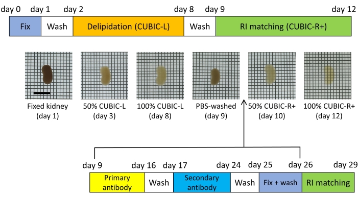
Figure 1: Tissue clearing and whole-mount immunofluorescent staining. Tissue clearing by CUBIC is a two-step process: delipidation (CUBIC-L) and refractive index (RI) matching (CUBIC-R+). Whole-mount staining is performed after the delipidation process. Scale bar = 10 mm. The image is reproduced from Hasegawa et al.9. Please click here to view a larger version of this figure.
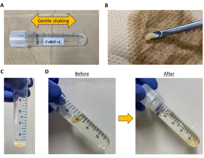
Figure 2: Images of the handling process. (A) The sample is immersed in 7 mL of 50% or 100% CUBIC-L in a 14 mL round bottom tube. The tube is kept horizontal with gentle shaking during the delipidation process. (B) A dispensing spoon is used for sample handling. (C) The amount of the staining buffer needed for one kidney is 500-600 µL. The tube is kept vertical during the staining process. (D) Although the sample floats in 50% CUBIC-R+ at the beginning of the RI matching, it sinks and becomes transparent in CUBIC-R+ at the end of the process. Please click here to view a larger version of this figure.
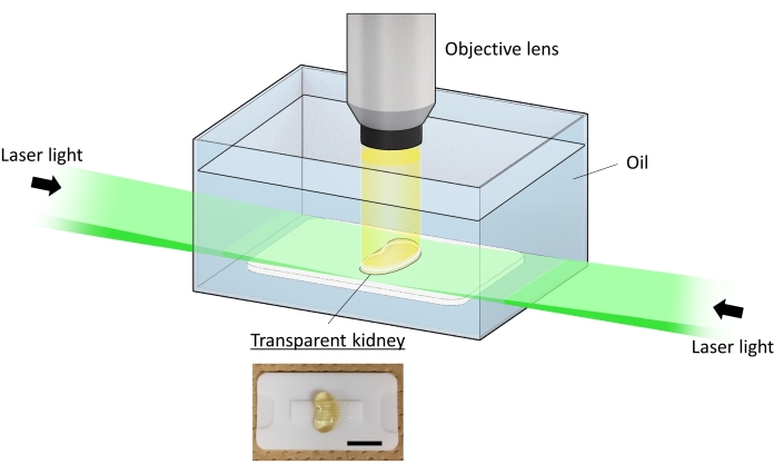
Figure 3: Light sheet fluorescent microscopy for whole-kidney 3D imaging. Light sheet fluorescent microscopy (LSFM) enables three-dimensional (3D) imaging of transparent samples. This image is reproduced from Hasegawa et al.9. Immersion oil is used during data acquisition. Light sheets from both sides illuminate the sample. The excitation signals are acquired through the objective lens located in a vertical position. The stage moves in the axial direction, and z-stack images are obtained. Scale bar = 10 mm. Please click here to view a larger version of this figure.

Figure 4. Whole-kidney three-dimensional imaging of sympathetic nerves and arteries. (A) Sympathetic nerves [anti-tyrosine hydroxylase (TH) antibody, green] and arteries [anti-α-smooth muscle actin (αSMA) antibody, magenta] are visualized in a whole kidney. (B) The enlarged x-y plane image of (A) is shown [anti-TH antibody, green; anti-αSMA antibody, magenta]. (C) Persistent denervation after renal ischemia-reperfusion injury (IRI) is visualized. (D) Sympathetic nerves in a spleen are also visualized in a whole spleen by the current protocol. This image is reproduced from Hasegawa et al.9. Please click here to view a larger version of this figure.
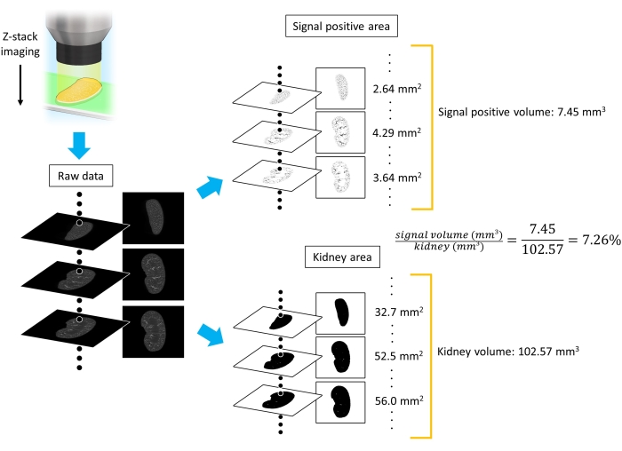
Figure 5: Quantification of three-dimensional immunofluorescent staining. The process of quantifying the ratio of signal-positive volume to total kidney volume is shown. Binary conversion is performed according to each threshold. The volumes are obtained by calculating the integral of the area of interest (voxel size: x = 6.45 µm, y = 6.45 µm, z = 7 µm). The image is reproduced from Hasegawa et al.9. Please click here to view a larger version of this figure.

Figure 6: Whole-kidney three-dimensional imaging of collecting ducts, proximal tubules, and glomeruli. (A) Collecting ducts [anti-aquaporin 2 (AQP2) antibody], S1 segment of proximal tubules [anti-sodium glucose cotransporter 2 (SGLT2) antibody], and glomeruli (anti-podocin antibody) are respectively visualized in a whole kidney. The image is reproduced from Hasegawa et al.9. (B) Glomerulomegaly is observed in the early stage of diabetic kidney disease (anti-podocin antibody), which is suppressed by drug administration. The image is reproduced from Hasegawa et al.11. Please click here to view a larger version of this figure.
Video 1: Rotating movie of the kidney. The rotating movie of the kidney in Figure 4A is shown. Sympathetic nerves [anti-tyrosine hydroxylase (TH) antibody, green] and arteries [anti-α-smooth muscle actin (αSMA) antibody, magenta] are visualized. Please click here to download this Video.
Discussion
The present protocol allowed whole-kidney 3D imaging of various structures such as sympathetic nerves, collecting ducts, arteries, proximal tubules, and glomeruli9,10,11. This staining method offered macroscopic observation and led to new biological discoveries, by visualizing the alteration in renal sympathetic nerves after ischemia-reperfusion injury9,10 and glomerulomegaly in the initial stage of diabetic kidney disease11.
Opacity is derived from optical inhomogeneity in a tissue5. The common principle of various tissue clearing methods is removing light scatterers/absorbers and fulfilling the space with RI-matching reagents. Compared to organic solvent-based methods such as 3D imaging of solvent-cleared organs (3DISCO/iDISCO)3,14, CUBIC is based on hydrophilic reagents, and thus is superior in fluorescent preservation5. This characteristic of CUBIC allows researchers to observe fluorescent signals from reporter mouse strains2 as well as signals from whole-mount immunofluorescent staining, providing more options for a comprehensive understanding of the 3D-organ structures.
The present CUBIC-based tissue clearing protocol can attain high transparency for 3D imaging compared with previous mouse kidney clearing research15. However, it must be noted that this clearing protocol is suitable for mouse kidneys. A stronger delipidation process is needed to clear human kidneys. For example, Zhao et al. recently presented an approach for intact human organ mapping by developing the efficient tissue clearing method named SHANEL (small-micelle-mediated human organ efficient clearing and labeling)16. As stronger delipidation damages samples, researchers need to choose an appropriate tissue clearing method according to their purpose.
Whole-mount organ staining is challenging because of the difficulty in antibody penetration; the present protocol has been established after trial and error. As the stability of staining is prioritized, the incubation time might be longer than the minimum required. As a result, this protocol has demonstrated high-quality whole-mount staining of the kidneys by the antibodies. However, there is a possibility that it may not work well, depending on the target antigens that we have not tried. A recent study has shown that two-step staining using primary and secondary antibodies like the current protocol is not optimal in some cases for the whole-mount staining of mouse brains17. This is because when the target amount is large, its primary antibody fills the sample, making it difficult for the secondary antibody to penetrate deep into the tissue17. Thus, the fluorescent-labeled primary antibody, or the complex of the primary antibody and secondary Fab fragment17, is worth trying if the penetration of antibodies is not efficient.
As for the imaging process, researchers need to choose an appropriate type of microscopy and image resolution according to their purpose. If researchers want to conduct whole-kidney 3D imaging as presented in this study, it is better to use light sheet fluorescent microscopy because it can quickly acquire wide z-stack images with a low level of photobleaching. As the file size of 3D imaging data is enormous, the image resolution should be reduced to the extent that whole-organ imaging can stand. In contrast, researchers can also use confocal or multiphoton microscopies when obtaining 3D imaging data with high resolution in narrow space.
In conclusion, the current study has developed whole-mount kidney staining for 3D imaging with the tissue clearing method CUBIC. The protocol allows visualization of 3D structures in a whole kidney, providing a macroscopic perspective for kidney research.
Disclosures
The authors have nothing to disclose.
Acknowledgements
Part of this work was conducted through collaboration with Prof. Hiroki R. Ueda (University of Tokyo), Prof. Etsuo A. Susaki (Juntendo University), Prof. Tetsuhiro Tanaka (Tohoku University), Prof. Masafumi Fukagawa, Dr. Takehiko Wada, and Dr. Hirotaka Komaba (Tokai University).
Materials
| Name | Company | Catalog Number | Comments |
|---|---|---|---|
| 14 mL Round Bottom High Clarity PP Test Tube | Falcon | 352059 | Tissue clearing, staining, wash |
| 2,3-dimethyl-1-phenyl-5-pyrazolone/antipyrine | Tokyo Chemical Industry | D1876 | CUBIC-R+ |
| 37%-Formaldehyde Solution | Nacalai Tesque | 16223-55 | Post fixation |
| 4%-Paraformaldehyde Phosphate Buffer Solution | Nacalai Tesque | 09154-85 | Kidney fixation |
| Alexa Flour 555-conjugated donkey anti-sheep IgG antibody | Invitrogen | A-21436 | Secondary antibody (1:100) |
| Alexa Flour 647-conjugated donkey anti-rabbit IgG antibody | Invitrogen | A-31573 | Secondary antibody (1:200) |
| Anti-aquaporin 2 (AQP2) antibody | Abcam | ab199975 | Primary antibody (1:100) |
| Anti-podocin antibody | Sigma-Aldrich | P0372 | Primary antibody (1:100) |
| Anti-sodium glucose cotransporter 2 (SGLT2) antibody | Abcam | ab85626 | Primary antibody (1:100) |
| Anti-tyrosine hydroxylase (TH) antibody | Abcam | ab113 | Primary antibody (1:100) |
| Anti-α-smooth muscle actin (α-SMA) antibody | Abcam | ab5694 | Primary antibody (1:200) |
| Blocker Casein in PBS | Thermo Fisher Scientific | 37528 | Staining buffer |
| Butorphanol Tartrate | Meiji | 005526 | Anesthetic |
| C57BL/6NJcl | Nippon Bio-Supp.Center | N/A | Mouse strain |
| Imaris | Bitplane | N/A | Imaging analysis software |
| Macro-zoom microscope | OLYMPUS | MVX10 | The observation unit of the custom-built microscope |
| Medetomidine Hydrochloride | Kyoritsu-Seiyaku | 008656 | Anesthetic |
| Midazolam | SANDOZ | 27803229 | Anesthetic |
| Mineral oil | Sigma-Aldrich | M8410 | Immersion oil |
| N-buthyldiethanolamine | Tokyo Chemical Industry | B0725 | CUBIC-L, CUBIC-R+ |
| Nicotinamide | Tokyo Chemical Industry | N0078 | CUBIC-R+ |
| Polyethylene glycol mono-p-isooctylphenyl ether/Triton X-100 | Nacalai Tesque | 12967-45 | CUBIC-L, PBST |
| Silicon oil HIVAC-F4 | Shin-Etsu Chemical | 50449832 | Immersion oil |
| Sodium azide | Wako | 195-11092 | Staining buffer |
References
- Velez-Fort, M., et al. The stimulus selectivity and connectivity of layer six principal cells reveals cortical microcircuits underlying visual processing. Neuron. 83 (6), 1431-1443 (2014).
- Susaki, E. A., et al. Whole-brain imaging with single-cell resolution using chemical cocktails and computational analysis. Cell. 157 (3), 726-739 (2014).
- Erturk, A., et al. Three-dimensional imaging of solvent-cleared organs using 3DISCO. Nature Protocols. 7 (11), 1983-1995 (2012).
- Tainaka, K., et al. Chemical Landscape for Tissue Clearing Based on Hydrophilic Reagents. Cell Reports. 24 (8), 2196-2210 (2018).
- Susaki, E. A., Ueda, H. R. Whole-body and whole-organ clearing and imaging techniques with single-cell resolution: toward organism-level systems biology in mammals. Cell Chemical Biology. 23 (1), 137-157 (2016).
- Keller, P. J., Dodt, H. U. Light sheet microscopy of living or cleared specimens. Current Opinion in Neurobiology. 22 (1), 138-143 (2012).
- Susaki, E. A., et al. Advanced CUBIC protocols for whole-brain and whole-body clearing and imaging. Nature Protocols. 10 (11), 1709-1727 (2015).
- Kubota, S. I., et al. Whole-body profiling of cancer metastasis with single-cell resolution. Cell Reports. 20 (1), 236-250 (2017).
- Hasegawa, S., et al. Comprehensive three-dimensional analysis (CUBIC-kidney) visualizes abnormal renal sympathetic nerves after ischemia/reperfusion injury. Kidney International. 96 (1), 129-138 (2019).
- Hasegawa, S., Inoue, T., Inagi, R. Neuroimmune interactions and kidney disease. Kidney Research and Clinical Practice. 38 (3), 282-294 (2019).
- Hasegawa, S., et al. The oral hypoxia-inducible factor prolyl hydroxylase inhibitor enarodustat counteracts alterations in renal energy metabolism in the early stages of diabetic kidney disease. Kidney International. 97 (5), 934-950 (2020).
- Gage, G. J., Kipke, D. R., Shain, W. Whole animal perfusion fixation for rodents. Journal of Visualized Experiments. (65), e3564(2012).
- Hasegawa, S., et al. Activation of sympathetic signaling in macrophages blocks systemic inflammation and protects against renal ischemia-reperfusion injury. Journal of the American Society of Nephrology. 32 (7), 1599-1615 (2021).
- Renier, N., et al. iDISCO: a simple, rapid method to immunolabel large tissue samples for volume imaging. Cell. 159 (4), 896-910 (2014).
- Klingberg, A., et al. Fully automated evaluation of total glomerular number and capillary tuft size in nephritic kidneys using lightsheet microscopy. Journal of the American Society of Nephrology. 28 (2), 452-459 (2017).
- Zhao, S., et al. Cellular and molecular probing of intact human organs. Cell. 180 (4), 796-812 (2020).
- Susaki, E. A., et al. Versatile whole-organ/body staining and imaging based on electrolyte-gel properties of biological tissues. Nature Communications. 11 (1), 1-22 (2020).
Reprints and Permissions
