Here, we describe the surgical procedure to perform Regenerative Peripheral Nerve Interface (RPNI) surgery for treating postamputation neuropathic pain in the context of an international, randomized controlled trial (RCT) (ClinicalTrials.gov, NCT05009394). The RCT compares RPNI with two other surgical techniques, namely, Targeted Muscle Reinnervation (TMR) and neuroma excision combined with intra-muscular transposition.
Method Article
Regenerative Peripheral Nerve Interface: Surgical Protocol for a Randomized Controlled Trial in Postamputation Pain
In This Article
Summary
Abstract
Surgical procedures, including nerve reconstruction and end-organ muscle reinnervation, have become more prominent in the prosthetic field over the past decade. Primarily developed to increase the functionality of prosthetic limbs, these surgical procedures have also been found to reduce postamputation neuropathic pain. Today, some of these procedures are performed more frequently for the management and prevention of postamputation pain than for prosthetic fitting, indicating a significant need for effective solutions to postamputation pain. One notable emerging procedure in this context is the Regenerative Peripheral Nerve Interface (RPNI). RPNI surgery involves an operative approach that entails splitting the nerve end longitudinally into its main fascicles and implanting these fascicles within free denervated and devascularized muscle grafts. The RPNI procedure takes a proactive stance in addressing freshly cut nerve endings, facilitating painful neuroma prevention and treatment by enabling the nerve to regenerate and innervate an end organ, i.e., the free muscle graft. Retrospective studies have shown RPNI's effectiveness in alleviating postamputation pain and preventing the formation of painful neuromas. The increasing frequency of utilization of this approach has also given rise to variations in the technique. This article aims to provide a step-by-step description of the RPNI procedure, which will serve as the standardized procedure employed in an international, randomized controlled trial (ClinicalTrials.gov, NCT05009394). In this trial, RPNI is compared to two other surgical procedures for postamputation pain management, specifically, Targeted Muscle Reinnervation (TMR) and neuroma excision coupled with intra-muscular transposition and burying.
Introduction
Chronic postamputation pain is a frequent occurrence following a major limb amputation1. Postamputation pain is a general term for unpleasant sensations that can manifest after amputation and is defined by the International Association for the Study of Pain as pain perceived in the residual limb (residual limb pain, RLP) or pain perceived in the missing extremity (phantom limb pain, PLP)2. The source of RLP is diverse and can stem from various causes, such as inflammation, infection, neuromas, heterotopic ossification, bursae, complex regional pain syndrome, and muscle and bone abnormalities3. For PLP, the precise origins remain incompletely elucidated, and it is thought to have a multifaceted etiology involving influences from both the peripheral and central nervous systems4,5.
When a peripheral nerve is injured, it will attempt to regenerate and reconnect with the relevant end organs6. In the situation of an amputation where the end organs are lost, abnormal sprouting of axons into surrounding scar tissue will take place and create a so-called neuroma7. The neuroma consists of a dense collagenous matrix where a disorganized tangle of axons, Schwann cells, endoneurial cells, and perineurial cells are trapped. Injured nociceptive fibers within the neuroma have a lower activation threshold, which results in the transmission of action potentials in the absence of external stimuli8. Furthermore, neuromas release inflammatory cytokines, which are associated with alterations in pain processing within the somatosensory cortex. This can lead to maladaptive changes in the central nervous system, further fueling the amplification and continuation of the neuropathic pain response9,10. There are complex and bidirectional interactions between the peripheral and central nervous systems that influence the chronification of pain. For example, individuals with sustained peripheral neuropathy might experience central sensitization, causing them to process new sensory input differently than those without chronic pain11. Painful neuromas are one of the known sources for both RLP and PLP; therefore, focusing on managing them is a crucial step toward minimizing the incidence and prevalence of postamputation pain.
Several surgical strategies have been suggested for the treatment and prevention of painful neuromas, and these strategies can generally be classified as reconstructive or non-reconstructive. Non-reconstructive methods commonly include excision of the neuroma without the intention for the severed nerve to reinnervate a physiologically appropriate target, such as nerve to bone or nerve to an already innervated muscle12. Whereas, reconstructive interventions are designed to facilitate the healthy, physiological regeneration of the donor nerve following neuroma excision. Several non-reconstructive methods include techniques such as nerve implantation within nearby tissue, nerve capping, applying proximal pressure, or using controlled thermal procedures on the distal nerve end13. One of the most common treatments is neuroma excision with implantation into nearby tissues such as muscle, bone, or vein14. However, according to neurophysiological principles, in all these aforementioned strategies, the freshly cut peripheral nerve will undergo axonal sprouting and elongation again. This process will likely result in the recurrence of the painful neuroma, as the regenerating axons will not have appropriate target end organs to reinnervate15. The outcomes of this technique have been diverse; some patients have experienced no, gradual, or full pain relief, while others have experienced pain relief shortly after surgical treatment but have developed pain after some time14,16. However, despite the fact that the technique has limited success in reducing pain, neuroma transposition with implantation and burying in muscle is still today widely used in amputation care and is, to a large extent, seen as the "gold standard" for surgical treatments of painful terminal neuromas12,17.
In the past decades, new developments in addressing painful neuromas have centered around a more proactive approach to treating the nerve ending after removing the neuroma, where the objective is to ensure the satisfaction of the nerve end and promote a more natural process of neuronal regeneration12,13. A novel intervention developed by Professor Paul Cederna's group at the University of Michigan, Ann Arbor, USA, is the regenerative peripheral nerve interface (RPNI). This technique includes excision of a neuroma, longitudinal dissection of the donor nerve stump into multiple fascicle groupings, and thereafter direct implantation of the fascicles into free denervated skeletal muscle grafts18,19. The implantation in devascularized, denervated muscle allows the nerve fascicles to reinnervate the free muscle graft after it revascularizes within its implanted wound bed20. Histological work has shown the innervation of free muscle grafts of different volumes; however, their viability and function have optimal dimensions21. Once the engrafted free muscle graft becomes revascularized and reinnervated, the RPNI thereby prevents the recurrence of painful neuromas. The procedure has been adopted in several clinics, mainly in the USA but also in places within Europe and Asia. However, this has given rise to variations in the procedure. Therefore, in this article, we propose a consensus on the technique among surgeons worldwide who practice it.
This article describes the step-by-step protocol for RPNI surgery, which is used in an international RCT (ClinicalTrials.gov, NCT05009394). The aim of this RCT is to assess the effectiveness of the two most used reconstructive techniques, RPNI and TMR, compared to the most used standard surgical treatment22. The purpose of this method paper is to standardize the technique for the centers involved in the RCT and to make the procedure available for everyone who would like to use it in amputation management.
Protocol
The RCT was approved in Sweden and Italy by the Swedish Ethics Review Authority, Etikprövningsmyndigheten, on 30 June 2021 with the application number 2021-0234622 and the Ethical Committee in the Region of Emilia Romagna, respectively. Further details on the RCT are outlined in the protocol22.
1. Presurgical preparations
- Diagnose the painful neuroma(s) following the international RCT protocol22.
- Plan the skin incisions depending on the results from step 1.1 and depending on the location of each painful neuroma.
NOTE: In principle, any skeletal muscle can be used; however, the muscle graft is most often harvested from the vastus lateralis muscle in the thigh.
2. Preparation of the recipient site
- Perform either regional or general anesthesia.
NOTE: The type of anesthesia varies depending on the site of the procedure. - Place the patient in a supine or prone position, depending on the site of the painful neuroma. For painful neuromas in the upper extremity, place the arm on a surgical arm board.
- Carry out the length and shape of the skin incision depending on the location of the painful neuroma.
- Identify the nerve with the painful neuroma under blunt dissection.
- Gently isolate the nerve and the neuroma with fine instruments. Use loupe magnification if needed.
NOTE: Isolation of the neuroma is optional when challenging. - Mobilize the nerve and transect the neuroma up to healthy neural fascicles using a commercial nerve cutting/preparation set.
NOTE: Resection of the neuroma is optional when challenging. - Make longitudinal intraneural dissections from the distal end of the nerve for about 2-3 cm with straight microscissors. The number of fascicles will depend on the amputation level and the size of the nerve. Ensure that the diameter of each fascicle is a maximum of 4-6 mm. For each nerve, a number of neural fascicles that should be prepared are suggested in Table 1.
| Amputation level | Nerve | Suggested number of neural fascicles |
| Shoulder disarticulation | Axillary | 2 |
| Musculocutaneous | 2 | |
| Median | 3 | |
| Ulnar | 2 | |
| Radial | 2 | |
| Transhumeral | Axillary | 2 |
| Musculocutaneous | 2 | |
| Median | 3 | |
| Ulnar | 3 | |
| Radial | 2 | |
| Transradial | Median | 3 |
| Ulnar | 2 | |
| Radial | 1* | |
| Hip disarticulation | Sciatic | 4 |
| Femoral | 3 | |
| Lateral Cutaneous Femoral | 1* | |
| Obturator | 1* | |
| Posterior Cutaneous Thigh | 2 | |
| Transfemoral | Sciatic | 3 |
| Tibial | 2 | |
| Deep peroneal | 1* | |
| Superficial peroneal | 1* | |
| Sural | 1* | |
| Saphenous | 1* | |
| Transtibial | Tibial | 2 |
| Deep peroneal | 2 | |
| Superficial peroneal | 1* | |
| Sural | 1* | |
| Saphenous | 1* | |
| * Use the nerve entirely | ||
Table 1: Suggested number of neural fascicles specified for each nerve at a specific amputation level.
3. Preparation of the donor site
- Identify a healthy native donor muscle as a source for obtaining a free muscle graft. Harvest the muscle graft as follows:
- Ensure that each graft has the dimensions 3 cm (length) x 1.5 cm (width) x 0.5 cm (thickness).
NOTE: The main axis should be parallel to the muscle fibers. - Dissect the muscle graft using delicate dissecting scissors following the main axis of the muscle fibers. Use magnifying loupes as needed.
- Gently excise adipose tissue and the muscle fascia from the muscle graft using dissecting scissors.
- Ensure that each graft has the dimensions 3 cm (length) x 1.5 cm (width) x 0.5 cm (thickness).
- Keep the harvested muscle in a moist gauze with 0.9% NaCl sterile until utilization in section 4.
- Repeat steps 3.1-3.2 for each muscle graft, based on the number of nerves and corresponding nerve fascicles to be wrapped with a prepared free muscle graft for an RPNI construct.
4. RPNI construct fabrication
- Expose the nerve, already isolated and divided in fascicles.
- Place the fascicle on the muscle graft so that the distal stump falls in the central or proximal third of the muscle graft while having its orientation parallel to the muscle fibers.
NOTE: A parallel orientation is preferred to optimize successful axonal reinnervation. - Secure the nerve stump in the center of the longitudinal axis of the muscle graft using 6-0 non-resorbable monofilament sutures. Add one more stitch to secure the nerve at the proximal edge of the muscle graft.
NOTE: Do not suture the nerve end into the muscle graft; the transected portion of the nerve remains free. - Fold the muscle graft around the fascicle and secure it with a 6-0 interrupted or continuous non-resorbable monofilament suture.
- Repeat steps 4.1-4.4 for each fascicle.
- Perform blunt dissection in the residual limb to provide a protected area where each RPNI can lie comfortably and out of weight-bearing surfaces of the limb. Where possible, offset the location of each RPNI in series.
- Close the surgical wounds in layers.
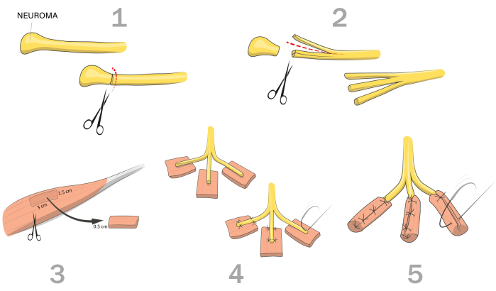
Figure 1: Schematic illustration of the Regenerative Peripheral Nerve Interface (RPNI) procedure. 1) Identify and isolate the nerve with the painful neuroma. Mobilize the nerve and transect the neuroma up to healthy neural fascicles; 2) Perform longitudinal intraneural dissections from the distal end of the nerve. The number of neural fascicles depends on amputation level and the size of the nerve; 3) Identify a healthy native donor muscle and harvest a muscle graft with the dimensions: 3 cm (length) x 1.5 cm (width) x 0.5 cm (thickness); 4) Place the fascicle in the muscle graft so the stump falls in the center of the muscle graft while having its orientation parallel to the muscle fibers. Secure the nerve within the muscle graft with sutures proximally and distally of the nerve stump; 5) Fold the muscle graft around the fascicle and secure. Please click here to view a larger version of this figure.
Results
The RPNI procedure has found application in treating postamputation pain in both upper and lower extremities (Table 2), as well as serving as a method for both treatment and prevention of painful neuroma development (Table 3)23,24. In a pilot study published in 2016, 16 patients were treated with RPNIs and were followed up for an average of 7.5months (range 3-15 months) post treatment23. The data was collected retrospectively between 2013 and 2016, and 71% of the patients reported a reduction in neuroma pain and 53% reduction in PLP. There was a statistically significant difference in the pain score pre and post-RPNI for both neuroma pain and PLP. In addition to pain relief, patients reported decreased (56%) or stable (44%) use of analgesics and a significant decrease in pain interference pre and post-RPNI. Surgical complications were reported in 5 patients, including wound healing delay, acute limb ischemia, deep venous thrombosis, hematoma, and neuroma at different sites23.
The procedure has also been used for treating painful neuromas after a partial hand or digit amputation25. In a study with retrospective data collection between 2014 and 2019, 30 RPNIs were performed in 14 patients. The patients were, on average, followed up for 37 weeks (range 6-128 weeks) after RPNI surgery, and 85% of the patients reported full pain relief or considerable pain reduction at the last follow-up visit. In terms of surgical complications, it is worth noting that 2 patients required two separate RPNI surgeries, and an additional 2 patients experienced infections following RPNI procedures. Also, there were no reported cases of delayed wound healing either on the volar aspect of the digit or at the muscle graft donor site. Furthermore, no occurrences of flexion contractures or difficulties with tendon gliding were identified after RPNI surgery25.
| Study | Study design | Amputation level (number of limbs) | RLP/NP reduction (%, p-value) | PLP reduction (%, p-value) |
| Woo et al. 201621 | Retrospective | Upper limb = 3 Lower limb = 14 | 71 %, p = 0.000001 | 53%, p = 0.009 |
| Hooper et al. 202023 | Retrospective | Upper limb = 17 | 85% | N/A |
| Lee et al. 202324 | Retrospective | 37 (amputation level not specified) | 77% | 61% |
| RLP, Residual limb pain; NP, neuroma pain; PLP, Phantom limb pain | ||||
Table 2: Studies investigating the effect of regenerative peripheral nerve interface (RPNI) as a treatment for postamputation pain in secondary amputations. High values of residual limb pain (RLP), neuroma pain (NP), and phantom limb pain (PLP) reduction indicate higher efficacy of RPNI as a treatment of postamputation pain.
Long-term data on the RPNI procedure as treatment and prevention of neuromas have been presented in a recently published abstract26. This data was retrospectively collected between 2014 and 2021, where 37 patients received RPNI for existing postamputation pain, and 40 patients received RPNI at the time of amputation. All patients had at least 1 year follow-up after RPNI, and the average time between surgery and the latest follow-up visit was 4.2 years. In the treatment group, upon the most recent follow-up, positive outcomes such as no reported symptoms or improved symptoms were observed in 77% of patients for neuroma pain and 61% for PLP. For the prophylactic group, 97% of the patients had no reported neuroma pain or PLP at the latest follow-up visit. Moreover, the prescriptions and consumables of opioids pre-surgery and at the latest follow-up visit were lower for both groups26.
Furthermore, two studies have presented retrospective data in which RPNI was conducted as a prophylactic measure against the development of painful neuromas at the time of amputation24,27. In the research conducted by Kubiak et al., a total of 90 patients were included, with 45 patients undergoing RPNI concurrently with primary amputation, while the remaining 45 received standard treatment. In the RPNI group, none of the patients developed painful neuromas, while in the control group, six patients experienced this condition. Furthermore, in the RPNI group, 23 patients developed PLP, as opposed to 41 patients in the control group. A higher rate of postoperative complications was reported in the control group (55.6%) compared to the treatment group (31.1%). These complications included both minor issues like delayed wound healing, surgical-site infection, and hematoma, as well as major complications such as deep infection requiring operating room (OR) washout, wound dehiscence necessitating OR closure, and delayed wound healing requiring OR debridement24. In a study conducted by Lin et al., RPNI was carried out during lower limb amputation for seven patients, and their outcomes were compared to those of seven patients who underwent traditional amputation. These patients were followed up at the 3-month and 6-month post-surgery marks. The study revealed significantly lower neuroma pain scores in the RPNI group compared to the traditional amputation group at both follow-up visits27.
| Study | Study design | Amputation level (number of limbs) | RLP/NP incidence (%, p-value) | PLP incidence (%, p-value) |
| Kubiak et al. 201922 | Retrospective | RPNI group Upper limb = 6 Lower limb = 46 Control group Upper limb = 48 Lower limb = 4 | RPNI group 0 % Control group 13.3 % p = 0.026 | RPNI group 51.1 % Control group 91.1 % p < 0.0001 |
| Lin et al. 202325 | Retrospective | RPNI group Lower limb = 7 Control group Lower limb = 7 | RPNI group 31 % Control group 69 % | N/A |
| Lee et al. 202324 | Retrospective | 40 (amputation level not specified) | 3% | 3% |
| RLP, Residual limb pain; NP, neuroma pain; PLP, Phantom limb pain | ||||
Table 3: Studies examining regenerative peripheral nerve interface (RPNI) as a prophylactic treatment for preventing postamputation pain at the time of primary amputation. Low percentage values of residual limb pain (RLP), neuroma pain (NP), and phantom limb pain (PLP) incidence indicate higher efficacy of RPNI as a prevention treatment.
A crucial point to note is the absence of reported prospective data on RPNI outcomes for pain relief28. This gap in information serves as one of the primary motivations behind conducting this RCT and developing this protocol. Furthermore, it's worth highlighting that most published studies have follow-up periods of less than 1 year, and there is significant variation in follow-up times within the same study population and across different studies.
Discussion
RPNIs have demonstrated their potential to serve as a treatment for postamputation pain as well as prevent the development of painful neuromas. The fundamental distinction between the RPNI procedure and alternative approaches to managing neuromas, such as nerve capping, applying proximal pressure, or employing thermal procedures on the distal nerve, lies in the primary goal of the severed nerve reinnervating a physiologically appropriate end organ. Additionally, an important contrast between RPNI and techniques like neuroma transposition and muscle implantation and burying, where the nerve's end target is also appropriate, is the use of denervated muscle targets. In cases where the muscle target is already innervated, each muscle fiber is already in physiological contact and occupied by a nerve fiber. This means that the freshly cut nerve cannot reinnervate the muscle and will thereby more likely redevelop a painful neuroma. Furthermore, in comparison to TMR surgery, where the freshly cut nerve end is coapted to a nearby expendable motor nerve and its accompanying motor end units of a target muscle, both techniques utilize a denervated target muscle. However, a distinction lies in the fact that RPNI employs a non-vascularized muscle graft, whereas in TMR, the nerve reinnervates a vascularized muscle. Furthermore, there are two other important differences with TMR related to the sizable caliber mismatch between donor and recipient nerves and the need to sacrifice otherwise healthy innervations. The size mismatch between donor and recipient nerves can potentially result in a neuroma-in-continuity, and the sacrificed nerves might develop painful neuromas. Moreover, the TMR procedure could be considered more complex than RPNI, as it incorporates techniques such as nerve transfers and coaptation. Whereas RPNI requires a longitudinal dissection to separate the never fascicles, the rest of the steps can be performed by a broader range of surgeons, including orthopedic surgeons, general surgeons, and others involved in amputations, rather than exclusively requiring the expertise of nerve surgeons, microsurgeons, or hand surgeons. Furthermore, there have been several combinations of both RPNI and TMR using key concepts of each technique. For example, nerve-to-nerve coaptation, including free muscle graft wrapping over the coaptation29 or splitting the nerve in two and performing coaptation with one part and RPNI constructs with the other30.
The procedure involves critical steps that must be carefully considered to ensure successful outcomes. Firstly, the muscle graft harvesting process should align with the muscle fiber axis to prevent disruption of individual muscle fibers, and the muscle graft should be trimmed off all connective tissue to optimize regeneration. The choice of the harvest site may vary depending on availability. In primary amputations, we recommend using the amputated part when possible. For transradial amputations, the brachioradialis muscle is a suitable donor site, while for transhumeral amputations, the triceps muscles can be utilized. In the case of lower extremity amputations, such as transradial and transfemoral, the ipsilateral proximal thigh, typically the vastus lateralis, serves as a suitable harvesting site. Furthermore, for transfemoral amputations, the sartorius and gracilis muscles are also viable donor options18. However, these mentioned harvest sites for each amputation level should be seen as recommendations. In RPNI surgery for pain relief, when the amputated part is not available, the harvest site could be from any of the aforementioned sites independently of the amputation level.
Moreover, it's vital to consider the ratio between the nerve stump and the muscle graft. Grafts that are excessively thick are susceptible to central necrosis, and grafts that are too thin or insufficiently denervated muscle fibers will result in neuroma formation within the RPNI construct. In this protocol, we recommend that the nerve stump is a maximum of 4-6 mm thick in diameter for a muscle graft with dimensions of 3 cm long, 1.5 cm wide, and 0.5 cm thick. The dimensions can be adjusted based on the nerve's thickness; for nerves with a diameter up 10 mm, the width of the nerve graft can be up to approximately 2 cm, but it should still facilitate complete wrapping of the nerve, extending at least 1 cm proximal to its end18. The nerve's circumference should be covered without causing any tension while also maintaining sufficient thinness to enable revascularization. In cases of thick nerves, such as the sciatic nerve, we recommend fascicular dissection, creating several RPNIs instead of creating one large RPNI (see Table 1).
The RPNI surgery is an easy, safe, straightforward, and reliable treatment; however, the technique has its drawbacks when compared to the conventional treatment. As previously documented in the literature by Dellon et al., this method involves additional surgical steps, necessitating the use of more Current Procedural Terminology (CPT) codes, such as incorporating a muscle graft. This, in turn, results in increased time needed in the surgical theater and thereby increased surgical expenses31. The additional surgical time of performing RPNI or TMR is highly dependent on the amputation level and the number of constructs. However, despite the associated increase in expenses, several vital long-term considerations come into play. Individuals experiencing chronic pain following amputation require continuous pain management, encompassing medication, rehabilitation, and specialized interventions. Additionally, postamputation pain often leads to heightened healthcare utilization, involving frequent visits to healthcare providers, emergency room trips, and hospital admissions. Surgical interventions like RPNI or TMR, designed to treat postamputation pain, have the potential to significantly extend the lifespan, promote mobility, gainful employment, and enhance the overall quality of life for individuals with postamputation pain. By alleviating suffering, facilitating improved functional outcomes, and fostering psychological well-being, these interventions offer invaluable benefits that extend far beyond mere financial considerations.
In addition to their role in neuroma management, RPNIs have also been employed in patients with limb loss to enhance motor and sensory prosthetic function30,32,33,34. By providing a stable and responsive interface between the residual nerve and prosthetic technology, RPNIs enable individuals with limb loss to achieve more natural and precise control over their prosthetic limbs. This advancement has the potential to greatly enhance their mobility, dexterity, and quality of life30. As a result, RPNIs represent a multifaceted approach that not only manages neuroma-related issues but also offers promising solutions for the broader needs of individuals with amputation, further underscoring their significance in the field of amputation rehabilitation.
Disclosures
The authors have no disclosures.
Acknowledgements
The authors would like to thank the funders of this project: Promobilia Foundation, the IngaBritt and Arne Lundbergs Foundation, and the Swedish Research Council (Vetenskapsrådet). The authors sincerely thank those who donated their bodies to science so that anatomical research could be performed. Results from such research can potentially improve patient care and increase mankind’s overall knowledge. Therefore, these donors and their families deserve our highest gratitude. Furthermore, the authors acknowledge Prof. Lucia Manzoli and Prof. Stefano Ratti for the precious collaboration of the Anatomy Center, Alma Mater Studiorum-University of Bologna to this project. Lastly, special recognition is extended to Carlo Piovani for his contribution to creating the illustrations.
Materials
| Name | Company | Catalog Number | Comments |
|---|---|---|---|
| 0.9% NaCl sterile solution | Thermo Fisher Scientific | Z1376 | The company and the catalog number is one example. |
| 6-0 Ethilon suture | Ethicon | 660H | The company and the catalog number is one example. |
| Dissecting scissors | Stille | 101-8172-23 | The company and the catalog number is one example. |
| Gauze | Mölnlycke | 152040 | The company and the catalog number is one example. |
| Loupes | Zeiss | Various | User can choose loupes according to personal preferences. |
| Nerve cutting set | Checkpoint Surgical | 9250 | The company and the catalog number is one example. |
| Straight microscissors | S&T | SAS-12 R-7 | The company and the catalog number is one example. |
References
- Hsu, E., Cohen, S. P. Postamputation pain: Epidemiology, mechanisms, and treatment. J Pain Res. 6, 121-136 (2013).
- Schug, S. A., Lavand, P., Barke, A., Korwisi, B., Rief, W. The IASP classification of chronic pain for ICD-11 chronic postsurgical or posttraumatic pain. Pain. 160 (1), 45-52 (2019).
- Davis, R. W. Phantom sensation, phantom pain, and stump pain. Arch Phys Med Rehabil. 74 (1), 79-91 (1993).
- Ortiz-Catalan, M. The stochastic entanglement and phantom motor execution hypotheses: A theoretical framework for the origin and treatment of Phantom limb pain. Front Neurol. 9, 748(2018).
- Flor, H. Phantom-limb pain: Characteristics, causes, and treatment. Lancet Neurol. 1 (3), 182-189 (2002).
- Rotshenker, S. Traumatic Injury to Peripheral Nerves. Nerves and Nerve Injuries. , Elsevier, Academic Press. (2015).
- Stokvis, A., Der Avoort, D. J. J. C., Van Neck, J. W., Hovius, S. E. R., Coert, J. H. Surgical management of neuroma pain: A prospective follow-up study. Pain. 151 (3), 862-869 (2010).
- Curtin, C., Carroll, I. Cutaneous neuroma physiology and its relationship to chronic pain. J Hand Surg Am. 34 (7), 1334-1336 (2009).
- Khan, J., Noboru, N., Young, A., Thomas, D. Pro and anti-inflammatory cytokine levels (TNF-α, IL-1β, IL-6 and IL-10) in rat model of neuroma. Pathophysiology. 24 (3), 155-159 (2017).
- Clark, A. K., Old, E. A., Malcangio, M. Neuropathic pain and cytokines: current perspectives. J Pain Res. 6, 803(2013).
- Costigan, M., Scholz, J., Woolf, C. J. Neuropathic pain: A maladaptive response of the nervous system to damage. Annu Rev Neurosci. 32, 1-32 (2009).
- Eberlin, K. R., Ducic, I. Surgical algorithm for neuroma management: A changing treatment paradigm. Plast Reconstr Surg Glob Open. 6 (10), e1952(2018).
- Ives, G. C., et al. Current state of the surgical treatment of terminal neuromas. Neurosurgery. 83 (3), 354-364 (2018).
- Dellon, A. L., Mackinnon, S. E. Treatment of the painful neuroma by neuroma resection and muscle implantation. Plast Reconstr Surg. 77, 427-438 (1986).
- Neumeister, M. W., Winters, J. N. Neuroma. Clin Plast Surg. 47 (2), 279-283 (2020).
- Guse, D. M., Moran, S. L. Outcomes of the surgical treatment of peripheral neuromas of the hand and forearm: A 25-year comparative outcome study. Ann Plast Surg. 71 (6), 654-658 (2013).
- Eftekari, S. C., Nicksic, P. J., Seitz, A. J., Donnelly, D. T., Dingle, A. M., Poore, S. O. Management of symptomatic neuromas: a narrative review of the most common surgical treatment modalities in amputees. Plast and Aesthet Res. 9 (7), 43(2022).
- Dean, R. A., Tsai, C., Chiarappa, F. E., Cederna, P. S., Kung, T. A., Reid, C. M. Regenerative peripheral nerve interface surgery: Anatomic and technical guide. Plast Reconstr Surg Glob Open. 11 (7), 5127(2023).
- Santosa, K. B., Oliver, J. D., Cederna, P. S., Kung, T. A. Regenerative peripheral nerve interfaces for prevention and management of neuromas. Clin Plast Surg. 47 (2), 311-321 (2020).
- Cedars, M. G. M. D., Miller, T. A. M. D. A review of free muscle grafting. Plast Reconstr Surg. 74 (5), 712-720 (1984).
- Hu, Y., et al. Regenerative peripheral nerve interface free muscle graft mass and function. Muscle Nerve. 63 (3), 421-429 (2021).
- Pettersen, E., et al. Surgical treatments for postamputation pain study protocol for an international , double - blind, randomised controlled trial. Trials. 24 (1), 304(2023).
- Woo, S. L., Kung, T. A., Brown, D. L., Leonard, J. A., Kelly, B. M., Cederna, P. S. Regenerative peripheral nerve interfaces for the treatment of postamputation neuroma pain: A pilot study. Plast Reconstr Surg Glob Open. 4 (12), e1038(2016).
- Kubiak, C. A., Kemp, S. W. P., Cederna, P. S., Kung, T. A. Prophylactic regenerative peripheral nerve interfaces to prevent postamputation pain. Plast Reconstr Surg. 144 (3), 421-430 (2019).
- Hooper, R. C., et al. Regenerative peripheral nerve interfaces for the management of symptomatic hand and digital neuromas. Plast Reconstr Surg Glob Open. 8 (6), e2792(2020).
- Lee, J. C., Kumar, N. G., Kemp, S. W. P., Cederna, P. S., Kung, T. A. S. P. 06 Regenerative peripheral nerve interface surgery and its four-year pain and medication intake outcomes for treatment or prevention of postamputation pain. Plast Reconstr Surg Glob Open. 11, 123-123 (2023).
- Lin, Z., Yu, P., Chen, Z., Li, G. Regenerative peripheral nerve interface reduces the incidence of neuroma in the lower limbs after amputation: a retrospective study based on ultrasound. J Orthop Surg Res. 18 (1), 619(2023).
- Mauch, J. T., Kao, D. S. Targeted muscle reinnervation and regenerative peripheral nerve interfaces for pain prophylaxis and treatment A systematic review. PM R. 15 (11), 1457-1465 (2023).
- Kurlander, D. E., et al. TMRpni: Combining two peripheral nerve management techniques. Plast Reconstr Surg Glob Open. 8 (10), e3132(2020).
- Zbinden, J., et al. Improved control of a prosthetic limb by surgically creating electro-neuromuscular constructs with implanted electrodes. Sci Transl Med. 15 (704), 3665(2023).
- Dellon, A. L., Aszmann, O. C. In musculus, veritas? Nerve "in muscle" versus targeted muscle reinnervation versus regenerative peripheral nerve interface: Historical review. Microsurgery. 40 (4), 516-522 (2020).
- Vu, P. P., et al. A regenerative peripheral nerve interface allows real-time control of an artificial hand in upper limb amputees. Sci Transl Med. 12 (533), 2857(2020).
- Vu, P. P., et al. Long-term upper-extremity prosthetic control using regenerative peripheral nerve interfaces and implanted EMG electrodes. J Neural Eng. 20 (2), 026039(2023).
- Ortiz-Catalan, M., et al. A higly integrated bionic hand with neural control and feedback for use in daily life. Sci Robot. 8 (83), (2023).
Reprints and Permissions
