This protocol describes the best practices for germ-free mouse transfer to and housing in experimental single-cage isolators (isocages) while maintaining sterile conditions. Methods for fecal transplant into germ-free mice and the collection of viable bacteria from these gut "humanized" mice for further applications are discussed.
A subscription to JoVE is required to view this content. Sign in or start your free trial.
Method Article
Preparation and Maintenance of Bioexclusion IsoPositive Cage Experiment for Human Fecal Transplantation into Germ-Free Mice
In This Article
Summary
Abstract
Germ-free mice are an important investigation tool for understanding the contribution of microorganisms in host health and disease, enabling assessment of the specific role of individuals, defined or complex groups of microorganisms in host response. Traditionally bred and reared in flexible-film or semi-rigid isolators, germ-free mouse husbandry and experimental manipulation are costly and require numerous trained staff and a large space footprint in animal housing facilities. The IsoPositive caging system allows for experimental manipulation of germ-free mice in individual, hermetically-sealed, positive-pressure isolator cages (isocages), reducing cost and enabling greater flexibility in experimental manipulations.
Here, a protocol is described for transferring germ-free mice from breeding isolators to isocages and subsequent fecal transfer from human donor stool into mice to create stable long-term gut "humanized" mice for future studies. The materials and preparation needed for the utilization of the isocage system are described, including the use of chlorine-dioxide sterilant chemical sterilant to clean cages, supplies, equipment, and personal protective equipment. The methods for confirming the germ-free status of transferred mice and how to determine contamination in the caging system are discussed. The procedure for husbandry, including bedding, food, and water supply, is further discussed. The protocol for human fecal slurry preparation and gavage into germ-free mice to create gut "humanized" mice, along with stool collection to monitor the microbial community composition of these mice, are described. An experiment illustrates that two weeks post-human fecal transplant allows for stable colonization of donor microbiota in the murine hosts, enabling subsequent experimental usage. Furthermore, the collection of humanized mouse feces in viability preservation media, enabling use in further functional experiments, is described. Overall, these methods allow for the safe and effective establishment of humanized mouse communities in experimental gnotobiotic cages for further manipulation.
Introduction
Germ-free mice are an essential tool in the repertoire of microbiome researchers, allowing one to dissect the contribution of the microbiota in host health and disease states. Germ-free mice are born completely sterile and remain axenic for their entire lives1. Colonization of germ-free mice with specific bacterial strains enables causative studies between those taxa and metabolic, immune, or other host functions2,3,4,5. Particularly advantageous is the ability to "humanize" germ-free mice at the level of the microbiota by transplanting feces obtained from human donors and, when housed in barrier conditions, prevent contamination from murine-derived microorganisms1. This approach has enabled many important discoveries in the field of microbiome, for instance, the effect of the human gut microbiome on cancer immunotherapy response6,7,8.
However, while humanized germ-free mice are invaluable to research efforts in the microbiome field, there are many limitations that have inhibited the wider adaptation of this approach. Germ-free mice are bred and maintained in semi-rigid or flexible-film large isolators, but functional experiments require separate mini-isolators to be set up, with one mini-isolator housing several cages but only under one experimental condition. This mini-isolator approach increases the space footprint and cost while severely limiting the number of experimental conditions that can be investigated in an experiment and the number of experiments that can be run in parallel. A promising solution is using an individual, modular caging system called the ISOcage P Bioexclusion System (here referred to as isocage system)9,10. The isocage system allows for experimental manipulation of germ-free mice in individual, hermetically-sealed, positive-pressure isolator cages, enabling separate experimental conditions between each cage rather than between each mini-isolator. With the proper aseptic technique, animals can be housed in isocages for up to 12 weeks under germ-free conditions or humanized by human fecal transplant for use in any compatible experimental approach (i.e., can be performed under aseptic conditions). Multiple independent experiments can be run in parallel using the isocage system, and the space footprint and cost are dramatically less than running multiple experiments across mini-isolators.
The purpose of breeding germ-free mice in flexible film breeding isolators is to carefully preserve axenic status11. Techniques used to monitor germ-free status include routine swabs of mouse body surfaces and oral cavities, as well as the aseptic collection of fecal samples, which are both cultured and tested by PCR-based commercial assays. Bacterial, serological, and fungal testing of these samples are all required to determine germ-free status11. When germ-free mice are transferred from breeding isolators to isocages for experimental usage, the mice are swabbed and tested to validate their germ-free status upon transfer. Isocage sterility checks are performed through aseptic collection of fecal samples, which are then cultured for detection of bacterial, viral, and fungal contaminants. Carefully collecting and recording the results of these sterility checks from birth to the end of an experimental protocol is necessary to validate the germ-free status of these mice.
The isocage system is composed of individual cages (Figure 1), transfer disks for transport out of breeding isolators (Figure 1), and the isocage rack, which houses the cages (Figure 2). Each isocage contains a cage-level high-efficiency particulate air (HEPA) filter installed on the supply air intake and a silicone gasket which makes an airtight seal when closed, ensuring no contaminants can enter the cage through the air (Figure 1A). This cage lid can be used as a sterile working surface when placed upside down within a sterilized biosafety cabinet (Figure 1A). A wire rack within the cage holds the food and water bottle (Figure 1B). Forceps autoclaved within the cage are used for all manipulations that require contact with interior cage surfaces. The cage itself has notches for a removable cage card holder to identify animals on the outside and air intake and export nozzles that dock into the isocage rack (Figure 1C-E). Safe closure clamps and a tab lock on the lid seal the cage when it is ready to be redocked on the rack system (Figure 1F). The suggested bedding is Alpha-dri, and an autoclavable enrichment hut is also recommended (Figure 1F). Transfer disks are used to move germ-free mice from breeding isolators to the isocages and contain a rotatable compartment lid with a triangular opening to allow for manipulation of animals (Figure 1G-H). Disks come in sizes small (21.6 cm diameter) and large (28 cm diameter), both of which have a capacity of eight mice. Autoclaved tape is used to create airtight seals on the circumference and air holes of the disk, which is performed prior to soaking with sterilant and transport in a sterilant-soaked bag (Figure 1I). The rack system itself has a screen to monitor the air blowers, rack-level HEPA filter status, and emergency battery power for the rack, which are all included features of the system (Figure 2A). An enclosed Magnehelic gauge displays the positive pressure maintained by the cage system, and an automatic visual docking indicator shows the docking status of the cages (yellow tab out means no cage is docked, or the dock was unsuccessful) (Figure 2B-D). Also necessary for the manipulation of isocages is a standard certified biosafety cabinet.
The protocol presented here describes the proper methods for the successful transfer of germ-free mice from breeding isolators under aseptic conditions to the isocages while maintaining germ-free status, the humanization of germ-free mice with human donor fecal slurry, and the collection of feces from mice housed in the isocage for either confirmation of germ-free status or viability preservation for further functional studies. In this example, germ-free mice are humanized with pooled fecal specimens from human subjects treated with immunotherapy for lung cancer and dichotomized as responders or non-responders to therapy. In this instance, the response phenotype to immunotherapy response was transferred by the gut microbiota humanization to the recipient mice, who could then be further inoculated with tumor cells and treated with immunotherapy. The human fecal slurry protocol can be readily adapted to any human donor feces or any disease preclinical model that the investigator wishes. Using this protocol, it is possible to transfer any human fecal donor microbiota into the germ-free host, enabling further investigation into the role of microbiota in health and disease.

Figure 1: Schematic diagram of isocage and transfer disks. (A) Top-down view of the underside of the cage lid, with labels indicating the location of the internal cage-level HEPA filter and the silicone gasket seal. (B) Top-down view of the interior of the cage, with labels indicating the wire bar lid, the internal water bottle, and spout, and the location in the wire rack to hold autoclavable chow. (C) Front view of cage showing notches for the cage card holder. (D) Top-down view of a full cage with the lid on top, showing how the HEPA filter is installed on the air intake nozzle. (E). Rear view of cage showing air intake and export nozzles which dock to the isocage rack system. (F) Lateral view of a full cage with the lid on top, with labels indicating the safe closure clamps in the open position, with white tabs on each clamp that lock them in place. The interior of the cage shows Alpha-dri bedding layered at the bottom and suggested enrichment hut placed in bedding. (G) Top-down view of transfer disks with lid on top. (H) Top down view of the interior of the transfer disk, showing the rotatable compartment lid with a triangular opening to allow for manipulation of animals. (I) Lateral view of fully assembled transfer disk showing placement of autoclaved tape, which creates an airtight seal during transfer from breeding isolator to isocage. Please click here to view a larger version of this figure.
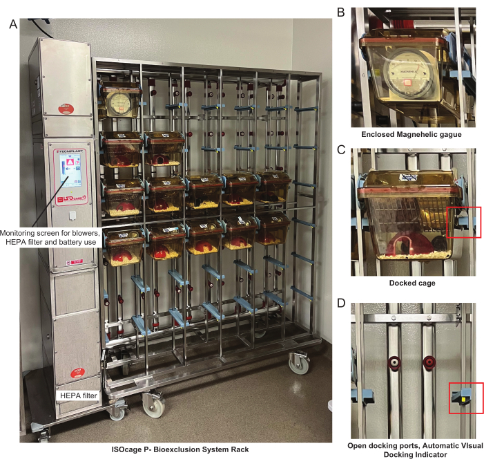
Figure 2: Schematic diagram of isocage rack system. (A) Complete isocage rack with cages docked and a label indicating the monitoring screen for air blower status, HEPA filter status, and emergency battery. On the bottom left side of the rack is the slot for the rack-level HEPA filter. (B) Enclosed Magnehelic gauge showing the positive pressure maintained by the rack. (C) A docked isocage with no visible yellow docking indicator, demonstrating a successful connection between the rack and the air nozzles. (D) An empty slot in the rack, with a visible automatic visual docking indicator indicating that no rack is in place and there is no connection of the air nozzles with an isocage. Please click here to view a larger version of this figure.
Access restricted. Please log in or start a trial to view this content.
Protocol
All animal experiments were approved by the Institutional Animal Care and Use Committee (IACUC) at the University of Florida (UF) and performed at UF Animal Care Facilities (IACUC Protocol #IACUC202300000005). Colonies of germ-free wild-type (GF WT; C57BL/6) mice were bred and maintained in isolators by UF Animal Care Services Germ-free Division. Mixed-gender GF WT mice were transferred from breeding isolators and placed into the ISOcage P Bioexclusion system to allow for microbial manipulation.
Human fecal samples were obtained from a prospective observational study that collected longitudinal stool samples from patients who received immune checkpoint inhibitor (ICI) treatment12. Informed consent was obtained from patients after study approval by Advarra IRB (MCC#18611, Pro00017235). Subjects received and completed a liquid dental transport medium (LDTM) stool collection kit meant to preserve bacterial viability for functional studies. Response assessment characterized n=4 samples as responders (R) and n=6 as non-responders (NR). The homogenized LDTM-preserved patient samples were individually thawed, each placed into an anaerobic chamber for no more than 90 s and pooled by response phenotype (R: n = 4, NR: n = 6). The pooled samples were then aliquoted and frozen at -80 °C for use in this protocol. To determine the anaerobic colony forming units (CFU) counts of the donor feces, the feces of each subject was serially diluted to 1 × 10⁻⁵, and 10 µL of each dilution was plated in duplicate on anaerobic brain heart infusion (BHI) and Luria Bertani (LB) agar plates and CFU counts per gram stool estimated. Equal CFU from each subject was pooled into fecal inoculum samples for gavage into mice.
1. Preparation of cages and autoclaving
- Isocage preparation
- Prefill cages with ~500 mL of 2018SX diet or any desired fortified autoclavable diet and layer the bottom with ALPHA-dri bedding. Place an autoclavable enrichment hut into the cage bed. Place an empty, unsealed water bottle and nozzle and long broad-tip forceps on top of the wire rack.
- Place a dual-species biological indicator into the food within one cage per autoclave cycle. Place a chemical integrator strip onto the outside of each cage.
- Autoclave the cages on a vacuum cycle for 45 min at 121 °C and a minimum of 15 PSI followed by 30 min dry time. Sterilize the cages using an international organization for standardization (ISO) decontamination rack that allows cages to remain sealed and steam to pass through the internal HEPA filter, which maintains the sterile environment until the cage is opened.
- Fill 1 L bottles with drinking water, seal with rubber caps, and place a chemical integrator strip on the surface of the bottle. Autoclave at 121 °C and a minimum of 15 PSI for 45 min, using the slow exhaust program for liquids.
- Visually check the chemical integrators affixed to each cage for immediate verification of appropriate autoclave parameters.
NOTE: The biological indicator must be removed upon the first opening of the isocage under sterile conditions. Incubate the biological indicator for 24 h at 37 °C and observe any color changes. A vivid color change from the original blue/purple clear solution to a yellow or turbid liquid indicates microbial growth is present. If the indicator remains clear and blue/purple in color, then this is confirmation of the complete sterility of the interior of the cages in that autoclave cycle.
2. Chlorine-dioxide sterilant preparation
CAUTION: Chlorine-dioxide sterilant is extremely corrosive once activated. Activated chlorine-dioxide sterilant expires 24 h from the mixing of the activator with the base. Chlorine-dioxide sterilant produces fumes, which can be irritating to mucosal surfaces and will cause irritation in contact with the skin. Ensure the room for sterilant preparation has access to a sink and proper ventilation. Don safety goggles, respirator, and chemical-resistant gloves when working with chlorine-dioxide sterilant in addition to the required personal protective equipment (PPE) for the animal housing facility.
- Mixing base and activator
- To make a standard 6 L volume of chlorine-dioxide sterilant, first measure 1 L of chlorine-dioxide sterilant base in a 1 L graduated cylinder and pour into a 20 L volume dunk tank.
- Using the same graduated cylinder, measure and pour 4 L of tap water into the dunk tank.
- Set aside the graduated cylinder used for the base and water. Use a new graduated cylinder to measure 1 L of chlorine-dioxide sterilant activator and pour it into the dunk tank.
- Once the activator has been added to the tank, use the graduated cylinder to mix the contents of the tank.
- Activation
- Place the lid onto the dunk tank and label it as chlorine-dioxide sterilant with the date and time the activator was added and the name of the staff who prepared it. If desired, the graduated cylinder used to mix the sterilant may be used to transfer 1 L to a spray bottle.
- Move the dunk tank and spray bottles to the animal housing room, where the Isocage rack and cages are located. The activated chlorine-dioxide sterilant must sit for at least 20 min prior to use in order to ensure complete activation.
NOTE: For manipulating large amounts of cages (>9), up to 12 L can be prepared at one time in the dunk tank. For manipulating 1 cage or in case of emergencies, 1.2 L of chlorine-dioxide sterilant can be prepared in a smaller container and then transferred into 2 1 L spray bottles. It is recommended that the sterilant be prepared at least 1 h prior to the anticipated transfer of germ-free mice.
3. Sterilization
- Don PPE
- As the primary cage manipulator, don safety goggles, a respirator, chemical-resistant gloves, a sterile surgical gown, a bouffant, sleeve covers, and shoe covers. Wear scrubs underneath the PPE as any contact of fabric with chlorine-dioxide sterilant will result in extensive staining.
- An assistant to the primary cage manipulator is recommended. Have the assistant don the same PPE as the primary cage manipulator, although they may wear a non-sterile surgical gown.
- Preparing the biosafety cabinet
- Place 10 wipes into the chlorine-dioxide sterilant dunk tank and ensure they are completely soaked.
- Move the soaked wipes into the biosafety cabinet and soak all surfaces of the cabinet in the following order from back to front: flat working surface, left side, back of the hood, right side, and front interior glass. Perform non-contact soaking of the non-protected surfaces of the biosafety cabinet by squeezing the wipes over these surfaces without touching them.
- After one round of cleaning, place the wipes back into the chlorine-dioxide sterilant dunk tank to soak.
- Preparing the isocages
- Prepare a large plastic bag by filling it with at least 100 mL of chlorine-dioxide sterilant using a graduated cylinder and shake the bag to ensure all interior surfaces are soaked. Place the bag on any flat surface.
- Remove a single isocage from the rack and place it into the chlorine-dioxide sterilant dunk tank so that every surface of the cage comes into contact with the liquid. Use the soaked wipes in the tank to further scrub the cage surfaces to ensure complete liquid contact.
- After soaking the cage, have the assistant open the soaked plastic bag. Place the cage into the bag, and have the assistant immediately close the opening. Spray the bag opening with chlorine-dioxide sterilant using the spray bottle. Follow this procedure for every cage to be used; up to four cages can fit into a single 36" 32" 48" bag.
- Submerse as many sterilized 1 L water bottles as needed (1 L water for 2 cages) in the chlorine-dioxide sterilant dunk tank, then place them into another soaked plastic bag. When all supplies have been placed into the bag, close the bag and spray the bag opening with chlorine-dioxide sterilant using the spray bottle.
- Once all cages and supplies are bagged, submerse the chemical-resistant gloves in chlorine-dioxide sterilant (as far up the gloves as possible without reaching the opening).
- 20 min sterilization period
- Complete sterilization requires a minimum of 20 min of liquid contact time. As soon as the last item has been sterilized and the plastic soaking bag is closed, have the assistant set a 20 min timer. Ensure that after the timer has started, the chemical-resistant gloves do not touch any surface not soaked with chlorine-dioxide sterilant.
- Perform the biosafety cabinet sterilization process (step 3.2) repeatedly until the timer indicates that 20 min has passed.
- Have the assistant frequently shake the outside surfaces of the soaked plastic bag to ensure consistent liquid contact with cage and bottle surfaces within.
- After the 20 min period, have the assistant open the soaked plastic bag to reveal the sterilized cages and water bottles, taking care to only touch the outside surfaces of the bag.
- Using the sterilized chemical-resistant gloves, move each cage and bottle to the sterilized biosafety cabinet. If there are too many cages to fit into the biosafety cabinet, leave the remaining cages in the plastic bags as they will remain sterile as long as the opening of the plastic bag is closed and soaked with chlorine-dioxide sterilant between each opening.
4. Germ-free mouse transfer
- Preparing the isocages
- To open the hermetically sealed isocage, lift up the white tabs on the two clamps on the sides of the lid and then pull out each clamp sideways. The lid must be free of the bottom portion of the cage in order to lift the lid off the cage. Place the lid upside down to the left of the cage and use this as a sterile workstation.
NOTE: An alternative to using cage lids or the interior of autoclaved instrument bags as a sterile work surface is to utilize autoclaved drapes, which provide a larger surface area and prevent unintentional contamination of the cage lid if an error is made. - Using the sterile forceps found inside the cage on top of the wire rack, remove the empty water bottle and set it on the inside of the lid. Open the 1 L water bottle by removing the rubber seal, and pour the water into the water bottle to fill it. Place the nozzle on the water bottle using the forceps and press down firmly to seal.
- Use sterile forceps to lift the wire rack and set it back several inches to allow for an opening to the cage bottom. Rest the sterile forceps on the wire rack, ensuring that the handles do not come in contact with cage surfaces.
- To open the hermetically sealed isocage, lift up the white tabs on the two clamps on the sides of the lid and then pull out each clamp sideways. The lid must be free of the bottom portion of the cage in order to lift the lid off the cage. Place the lid upside down to the left of the cage and use this as a sterile workstation.
- Use of transfer disk
NOTE: Trained germ-free staff are responsible for the care and maintenance of the breeding isolators. Given the risks associated with the opening of breeding isolators, these staff perform the transfer disk sterilization, preparation, and transfer of germ-free mice from isolators to isocages. To briefly describe the process, transfer disks are prepared in an autoclavable cylinder to allow for sterilization. Biological indicators are used to verify their sterility. Tape to seal the transfer disks is autoclaved inside the cylinder as well. The sterilized cylinder enclosing these materials is attached via a transfer sleeve to the isolator, and mice are moved from the cages to the disk. The lid is then placed on the disk, and autoclaved tape is used to create an airtight seal on the circumference of the disk and the air holes. The sealed disk is immediately placed in the exit port of the isolator. The home isolator's port cap is then closed, and the outside of the disk is thoroughly swabbed with sterilant and placed into a sterilant-soaked bag, monitored for 20 min to ensure complete decontamination. Germ-free breeding staff then deliver these disks to study staff. Mice cannot be kept in the sealed disk longer than 30 min from the time the transfer disk is sealed, so it is essential that all previous steps in this protocol have been completed with ample time prior to the arrival of the transfer disk.- Upon receipt of the transfer disk, have the assistant hold and partially unwrap the plastic cover so that the soaked surface of the transfer disk is exposed but not touched by the assistant.
- Upon receipt of the transfer disk, have the assistant hold and partially unwrap the plastic cover so that the soaked surface of the transfer disk is exposed but not touched by the assistant.
- Wearing the chemical resistant gloves soaked in chlorine-dioxide sterilant, remove the transfer disk from the plastic wrapping, taking care not to touch any surface not soaked in sterilant . Then, place the transfer disk on the flat surface of the sterilized biosafety cabinet.
- To open the transfer disk, peel off the tape, seal the disk circumference, and discard it outside the biosafety cabinet. Remove the lid of the transfer disk and discard it outside the biosafety cabinet.
- Inside the transfer disk is a rotatable compartment lid with a single opening. Use the sterile forceps previously rested on the wire rack of the cage to manipulate this compartment lid to move the opening to the mouse needed for transfer.
- Transfer of mice from disk to isocage
- Using the forceps, grasp the base of the tail of the mouse through the opening in the plastic disk cover, and lift and transfer the mouse into the isocage through the space opened previously between the wire rack and the cage. Repeat for all mice destined for that cage.
- Once all mice have been transferred to that isocage, replace the wire rack using the forceps. Then, lift the cage lid and place it back on top of the cage using the forceps.
- Lift each clamp of the cage lid up and carefully lower over the sides of the cage, followed by pushing down the white tabs to seal the cage lid.
- Once the cage is sealed, have the assistant spray each nozzle of the docking site on the cage rack with chlorine dioxide sterilant. Then, remove the cage from the hood and pass it to the assistant, who can then dock the cage on the rack.
- Repeat these steps for each mouse in the transfer disk.
- Cleaning up the biosafety cabinet
- Upon completion of all mouse transfers into isocage, empty the hood of any debris and completely wipe down with chlorine-dioxide sterilant-soaked wipes.
- Wipe the hood with isopropyl alcohol to remove residual chlorine-dioxide sterilant. The space underneath the working surface of the hood collects a large volume of sterilant from the sterilization process. Remove this via absorption with dry wipes and wipe with isopropyl alcohol.
- Dispose of liquid chlorine-dioxide sterilant via sink drain 24 h post-activation. Dispose of solid materials contaminated with sterilant as regular waste.
NOTE: Germ-free mice transferred to isocage conditions are left for 1 week to acclimate to their new environment prior to any intervention. This reduces the stress experienced by the animals, which could interfere with study results. At the end of this 1 week acclimation period, collect feces as described in step 6 to confirm germ-free status prior to any intervention.
5. Oral gavage of human fecal slurry into germ-free mice
- Preparing autoclaved gavage supplies
- Place oral gavage needles in self-sealing sterilization pouches (1 per mouse) and sterile 1 mL syringes in self-sealing sterilization pouches (1 per mouse) 1 day prior to the oral gavage procedure. Place these and 600 mL polypropylene beakers (1 per mouse) and a pair of long forceps into an autoclave-safe bag and sterilize via autoclave.
- Immediately upon removal of the bag from the autoclave, seal the bag with packaging tape and store it until the next day.
- Preparing human fecal slurry
- On the day of gavage, transfer human feces homogenized and stored in anaerobic preservation media (in this instance, Liquid Dental Transport Media) from the -80 °C freezer to the anaerobic chamber. Dilute homogenized fecal material approximately 1:10 in sterile, anaerobic saline solution in a 10 mL conical tube.
- Seal the tube containing the human fecal slurry with parafilm, homogenize by vortex, and then centrifuge at 200 g for 5 min to settle particulates.
- Place the tube back into the anaerobic chamber and transfer the supernatant to another 10 mL conical tube. Seal the tube containing the human fecal supernatant with parafilm, remove it from the anaerobic chamber, and place it into a secondary leak-proof container. Transport the container along with the autoclaved bag of gavage supplies to the animal housing location.
NOTE: It is essential to estimate the total CFU/mL of the human feces intended for gavage for reporting purposes. It is unclear what the minimum CFU load is to ensure adequate colonization, but higher CFUs lead to better engraftment of donor stool13. If the CFU load of human fecal material results in low engraftment rates, recollect human fecal specimens. The use of a viability preservation media will enhance CFU recovery from collected fecal specimens. To determine the anaerobic/aerobic CFU counts of the donor feces, serially dilute the sample to 1 x 10⁻⁵ and plate 10 µL of each dilution in duplicate on aerobic and anaerobic BHI and LB agar plates. After 24 h (aerobic) and 48 h (anaerobic), count the CFU per gram of stool.
- Preparing the isocages
- Repeat steps 2 and 3, with the only difference now being that there are mice housed in these cages, and care should be taken to ensure that within 30 min of rack removal, each isocage is placed into the sterilized hood and the lid vented to allow for airflow to the mice.
- Sterilize the autoclaved gavage material bag and human fecal slurry tube from steps 5.1 and 5.2 via chlorine-dioxide sterilant saturation in a similar manner to the water bottles (i.e., submerge them in sterilant and then place them into a sterilant-soaked bag).
- Transfer the isocages and oral gavage supplies to the biosafety cabinet following completion of the 20 min sterilization period. Puncture the autoclaved supply bag by pushing the bag against the long forceps contained within, and then remove the supplies and discard the bag outside of the hood.
- Gavage
- Don a new sterile surgical gown and sterile surgical gloves in place of the chemical resistant gloves in order to prevent residual chlorine-dioxide sterilant from coming into contact with mice. Have the assistant help in this process if needed.
- Prepare the gavage needles by unwrapping each sterilization pouch and use the interior of the pouch as a dry, sterile resting surface. Connect the gavage needle to each syringe, open the fecal slurry tube, and pull up 200 µL of fecal slurry into each syringe.
- Using forceps, grasp the base of the tail of a single mouse in the cage and place it on the wire rack. Gently restrain the mouse by scuffing, and while holding the mouse in an upright vertical position, insert the needle and gently inject the fecal slurry, followed by immediate removal of the needle.
- Place the mouse directly into one of the sterilized cups for observation. Repeat the process for every mouse in the cage. After all mice have received the gavage, use the forceps to move each mouse back into the cage bed and seal the cage as described in steps 4.3.2-4.3.4.
NOTE: In cases where two or more separate fecal slurries are being used, complete re-sterilization of the biosafety cabinet and required cages and materials are required. In cases where a group of mice remains germ-free, it is recommended that these mice receive their control gavages before any other group is treated. - Follow the procedure in step 4.4 to dispose of chlorine-dioxide sterilant and sterilant-soaked materials. Treat any materials contaminated with human fecal slurry as biomedical waste and dispose of them according to the environmental health and safety procedures.
6. Stool collection from humanized mice for viability preservation
- Prepare autoclaved supplies
- Place short broad-tip forceps in self-sealing sterilization pouches (1 per mouse), 600 mL polypropylene beakers (1 per mouse), and a pair of long forceps into an autoclave-safe bag and sterilize via autoclave 1 day prior to stool collection procedure.
- Immediately upon removal of the bag from the autoclave, seal the bag with packaging tape and store it until the next day.
- Preservation media tube preparation
- Select an anaerobic viability preservation media (here Cary Blair was used). Aliquot 1 mL of preservation media into sterile, screw cap 2 mL tubes in a biosafety cabinet. A ratio of 1:10 feces:media is recommended for optimal preservation.
- Pre-label each tube with a permanent marker, but be aware that exposure to chlorine-dioxide sterilant can remove permanent marker labels from plastic surfaces. Another method is to leave the tubes unlabeled and to have the assistant label tubes immediately post-collection before freezing.
- Place these tubes in a standard polypropylene tube rack to allow the tubes to be stored upright while still enabling sterilization by contact with chlorine-dioxide sterilant.
- Sterilizing cages and biosafety cabinet
- Repeat steps 2 and 3 to sterilize isocages and the biosafety cabinet. Again, take care to ensure that within 30 min of rack removal, each isocage is placed into the sterilized hood, and the lid is vented to allow for airflow to the mice.
- Additionally, sterilize the autoclaved supply bag and rack containing prepared tubes via chlorine-dioxide sterilant saturation and place them into the soaked plastic bag containing the cages. The goal is complete liquid contact with all surfaces of each tube and the rack itself.
- Fecal sample collection and storage
- Transfer the isocages, autoclaved supplies, and tube rack to the biosafety cabinet following completion of the 20 min sterilization period. Puncture the autoclaved supply bag by pushing the bag against the long forceps contained within, remove the supplies and discard the bag outside of the hood.
- Don a new sterile surgical gown and sterile surgical gloves in place of the chemical resistant gloves in order to prevent residual chlorine-dioxide sterilant from coming into contact with mice. Have the assistant assist in this process if desired.
- Prepare the blunt tip forceps by unwrapping the pouches and using the interior pouch surface as a sterile area. Place the tube rack on the surface of the biosafety hood.
- Using long forceps, grasp the base of the tail of a single mouse in the cage and place them directly into one of the sterilized cups for observation. Repeat this process for every mouse in the cage.
- Observe the mice until at least two freshly passed fecal pellets have been produced.
- Using the blunt tip forceps, pick up the fecal pellets and place them directly into the tube. Immediately seal the screw cap lid and pass it to the assistant.
- Have the assistant label the tube and immediately homogenize the stool via vortexing. Once homogenous, snap freeze the tube in liquid nitrogen and store long term at -80 °C.
- Repeat the stool collection process for each mouse in the cage. Replace each mouse into the home cage after fecal collection, and redock the cages on their rack. Repeat step 4.4 to clean the hood and dispose of waste.
Access restricted. Please log in or start a trial to view this content.
Results
Human fecal samples, pooled by ICI responder and non-responder phenotype (previously described in the protocol), were gavaged into mixed gender GF-WT mice housed in 3 isocages per group (n = 1-2 mice/cage, n =6 for responder and n = 5 for non-responder). Mice were allowed to acclimate for 1 week post-transfer. Fecal samples were then collected from these mice (germ-free conditions). Mice were then gavaged with 1 × 10⁷ CFU of either responder or non-responder pooled human feces. The stool was then collected 1, ...
Access restricted. Please log in or start a trial to view this content.
Discussion
The protocol described here provides a reproducible, highly detailed method for the humanization of germ-free mice housed in experimental isocages. The ability to exclusively transplant fecal communities from human subjects into murine hosts is invaluable to microbiome research. Without contamination from mouse-specific commensal microbiota, one can study the impact of human-resident bacteria on a variety of health and disease states or the impact of interventions such as diet or drug administration on human microbiota
Access restricted. Please log in or start a trial to view this content.
Disclosures
The authors have no conflicts of interest.
Acknowledgements
The authors are grateful to the Germ-Free Services Division of UF Animal Care Services for the assistance with gnotobiotic husbandry, to Dr. Brooke Bloomberg and Dr. Laura Eurell for veterinary and IACUC assistance, and Josee Gauthier for the assistance with 16S rRNA gene sequencing. This research was supported, in part, by the UF Health Cancer Center Funds (C.J.) and the UF Department of Medicine Gatorade Fund (C.J.). R.Z.G. was supported by UF Health Cancer Center funds. R.C.N. was supported by the National Institutes of Health TL1 Training Grant at the University of Florida (TL1TR001428, UL1TR001427), the National Cancer Institute of the National Institutes of Health Team-Based Interdisciplinary Cancer Research Training Program award T32CA257923 and the UF Health Cancer Center. Research reported in this publication was supported by the UF Health Cancer Center, supported in part by state appropriations provided in Fla. Stat. § 381.915 and the National Cancer Institute of the National Institutes of Health under Award Number P30CA247796. The content is solely the responsibility of the authors and does not necessarily represent the official views of the National Institutes of Health or the State of Florida. The funders had no role in study design, data collection and analysis, decision to publish, or preparation of the manuscript.
Access restricted. Please log in or start a trial to view this content.
Materials
| Name | Company | Catalog Number | Comments |
|---|---|---|---|
| 1 mL BD Slip Tip Syringe sterile, single use | Fisher Scientific | 309659 | |
| 2.0 mL Screw Cap Tube, NonKnurl,Skirted,Natural, E-Beam Sterile tube w/ attached cap | Fisher Scientific | 14-755-228 | |
| 36 x 32 x 48" 3 Mil Gusseted Poly Bags | Uline | S-13455 | |
| 5 gallon tank of Exspor chlorine-dioxide sterilant activator | Ecolab | 6301680 | |
| 5 gallon tank of Exspor chlorine-dioxide sterilant base | Ecolab | 6301194 | |
| 600 mL polypropylene beakers | Fisher Scientific | S01914 | |
| ALPHA-dri bedding | Shepherd Specialty Papers | ||
| Anaerobic chamber | Coy Lab Products | Type B | |
| Biosafety cabinet class 2 | Nuaire | ||
| Certified IsoCage autoclavable HEPA filter XT Extreme Temperature | Tecniplast | 1245ISOFHXT | |
| Clear Lens LPX IQuity Safety Goggles | Fastenal | 922205455 | |
| DuPont Tyvek Sleeve - 18" | Uline | S-13893E | |
| DWK Life Sciences DURAN 45 mm Push-on Natural Rubber Cap | Fisher Scientific | 01-258-107 | Rubber cap for 1 L autclave bottles |
| Dynalon Quick Mist HDPE Sprayer Bottles | Fisher Scientific | 03-438-12B | |
| Fisherbran Polypropylene Graduated Cylinders | Fisher Scientific | 03-007-44 | |
| Fisherbran Dissecting Blunt-Pointed Forceps | Fisher Scientific | 08-887 | |
| Fisherbrand Instant Sealing Sterilization Pouches | Fisher Scientific | 01-812-51 | |
| Fisherbrand Straight Broad Strong Tip General Application Forceps | Fisher Scientific | 16-100-107 | |
| Fisherbrand lead Free Autoclave Tape | Fisher Scientific | 15-901-110 | |
| Gavage needle, reusable stainless steel. Straight. 22 gauge needle, tip diameter 1.25 mm, length 38 mm or 1.5 inches(doz) | Braintree Scientific | N-PK 020 | |
| H-B Instrument Durac Timer | Fisher Scientific | 13-202-015 | |
| IsoPositive Cages and Rack (i.e. isocages) | Tecniplast | ISO30P | 30 cages (6 w x 5 h), single sided |
| Nitrile Chemical Resistant Gloves Size S (7), M (8) or L (9) 18” long, 22 mil, Ansell | Grainger | 4T426 | |
| Nitrile Exam Gloves, Medium, Non-Sterile, Powder-Free | MedSupply Partners | KG-1101M | |
| Olive / Magenta Bayonet Gas & Vapor Cartridges / Particulate Filter 2Ct | 3M/Fastenal | 50051138541878 | |
| Polycarbonate RadDisk Mini for Mice 8-75 x 4 | Braintree Scientific | IRD-P M | |
| Polypropylene Bouffant Caps - 24", Blue | Uline | S-10480BLU | |
| Puritan Cary-Blair Medium, 5 mL | Fisher Scientific | 22-029-646 | |
| S, M and L Blue Silicone Dual-Mode Head Harness Half Mask Respirator | 3M/Fastenal | 50051131370826 | |
| Sgpf Series Sterile Powder Free Latex Gloves, CT International, Thickness = 6.5 mm, Length = 30.5 cm (12), Glove Size = 8.5, Glove Color = White | Fisher Scientific | 18-999-102F | |
| Skid Resistant Shoe Cover | Uline | S-25639 | |
| Surgical Gown, Towel, Sterile, Large, 32/cs | Thomas Scientific | KIM 95111 | |
| Teklad Global 18% protein extruded rodent diet (sterilizable) | Inotiv | 2018SX | |
| Thermo Scientific Nalgene Heavy-Duty Rectangular LLDPE Tank with Cover (20 L volume) | Thermo Scientific | 14-831-330J | |
| VERIFY Dual Species Self Contained Biological Indicators | Steris Healthcare | S3061 | |
| WypAll L40 1⁄4 Fold Wipers | Uline | S-8490 |
References
- Park, J. C., Im, S. -H. Of men in mice: the development and application of a humanized gnotobiotic mouse model for microbiome therapeutics. Exp Mol Med. 52 (9), 1383-1396 (2020).
- Li, F., et al. Microbiome remodelling leads to inhibition of intestinal farnesoid X receptor signalling and decreased obesity. Nat Commun. 4, 2384(2013).
- Schwabe, R. F., Jobin, C. The microbiome and cancer. Nat Rev Cancer. 13 (11), 800-812 (2013).
- Wen, L., et al. Innate immunity and intestinal microbiota in the development of Type 1 diabetes. Nature. 455 (7216), 1109-1113 (2008).
- Gray, S. M., et al. Mouse adaptation of human inflammatory bowel diseases microbiota enhances colonization efficiency and alters microbiome aggressiveness depending on recipient colonic inflammatory environment. Microbiome. 12 (1), 147(2024).
- Gopalakrishnan, V., et al. Gut microbiome modulates response to anti-PD-1 immunotherapy in melanoma patients. Science. 359 (6371), 97-103 (2018).
- Matson, V., et al. The commensal microbiome is associated with anti-PD-1 efficacy in metastatic melanoma patients. Science. 359 (6371), 104-108 (2018).
- Routy, B., et al. Gut microbiome influences efficacy of PD-1-based immunotherapy against epithelial tumors. Science. 359 (6371), 91-97 (2018).
- Paik, J., et al. Potential for using a hermetically-sealed, positive-pressured isocage system for studies involving germ-free mice outside a flexible-film isolator. Gut Microbes. 6 (4), 255-265 (2015).
- Hecht, G., et al. A simple cage-autonomous method for the maintenance of the barrier status of germ-free mice during experimentation. Lab Anim. 48 (4), 292-297 (2014).
- Dremova, O., et al. Sterility testing of germ-free mouse colonies. Front Immunol. 14, 1275109(2023).
- Newsome, R. C., et al. Interaction of bacterial genera associated with therapeutic response to immune checkpoint PD-1 blockade in a United States cohort. Genome Med. 14 (1), 35(2022).
- Le Roy, T., et al. Comparative evaluation of microbiota engraftment following fecal microbiota transfer in mice models: age, kinetic and microbial status matter. Front Microbiol. 9, 3289(2018).
- Lebeuf, M., et al. Contaminants and where to find them: microbiological quality control in axenic animal facilities. Front Microbiol. 12, (2021).
- He, Z., et al. Campylobacter jejuni promotes colorectal tumorigenesis through the action of cytolethal distending toxin. Gut. 68 (2), 289-300 (2019).
- Wu, G. D., et al. Linking long-term dietary patterns with gut microbial enterotypes. Science. 334 (6052), 105-108 (2011).
- Ross, F. C., et al. The interplay between diet and the gut microbiome: implications for health and disease. Nat Rev Microbiol. 22 (11), 671-686 (2024).
- Maier, L., et al. Extensive impact of non-antibiotic drugs on human gut bacteria. Nature. 555 (7698), 623-628 (2018).
- Walter, J., Armet, A. M., Finlay, B. B., Shanahan, F. Establishing or exaggerating causality for the gut microbiome: Lessons from human microbiota-associated rodents. Cell. 180 (2), 221-232 (2020).
- Berland, M., et al. High engraftment capacity of frozen ready-to-use human fecal microbiota transplants assessed in germ-free mice. Sci Rep. 11 (1), 4365(2021).
- Choo, J. M., Rogers, G. B. Establishment of murine gut microbiota in gnotobiotic mice. iScience. 24 (2), 102049(2021).
- Bokoliya, S. C., Dorsett, Y., Panier, H., Zhou, Y. Procedures for fecal microbiota transplantation in murine microbiome studies. Front Cell Infect Microbiol. 11, 711055(2021).
- Li, Y., Cao, W., Gao, N. L., Zhao, X. -M., Chen, W. -H. Consistent alterations of human fecal microbes after transplantation into germ-free mice. Genomics Proteomics Bioinformatics. 20 (2), 382-393 (2022).
- Turnbaugh, P. J., Ridaura, V. K., Faith, J. J., Rey, F. E., Knight, R., Gordon, J. I. The effect of diet on the human gut microbiome: a metagenomic analysis in humanized gnotobiotic mice. Sci Transl Med. 1 (6), 6ra14(2009).
- Staley, C., et al. Stable engraftment of human microbiota into mice with a single oral gavage following antibiotic conditioning. Microbiome. 5 (1), 87(2017).
- Lebeuf, M., Turgeon, N., Faubert, C., Robillard, J., Paradis, É, Duchaine, C. Managing the bacterial contamination risk in an axenic mice animal facility. Can J Microbiol. 67 (9), 657-666 (2021).
- Basic, M., et al. Monitoring and contamination incidence of gnotobiotic experiments performed in microisolator cages. Int J Med Microbiol. 311 (3), 151482(2021).
- Amorim, N., et al. Refining a protocol for faecal microbiota engraftment in animal models after successful antibiotic-induced gut decontamination. Front Med. 9, 770017(2022).
Access restricted. Please log in or start a trial to view this content.
Reprints and Permissions
