We establish a standardized in vitro model for studying cannabis vapor exposure at the air-liquid interface. This model provides a systematic approach to examine the impacts of vaporized cannabis, addressing the growing interest in cannabis vaporizers as an alternative to smoking.
Method Article
In vitro Cannabis Exposures of Lung Epithelial Cells at the Air-Liquid Interface
In This Article
Summary
Abstract
Cannabis is used by an estimated 192 million people around the world. Most people use cannabis through the inhalation of cannabis smoke, which contains combustion by-products that can negatively affect lung health. Knowledge of these risks has led to a growing interest in cannabis vaporizers, which heat the dry cannabis flower without burning. Vaporizing cannabis still releases cannabinoids for inhalation but heats the plant material at a lower temperature. There is currently no standardized in vitro model for assessing the effects of dry cannabis vapor. Therefore, we established a model for the exposure of lung cell cultures at an air-liquid interface (ALI), whereby cells are apically exposed to vaporized cannabis, thereby more accurately simulating lung epithelial cell physiology. This protocol ensures consistent and reproducible delivery of cannabis vapor to the cell surface, providing a reliable platform for investigating the cellular and molecular impacts of vaporized cannabis. This work is the first to standardize an in vitro cannabis vapor delivery method, which can serve as a benchmark for future preclinical cannabis research.
Introduction
Cannabis is typically used by inhaling smoke from burning the flower of the plant in the form of a joint, whether recreationally or for medicinal purposes. The principal biological activities of cannabis are due to cannabinoids such as Δ9-tetrahydrocannabinol (Δ9-THC) and cannabidiol (CBD), which are released into the body upon inhalation. Cannabis smoke also contains pyrogenic compounds, including carcinogens, mutagens, and teratogens, which have the potential to cause adverse health outcomes1. Given these concerns, there is interest in turning to cannabis vaporizers that heat the cannabis flower without burning, in the hope that this allows the release of cannabinoids for subsequent inhalation but without combustion products. Some work has been done to characterize the chemical components of cannabis vapor, showing the presence of cannabinoids but also various respiratory toxicants, challenging the notion that these devices are safe2,3. There are significant gaps in knowledge as to how vaporized cannabis impacts the lungs and other organs.
Cell culture studies are essential in toxicology to isolate and understand the specific effects of inhaled substances at the cellular level, enabling high-throughput, cost-effective, and mechanistic insights that are relevant to human biology. The method of culturing the cells is also an important determinant of the overall results obtained from an in vitro experiment. Most cell culture studies are performed using a submerged setting, where cells are grown on plastic and exposed apically to aqueous media. Airway epithelial cells, however, present a unique challenge in this regard. Throughout the respiratory system, the apical surface of epithelial cells is in contact with air. Within the alveoli, epithelial cells produce surfactant, whereas other airway epithelial cells produce mucus; collectively, this protects and maintains lung function. Submerged cultures fail to replicate apical air exposure, limiting their ability to model the in vivo environment accurately. An alternative to submerged culture models is culturing at the air-liquid interface (ALI), where respiratory epithelial cells are grown on a collagen-coated membrane with media on the basolateral side while being apically exposed to air. Epithelial cells cultured at ALI are more accurate models of the respiratory epithelium due to their ability to polarize, produce surfactant or mucus, and form junctions in this environment4.
An additional consideration that traditional submerged cell culture methods face is that hydrophobic compounds like cannabinoids solubilize poorly in aqueous media5. Ethanolic extracts are a common solvent to study hydrophobic compounds in submerged cultures6,7,8,9,10,11,12. These extracts allow both hydrophobic and hydrophilic compounds to be dissolved in an aqueous media. However, a limitation is the potential for compound agglomeration as well as compounds interacting with the media, both of which can alter bioavailability13,14. There have recently been developments to allow for aerosol phase compounds to be delivered to cells in the absence of a solvent or media using advanced exposure systems such as the expoCube15 (ALI/Transwell in vitro/ex vivo exposure system), as well as others. Direct deposition of aerosols and particle suspensions using an advanced exposure system eliminates the collection-resuspension process crucial for submerged exposures, which is known to substantially alter particle physicochemical properties16. Here, we describe a standardized protocol for the exposure of an alveolar epithelial cell line at ALI.
Protocol
1. Culture lung epithelial cells at ALI
NOTE: Here, a protocol for culturing A549 cells at ALI is described. Please review the literature for the cell type to be used in the study. Different cell lines need a different amount of time at ALI to differentiate, form tight junctions, and produce surfactant/mucus.
- Cell culture
- Prepare media by adding 50 mL of heat-inactivated fetal bovine serum (FBS), 5 mL of Glutamax, 5 mL of antibiotic-antimycotic, and 500 µL of Gentamycin to a 500 mL bottle of DMEM.
- Place prepared media in a water bath for 10-15 min to warm to 37 °C.
- Spray the biosafety cabinet with 70% ethanol and wipe it down. Once the medium is warmed, place it under the sterilized biosafety cabinet.
- Under the culture hood, pipet approximately 10 mL of media into a 15 mL tube.
- Retrieve cells from liquid nitrogen and thaw by gently agitating in a water bath.
- Under the culture hood, pipet the thawed cells of the cryovial into the 15 mL tube.
- Centrifuge the 15 mL tube at 300 g for 5 min.
- Carefully remove the supernatant without disturbing the pellet.
- Mix the pellet with media and transfer it to the T75 flask.
- Check the T75 flask under a microscope.
- Incubate the cells at 37 °C, 5% CO2 with saturating humidity.
- Refresh cell culture media every 2-3 days.
- Passage and grow cells until the desired cell count is reached. To seed one 12 well plate, 3 × 10⁶ cells are required.
- Collagen coating of transwells
- Preparation of collagen solution
- Dissolve 10 mg of collagen Type IV in 20 mL of ddH2O with 50 µL of glacial acetic acid.
- Incubate at 37 °C for 30 min, swirling occasionally, until fully dissolved.
- Sterilize the solution by filtering through a 0.22 µm filter.
- Aliquot 1.5 mL of the solution into 15 mL tubes and store at -20 °C.
- Coating transwell filters with Collagen Type IV
- Thaw one tube of collagen type IV (50 µg/mL) from -20 °C.
- Add 150 µL of collagen to each Transwell filter.
NOTE: Only 12-well plates are compatible with the exposure system. - Leave plates with open lids in the biosafety cabinet (BSC) for 18 h to dry.
- Sterilize by exposing to ultraviolet (UV) light for 30 min.
- Store plates at 4 °C, wrapped in parafilm, for up to 30 days.
- Before plating cells, rinse both sides of the filters with sterile phosphate-buffered saline (PBS).
- Preparation of collagen solution
- Seeding cells in inserts and transitioning to ALI
- Preparation of plate and cell suspension
- Pipette 1.5 mL of prewarmed prepared media into each well of the 12-well plate.
- Place the plate into the incubator at 37 °C and 5% CO2.
- Prepare a cell suspension with a concentration of 5 × 10⁵ cells/mL in prepared media.
- Seeding cells in inserts
- Remove the prepared cell culture plate from the incubator and place it under a sterilized biosafety cabinet.
- Pipette 0.5 mL of the cell suspension (2.5 × 10⁵ cells/insert) onto the top of the membrane in the insert.
- Cover the plate with its plate cover and place it back into the incubator at 37 °C and 5% CO2.
- Establish ALI
- Remove apical media 24 h after seeding cells.
- Replace the basolateral media with 0.6 mL of prepared media. At this point, the cells are apically exposed to air, establishing the ALI.
- Culture the A549 cells at ALI for up to 2 weeks, changing media every 2 days to allow the cells to differentiate as has been characterized previously4.
- Preparation of plate and cell suspension
2. Dosimetry assessment of cannabis vapor exposure using LC/MS
NOTE: In this part of the protocol, cell-free methods were used to characterize the dose of cannabis vapor deposited within each well by analyzing Δ9-THC concentrations. Δ9-THC adheres to plastic materials, so only glass vials should be used for sample collection. This method requires that the cannabis cultivar chosen contains Δ9-THC.
- Preparation and setup of the exposure system
- Set up the tubing for the exposure system according to the specified flow path (Figure 1A, B): ENDS Unit > Condenser > Puffing Pump > Isometric Sampling Assembly with Aeroneb pump > expoCube > Humidity Sensor with Filter > expoCube Exposure Flow Port.
- Switch on temperature controls to heat the system to 35 °C and 50 °C.
- Perform flow tests to ensure the system is leak-free and pressures are correctly set (refer to SCIREQ Technote A Guide to Troubleshooting Uneven Flows in the expoCube).
- Start the Aeroneb pumps to achieve greater than 75% relative humidity.
- Dosimetry testing and vaporizer preparation
- Cannabis vaporizer preparation
- Grind the cannabis flower using an herb grinder.
- Weigh out 0.25 g aliquots, using a pill separator to organize each portion.
- Place stainless steel inserts into the exposure row of the 12-well transwell plate within the exposure system.
- Cannabis vaporizer preparation
- Exposure of stainless-steel inserts to cannabis vapor
- Begin exposure
- Pack the vaporizer oven with 0.25 g of cannabis.
- Attach the cannabis vaporizer to the ENDS Unit of the exposure system with a secure gasket and begin the desired puffing regimen. Set the vaporizer to the Smart Path Level 2 temperature setting.
NOTE: The exposure system used in this study has four smart path levels to choose from, including Level 1 (176-188°C), Level 2 (188-199 °C), Level 3 (199-210 °C), and Level 4 (210-221 °C). - Create a puff profile in the flexiWare software (data acquisition software; refer to SCIREQ Technote: A Guide to Creating Profiles for flexiWare 8). For this protocol, use a 70 mL puff volume with a frequency of 4 puffs/min.
- Run the dry herb vaporizer for 5 min before switching cannabis by removing the cannabis flower from the vaporizer oven and adding a fresh 0.25 g of cannabis to the vaporizer. Alternate between two vaporizers for continuous exposure.
- Continue steps 2.3.1.1-2.3.1.4 until the desired exposure time is reached.
- After each exposure, carefully remove the inserts from the exposure system using tweezers and prepare them for sample collection as described below.
- Begin exposure
- Collection and preparation of samples for LC/MS analysis
- Using tweezers, place filters corresponding to each exposure into a labeled glass vial containing 10 mL of LC/MS-grade methanol (e.g., Exposure 1, Exposure 2, Exposure 3).
- Wash the insert.
- Place each exposed stainless-steel insert into a labeled glass vial containing 10 mL of LC/MS-grade methanol.
- Label vials as per exposure (e.g., Exposure 1 Well 1-4).
- Extract Δ9-THC
- Use a sonicator bath to sonicate the vials with inserts for 10 min to extract Δ9-THC.
- Transfer the inserts to a second set of glass vials with 10 mL of fresh methanol and sonicate for an additional 10 min.
- Submit all prepared samples (wells, wash, and filter) for LC/MS analysis to quantify Δ9-THC levels.
- Dosimetry calculations
- Using the LC/MS analysis results, calculate the amount of deposited Δ9-THC to determine the dosimetry of the exposure flow (amount deposited in the filter) and the dosimetry in the well.
- Calculate the ratio between the exposure flow filter and the well deposition for Δ9-THC.
- Use this ratio to determine the dose that will be delivered to the cells during exposures.
3. Dosimetry assessment of cannabis vapor exposure using a quartz crystal microbalance
NOTE: This method details the setup and calibration of a Quartz Crystal Microbalance (QCM) to assess the mass of cannabis vapor particles deposited during exposures. Each QCM provides real-time measurements, enabling precise dosimetry of cannabis vapor exposure.
- Preparation and setup of the QCM in the exposure system
- Configure the exposure system as described previously.
- Attach each QCM to its electronics module using the clip connector, and connect the module to the inExpose (inhalation exposure platform) unit inputs with a 7-pin connector.
- Place QCM insert(s) into a well plate on the exposure side of the exposure system, ensuring the QCM is well aligned.
- Preheat the exposure system with attached QCMs until the system stabilizes at the target temperatures of 35 °C and 50 °C.
- Activate microflows and exposure flows according to the experimental settings.
- Calibrating the QCM
- Use the data acquisition software calibration wizard to calibrate the QCM before each exposure run.
- Wait for the blue calibration bar to stabilize, then select Next to record the QCM resonant frequency for accurate mass calculations.
NOTE: Expected calibration range is -15% to 15%. Deviations may indicate issues like a dirty QCM or disconnected LEMO connector.
- Real-time monitoring of QCM signals
- Monitor the QCM outputs; the system outputs the accumulated mass deposited. The value is displayed in data acquisition software both as part of a graph and the actual present value.
NOTE: For each experimental run, the maximum and minimum deposition are also reported in the software.
- Monitor the QCM outputs; the system outputs the accumulated mass deposited. The value is displayed in data acquisition software both as part of a graph and the actual present value.
- Cleaning the QCM
- To maintain accuracy, clean the QCM after each experiment.
- Add 1 mL of 70% isopropyl alcohol inside each QCM insert.
- Pipette the alcohol solution up and down 2-3 times to dissolve residues.
- Remove the alcohol solution and allow the QCM to air dry completely in a dust-free environment before the next use.
4. Outcomes of exposing cells to cannabis vapor
NOTE: This protocol is designed to evaluate the effects of cannabis vapor on respiratory cells cultured at ALI using the expoCube system. Post-exposure assays, including 3- (4, 5-dimethylthiazol-2-yl)-2,5-diphenyltetrazolium bromide (MTT) and lactate dehydrogenase (LDH), provide insights into dose-dependent cytotoxicity and metabolic activity.
- Cannabis vapor delivery
- Set up the exposure system as described above: Ensure the system has reached 35 °C and 50 °C temperature and relative humidity levels above 75%.
- If using a QCM, attach the device to its electronics module using the clip connector, and connect the module to the inhalation exposure unit inputs with a 7-pin connector. Place the singular QCM insert into a well plate on the exposure side of the expoCube, ensuring the QCM is well aligned.
- Open the exposure system and place the cell culture plate inside.
- Begin the puff regimen for the desired duration based on section 2. Using the QCM will allow for real-time tracking of total vapor deposited.
- Post-exposure, return cells to the incubator until the desired assay timepoint.
- Collect samples as needed from various compartments, including the basolateral compartment, the apical compartment wash (for surfactant or mucus), and the cell lysate.
- Dose Selection Assays: Use LDH and MTT assays to help determine optimal dose and potential cytotoxicity.
- LDH assay for cells at ALI
- Collect media: At the desired time point post-exposure, collect media from the basolateral compartment.
- Transfer 100 µL of collected media to a clear 96-well microplate.
- Add 100 µL of freshly prepared LDH reaction mixture to each well.
- Incubate the plate at room temperature (15-25 °C) for 30 min, protecting it from light.
- Measure absorbance at 490-492 nm, using a reference wavelength over 600 nm.
- Calculate cytotoxicity using the following formula:
Cytotoxicity (%) = (experimental value-negative control)/(positive control-negative control) x 100
- MTT assay for cells at ALI
- Culture A549 cells at ALI as previously established.
- After exposure delivery in the exposure system, return cells to the incubator until the desired assay timepoint.
- Dissolve 1 mg of MTT in 1 mL of PBS to make the MTT solution.
- Add 500 µL of MTT solution to the apical side of the cells and 500 µL of MTT solution to the basolateral compartment.
- Return cells to the 37 °C incubator for 1 h.
- Carefully remove all media.
- Add 500 µL of dimethyl sulfoxide (DMSO) to the apical side and 1,000 µL of DMSO to the basolateral side.
- Incubate for 1 h to dissolve formazan crystals.
- Collect and combine the solution from both the apical and basolateral sides.
- Measure absorbance at 540 nm (OD540) with a reference at 690 nm (OD690).
- Calculate cell viability using the following formula:
Cell viability (%) = 100 × (Average OD_exposure/Average OD_negative control)
Results
During experiments with the expoCube system, four wells receive the cannabis vapor exposure, four wells are exposed to conditioned air as vehicle controls, and the remaining four wells are fully enclosed as negative controls (Figure 1C)17. This setup enables a precise comparison between treated, vehicle, and negative conditions. The system is humidified to create an environment suitable for cell cultures (Figure 2), and temperature regulation within the system enhances particle deposition through thermophoresis, allowing for efficient delivery of vaporized compounds to the cells18. To quantify the exposure, we measure the amount of Δ9-THC as a marker of cannabis vapor deposition in the cell exposure site. This quantification is crucial for optimizing exposure times and doses, enabling researchers to adjust active exposure periods to achieve desired doses in cell culture experiments (Figure 3). By calculating the ratio of Δ9-THC collected in the exposure flow filter to the amount deposited in individual wells, we can estimate the actual dose delivered to the cells (Table 1). This calculated dose informs the desired exposure range, aiding in the determination of optimal parameters for studying cellular responses to cannabis vapor in a consistent and measurable manner. This can be performed for other cannabinoids, depending on the cannabinoid profile of the selected cannabis strain. Additionally, QCM analysis can be performed to measure real-time cannabis vapor deposition. Figure 4 shows the cannabis vapor deposition over a 30-min time course in which the vaporizer and cannabis material were changed every 5 min. Each well displayed a consistent increase in mass over time, indicating effective and continuous deposition of whole cannabis vapor particles.
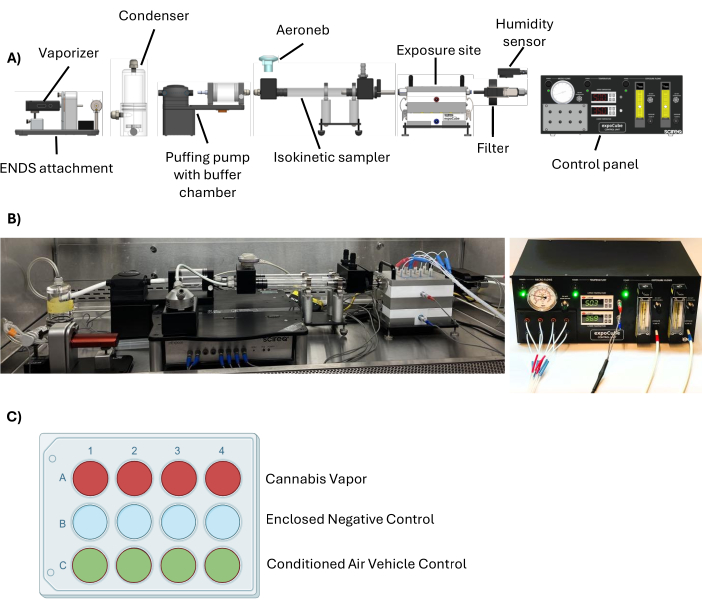
Figure 1: Schematic of the ALI/Transwell in vitro/ex vivo exposure system for cannabis vapor exposure at the ALI. (A) DaVinci MIQRO vaporizer was used to generate cannabis vapor. This sample is humidified and shuttled through the isokinetic sampler and into the exposure site. The control unit allows adjustments to airflow outputs and temperature gradients. (B) Photographs of the exposure system setup. (C) The inside of the expoCube is compartmentalized into three rows: a test row for cannabis vapor exposure, a vehicle control row of conditioned air, and the enclosed negative control row that remains untouched by any airflow. Please click here to view a larger version of this figure.
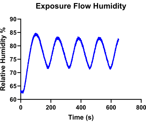
Figure 2: Relative humidity (%) profile recorded over time. This data shows the relative humidity levels when the Aeronebs are activated, reaching and maintaining an average humidity of 75%. Please click here to view a larger version of this figure.
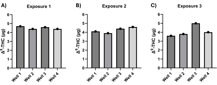
Figure 3: Quantification of Δ9-THC deposition via LC/MS analysis following 30-min exposures to cannabis vapor. (A-C) Δ9-THC deposition was measured in each well after three independent 30-min vapor exposures, each using 1.5 g of a high-THC cannabis cultivar. Δ9-THC levels were quantified by LC/MS. Please click here to view a larger version of this figure.
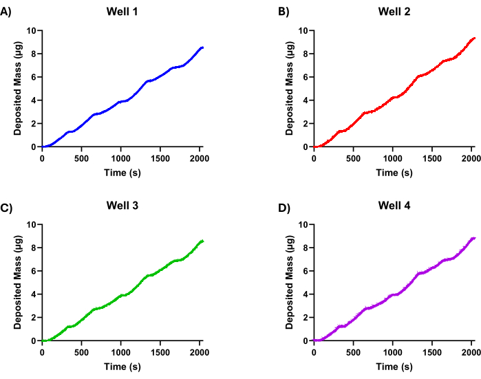
Figure 4: QCM measurements of cannabis vapor deposition in the ALI/Transwell in vitro/ex vivo exposure system over a 30-min exposure regimen. (A-D)Each graph represents the cumulative deposition of cannabis vapor particles in wells 1-4, with mass measured in micrograms (µg) over time. The deposition rate demonstrates a steady accumulation across all wells. Please click here to view a larger version of this figure.
| Primary Flow Filter Deposition (μg) | Well Depositions (μg) | Percent of Primary Flow Deposited in Well (%) | |
| Exposure 1 | 2142.3 | 4.7 | 0.22 |
| 4.4 | 0.2 | ||
| 4.6 | 0.22 | ||
| 4.4 | 0.21 | ||
| Exposure 2 | 2726.5 | 4.1 | 0.15 |
| 3.9 | 0.14 | ||
| 4.4 | 0.16 | ||
| 4.6 | 0.17 | ||
| Exposure 3 | 2400 | 3.6 | 0.15 |
| 3.8 | 0.16 | ||
| 5 | 0.21 | ||
| 4 | 0.17 | ||
| Average | 2422.9 | 4.3 | 0.18 |
| Standard Deviation | 292.8 | 0.4 | 0.03 |
Table 1. Comparison of Δ9-THC deposition in the ALI/Transwell in vitro/ex vivo exposure system between the exposure flow filter and the cell exposure site.
Discussion
Culturing cells at the ALI is an important method in respiratory research due to its ability to more accurately represent in vivo airway conditions compared to traditional submerged cultures19. Culturing at ALI allows lung epithelial cells to be exposed directly to the air on their apical surface while maintaining essential nutrients from the culture medium on the basolateral side. This setup enables cells to produce surfactant or mucus, key components of the respiratory defense mechanism that are critical for trapping and removing inhaled particles and pathogens20,21,22. A549 cells are commonly employed for in vitro studies but are just one of the many respiratory epithelial cell cultures that can be used for ALI. Silva et al. have reviewed cell lines and primary cultures of the respiratory epithelium for ALI from the nasal cavity to the alveoli22. The flexibility of the presented ALI exposure system allows it to be adapted for different cell lines and primary cells.
ALI cultures are increasingly regarded as the "gold standard" for in vitro inhalation studies, particularly for research on air pollutants, particulate matter, and other inhalable agents23. A primary advantage of ALI models lies in their ability to provide insights that are more translatable to in vivo human effects. However, a notable drawback is the lack of standardized protocols for ALI exposures, which affects reproducibility and comparability across studies. One of the gaps is a standardized exposure workflow. By exposing cells to cannabis vapor at the ALI, the impacts of cannabis on cellular function, viability, and cytotoxicity can be evaluated. These exposures allow for a detailed assessment of biological outcomes, including markers of cytotoxicity, inflammation, and oxidative stress. The precise quantification of deposited cannabinoids enables reproducible studies on the cellular effects of cannabis vapor (and other forms of cannabis products), advancing our understanding of its potential health impacts in a controlled laboratory environment.
Advanced exposure systems address limitations in traditional submerged cultures by enabling direct particle deposition onto the cell surface, thereby providing more physiologically relevant exposures. This also reduces the potential effects of the solvents or carriers. By allowing for direct particle deposition without the need for these solvents, exposure systems help minimize vehicle-related effects. Nonetheless, some vehicle-related considerations remain. For example, uncontrolled humidity levels can cause unwanted cell stress24, potentially impacting experimental results. Ensuring that humidity is carefully regulated to reach optimal levels is essential to maintain cell stability and accuracy in results. Additionally, each particle, aerosol, or gas mixture has its own unique physicochemical properties, which could alter deposition on the cell surface25. It is, therefore, important to characterize an exposure regimen for each unique product. The factors that could impact particle deposition include thermophoresis, the movement of particles from a region of higher to lower temperature26. Thermophoresis aids in directing particles toward the cells and enhances deposition efficiency.
Dosing consistency and precision are additional challenges when working with exposure systems at the ALI, particularly when aerosols or particles are involved. In vivo particle deposition in the lung is a complex process influenced by particle size, shape, density, and solubility, as well as the specific region of the lung being exposed27. These factors result in varied deposition rates that can be difficult to replicate in vitro. Mathematical models, such as multi-path particle dosimetry (MPPD), are useful tools for estimating in vivo deposition rates and guiding in vitro dose translation28. However, matching in vitro dosing to in vivo exposure scenarios remains a challenge, particularly for complex aerosols with heterogeneous compositions.
For cell-free dose characterization, it is important to note that stainless steel insert washes tend to overestimate deposition, as compounds can adhere to both the walls of the insert and the base where cells would typically be located. The QCM offers an advantage here by measuring only the mass deposited directly at the cell culture location, providing a more accurate assessment of deposition specifically relevant to cell exposure.
As toxicology moves toward more physiologically relevant models, the adoption of unified ALI exposure protocols will be fundamental in bridging the gap between in vitro findings and in vivo human responses, ultimately supporting more reliable assessments of respiratory toxicants and their impacts on health.
Disclosures
PG and BU are employed at SCIREQ - Scientific Respiratory Equipment, Inc. ETW completed an internship at SCIREQ - Scientific Respiratory Equipment, Inc. through the Mitacs Accelerate Program. The other authors have no conflicts to disclose
Acknowledgements
This work was supported by the Canadian Institutes for Health Research (CIHR) Project Grant 162273. CJB was supported by the Fonds de Recherche du Québec -Santé (FRQS). This study was also supported by a Mitacs grant #IT34076 to ETW.
Materials
| Name | Company | Catalog Number | Comments |
|---|---|---|---|
| 15 mL Centrifuge tube, Rack (Polystyrene Rack), Sterile | Montreal Biotech Inc. | MBI601051C | |
| 30 mL glass vials | Thomas Scientific | 1755F32 | |
| A549 cells | ATCC | CCL-185 | |
| Antibiotic-antimycotic (100x) | Wisent | 450-115-EL | |
| Cannabis herb grinder | Genchi | https://www.amazon.ca/Genchi-Grinder-Crystal-Threads-Premium/dp/B094WX443Y/ref=sr_1_10?crid=A1EV93N8PW1V&dib=eyJ2Ijo iMSJ9.WX_vMBLeAHzc6JplZiba8i w7k_cGpHrEm0VunfiDZqO0U1mT NA0Tr8j5nGMqrGq1BW75hAV8Fr3 SDCGH7O7zwsmmCWOxvdAEN4 97Npj07w0FHMuNDtuKZ3hKv9d8 vauHRtueWaDqYEPOCIAy956bGf PwXu2Gp9nCWVhFCo1gHqgeh1N nrWwD3lJ_r_wx2GoauoNdVw9eH A2QYc1tx3XKMidd2YkVRF_ABYL ZJd0EIDxi22hBnzaM4KklLO6Ep0v OGb1QifhM062KUvTTY9PAOUn8F -O1V0qNYQiRk6SQo2WId2wOHS Mz_zo4YCbkDQf2rNGy81iXhuBzV qu9LhvOcI1xnPUd-JPLx_4rZdSC92 Q_QZODiSxBhH-ellca78LW2iV4XXU vIr-fVWIKdhrS49MD7KV1lCjdBcT_ K3l4bT0mBgZB1_Qpgf_5tmrj-kf.-ctY GDLXKn-ovr6On-5g1G0_oK5mrHdj CN3LXumyu8Q&dib_tag=se& keywords=herb%2Bgrinder&qid=1736346511& sprefix=herb%2Bgrinder%2Caps% 2C168&sr=8-10&th=1 | |
| Collagen from human placenta, type IV | Sigma-Aldrich | C7521 | |
| Costar 12 mm transwell, 0.4 µm pore polyester membrane Inserts | Stemcell Technologies | 38023 | |
| Cytotoxicity detection kit (LDH) | Roche | 11644793001 | |
| Davinci MIQRO-C dry herb vaporizer | DAVINCI | https://davincivaporizer.com/products/davinci-miqro-c | |
| Dimethyl sulfoxide | Sigma-Aldrich | 34869 | |
| DMEM medium | Wisent | 319-005-CL | |
| Dried cannabis flower | Société québécoise du cannabis | https://www.sqdc.ca/en-CA/dried-cannabis?&fn1=CannabinoidBalanceLookup Value&fv1=THC | Cannabinoid Content: 17–23% THC < 0.1% CBD |
| Fetal bovine serum | Wisent | 080-150 | FBS |
| flexiWare | SCIREQ | https://www.scireq.com/downloads/ | Version 8.4.1 |
| Gentamycin | Wisent | 450-134-XL | |
| Glacial acetic acid | Sigma-Aldrich | 100063 | |
| Glutamax | ThermoFisher Scientific | 35050061 | |
| inExpose expoCube in vitro exposure package | SCIREQ | IX-XCP | |
| Infinite 200 PRO | TECAN | 30125944 | |
| LC/MS-grade methanol | Sigma-Aldrich | 106035 | |
| Phosphate-buffered saline | ThermoFisher Scientific | 10010-023 | |
| T175 EasYFlask, TC Surface, Filter Cap | ThermoFisher Scientific | 159910 | |
| T75 EasYFlask, TC Surface, Filter Cap | ThermoFisher Scientific | 156499 | |
| Thiazolyl blue tetrazolium bromide | Sigma-Aldrich | M 2128 | MTT |
| Triton X-100 | Sigma-Aldrich | 108603 | |
| Ultrasonic cleaner bath | VWR | 18303 | Sonicator Bath |
| Weekly pill organizer | HealthSmart | 640-8223-0000 |
References
- Graves, B. M., et al. Comprehensive characterization of mainstream marijuana and tobacco smoke. Sci Rep. 10 (1), 7160(2020).
- Lanz, C., Mattsson, J., Soydaner, U., Brenneisen, R. Medicinal cannabis: in vitro validation of vaporizers for the smokefree inhalation of cannabis. PLoS One. 11 (1), e0147286(2016).
- Arlen, M., et al. Cannabis vaping elicits transcriptomic and metabolomic changes in inflammatory, oxidative stress and cancer pathways in human bronchial epithelial cells. Am J Physiol Lung Cell Mol Physiol. , (2025).
- Wu, J., et al. Characterization of airliquid interface culture of A549 alveolar epithelial cells. Braz J Med Biol Res. 51 (2), e6950(2017).
- Kaur, K., et al. Comparison of biological responses between submerged, pseudoairliquid interface, and airliquid interface exposure of A549 and differentiated THP1 cocultures to combustionderived particles. J Environ Sci Health A Tox Hazard Subst Environ Eng. 57 (7), 540-551 (2022).
- Adekola, M. B., et al. Invitro antioxidant and antiinflammatory activities of ethanol stembark extract of Blighia sapida K.D. Koenig. J Pharm Anal. 12 (2), 350-354 (2022).
- Final report on the safety assessment of capsicum annuum extract, capsicum annuum fruit extract, capsicum annuum resin, capsicum annuum fruit powder, capsicum frutescens fruit, capsicum frutescens fruit extract, capsicum frutescens resin, and capsaicin. Int J Toxicol. 26 (Suppl 1), 3-106 (2007).
- Daltro, S. R. T., et al. In vitro and in vivo immunomodulatory activity of Physalis angulata concentrated ethanolic extract. Planta Med. 87 (1-02), 160-168 (2021).
- Araujo, J., et al. Antioxidant and in vitro cytogenotoxic properties of Amburana cearensis (Allemão) A.C.Sm. leaf extract. Drug Chem Toxicol. 46 (1), 104-112 (2023).
- Spósito, L., et al. In vitro and in vivo antihelicobacter pylori activity of Casearia sylvestris leaf derivatives. J Ethnopharmacol. 233, 1-12 (2019).
- Ngulde, S. I., et al. Ethanol extract of Securidaca longipedunculata induces apoptosis in brain tumor (U87) cells. Biomed Res Int. 2019, 9826590(2019).
- Phuwajaroanpong, A., et al. In vitro and in vivo antimalarial activities and toxicological assessment of Pogostemon cablin (Blanco) Benth. J Evid Based Integr Med. 25, (2020).
- Upadhyay, S., Palmberg, L. Airliquid interface: relevant in vitro models for investigating air pollutantinduced pulmonary toxicity. Toxicol Sci. 164 (1), 21-30 (2018).
- Lenz, A. G., et al. Inflammatory and oxidative stress responses of an alveolar epithelial cell line to airborne zinc oxide nanoparticles at the airliquid interface: a comparison with conventional, submerged cellculture conditions. Biomed Res Int. 2013, 652632(2013).
- Ritter, D., et al. In vitro inhalation cytotoxicity testing of therapeutic nanosystems for pulmonary infection. Toxicol In Vitro. 63, 104714(2020).
- Loret, T., et al. Airliquid interface exposure to aerosols of poorly soluble nanomaterials induces different biological activation levels compared to exposure to suspensions. Part Fibre Toxicol. 13 (1), 58(2016).
- Detlef, R., et al. In vitro inhalation cytotoxicity testing of therapeutic nanosystems for pulmonary infection. Toxicol In Vitro. 63, 104714(2020).
- Ritter, D., et al. Development and evaluation of an in vitro test system for toxicity screening of aerosols released from consumer products and first ap-plication to aerosols from a hair straightening process. Appl In Vitro Toxicol. 4 (2), 180-192 (2018).
- Silva, S., Bicker, J., Falcão, A., Fortuna, A. Airliquid interface (ALI) impact on different respiratory cell cultures. Eur J Pharm Biopharm. 184, 62-82 (2023).
- Fahy, J. V., Dickey, B. F. Airway mucus function and dysfunction. N Engl J Med. 363 (23), 2233-2247 (2010).
- Kingma, P. S., Whitsett, J. A. In defense of the lung: surfactant protein A and surfactant protein D. Curr Opin Pharmacol. 6 (3), 277-283 (2006).
- Soraia, S., Joana, B., Amílcar, F., Ana, F. Airliquid interface (ALI) impact on different respiratory cell cultures. Eur J Pharm Biopharm. 184, 62-82 (2023).
- Graf, J., TrautmannRodriguez, M., Sabnis, S., Kloxin, A. M., Fromen, C. A. On the path to predicting immune responses in the lung: modeling the pulmonary innate immune system at the airliquid interface (ALI). Eur J Pharm Sci. 191, 106596(2023).
- Zavala, J., et al. Regulating temperature and relative humidity in airliquid interface in vitro systems eliminates cytotoxicity resulting from control air exposures. Toxicol Res (Camb). 6 (4), 448-459 (2017).
- Speen, A. M., et al. Benchmark dose modeling approaches for volatile organic chemicals using a novel airliquid interface in vitro exposure system. Toxicol Sci. 188 (1), 88-107 (2022).
- Seidel, S. A., et al. Microscale thermophoresis quantifies biomolecular interactions under previously challenging conditions. Methods. 59 (3), 301-315 (2013).
- Morawska, L., Buonanno, G., Mikszewski, A., Stabile, L. The physics of respiratory particle generation, fate in the air, and inhalation. Nat Rev Phys. 4 (11), 723-734 (2022).
- Kuempel, E. D., Sweeney, L. M., Morris, J. B., Jarabek, A. M. Advances in inhalation dosimetry models and methods for occupational risk assessment and exposure limit derivation. J Occup Environ Hyg. 12 (Suppl 1), S18-S40 (2015).
Reprints and Permissions
