This protocol presents techniques and methodology necessary for targeted hyperthermic therapy in solid tumor models. The approach leverages the photothermal conversion of near-infrared light by intratumorally injected gold nanorods to induce localized heating within the tumor microenvironment.
Method Article
Inducing Targeted Mild Hyperthermia in Murine Tumor Models through Photothermal Conversion of Near-infrared Light by Intratumoral Gold Nanorods
In This Article
Summary
Abstract
Mild hyperthermia (42-48 °C) is a well-established therapeutic modality that can induce controlled tumor cell death and stimulate anti-cancer immune responses. However, delivering heat precisely to tumor tissue while sparing surrounding healthy tissue remains a significant challenge. Traditional hyperthermia methods, such as isolated limb perfusion, require complex, invasive procedures and carry substantial risk of local toxicity and patient morbidity. In contrast, photothermal therapy using gold nanoparticles activated by near-infrared (NIR) light has emerged as a promising, less invasive strategy for achieving localized hyperthermia. Gold nanorods (GNRs), in particular, exhibit tunable optical properties and high photothermal conversion efficiency, making them ideal candidates for precise thermal modulation of tumor sites. Although this technique has shown considerable promise, especially for superficial and accessible tumors, reproducible delivery, spatial confinement, and safety remain active areas of refinement. In this protocol, we present our validated and optimized method for achieving localized, mild hyperthermia using intratumoral injection of biocompatible GNRs followed by short-duration, targeted NIR laser exposure. This approach enables rapid, controllable heating within the therapeutic range, promoting immunogenic tumor cell death and stimulating innate immune responses, mechanisms particularly relevant for immunologically "cold" tumors. Real-time temperature monitoring and local delivery ensure reproducibility, safety, and minimal systemic exposure. This streamlined protocol offers a robust and accessible platform for preclinical studies, supporting broader efforts to harness mild hyperthermia in cancer immunotherapy.
Introduction
Significant challenges remain in cancer treatment, including drug resistance, immune evasion, and off-target toxicity, all of which undermine the long-term success of conventional therapies. These limitations have driven the development of complementary therapeutic strategies designed to overcome resistance mechanisms while minimizing collateral damage to healthy tissue. Mild hyperthermia, defined as the elevation of tissue temperatures to 42-48 °C, has long been recognized as an effective adjunct to standard cancer therapies1. Historically, mild hyperthermia has been used in combination with surgery, chemotherapy, or radiotherapy to enhance the efficacy of these therapies by increasing tumor perfusion, disrupting stromal architecture, and sensitizing malignant cells to cytotoxic agents1. More recently, with the rising success of immunotherapy in oncology, there is growing interest in leveraging mild hyperthermia's inherent immunomodulatory potential to improve antitumor immune responses1,2,3.
Despite its demonstrated clinical benefits, the broader application of tumor hyperthermia remains limited due to challenges in achieving localized, controllable, and reproducible heating of tumors while avoiding off-target tissue damage4. Current clinical and experimental hyperthermia techniques, such as regional limb perfusion or systemically delivered nanoparticle-based approaches, often suffer from limited spatial precision, variable tumor uptake, and the potential for systemic toxicity2,5,6,7,8,9. Targeted hyperthermic therapy is an emerging modality in this field of cancer treatment, designed to selectively elevate tumor temperatures while minimizing damage to surrounding healthy tissue. More specifically, the targeted delivery of gold nanorods (GNRs) is a promising method for overcoming the current limitations of mild tumor hyperthermia precision and toxicity. GNRs are particularly well-suited for photothermal applications due to their tunable surface plasmon resonance in the NIR region, high photothermal conversion efficiency, and favorable biodistribution profiles compared to other gold nanostructures10,11. Their responsiveness to NIR wavelengths enables efficient light absorption and localized heating even in deeper tissues, with minimal damage to surrounding structures. As such, GNRs can provide improved efficacy, targeting, and safety in hyperthermic therapies.
The goal of this method is to provide a highly efficient, low-resource-intensive, and reproducible protocol for achieving mild, gold-nanorod-mediated hyperthermia in solid tumors. This technique, termed GNR-mediated targeted hyperthermic therapy (THT), addresses key limitations of conventional hyperthermia approaches by combining direct intra-tumoral GNR injection with external NIR light activation. This ensures high local nanoparticle concentration, improving reproducibility and safety, particularly for small or poorly vascularized tumors that may not efficiently accumulate systemically delivered agents. Additionally, employing direct intratumoral injection of GNRs followed by exposure to near-infrared (NIR) light ensures precise, controlled, and localized heating of tumors, while minimizing systemic exposure, toxicity, and off-target effects associated with conventional regional hyperthermia techniques.
Many cancers portray complex and diverse mechanisms of immune escape, allowing them to resist immunotherapy through various means, most evidently, through poor immune cell infiltration within the tumor microenvironment2,4,5. Mild hyperthermia offers a synergistic solution when used in combination with immunotherapies. In addition to direct cytotoxic effects, mild hyperthermia preferentially induces immunogenic cell death (ICD), leading to the release of tumor-associated antigens (TAAs) and damage-associated molecular patterns (DAMPs)1,2. These signals activate innate immune pathways, enhance tumor antigen presentation, promote dendritic cell maturation, and enhance T cell priming, ultimately increasing immune cell infiltration into the tumor microenvironment1,6,12. In our own preclinical models, this GNR-mediated THT technique functioned to activate local immune responses and enhance tumor immune infiltration13. When combined with intra-tumoral interleukin-2 (IL-2) therapy or systemic anti-PD1 checkpoint inhibition, this GNR-mediated THT technique further augmented anti-tumor immunity and outperformed either monotherapy alone in breast cancer and melanoma models13. These results are consistent with a growing body of evidence supporting the synergy between nanoparticle-mediated hyperthermia and immunotherapy in solid tumors, including breast cancer14,15,16 and brain tumors17,18. In short, by converting immunologically 'cold' tumors into 'hot' ones, GNR-mediated THT can improve responsiveness to immunotherapies, including checkpoint inhibitors1,2,12.
The GNR-mediated THT protocol presented here is low-resource intensive, technically straightforward, and broadly adaptable to subcutaneous or superficial tumors. This technique is particularly appropriate for researchers working with murine models of subcutaneous or superficial tumors, including melanoma and breast cancer. However, with minor modifications, it may be extended to other tumor types and anatomical sites. Additionally, this technique offers a versatile platform for integrating spatially controlled hyperthermia into preclinical cancer studies, particularly those investigating immunotherapy-enhancing strategies, and looking to explore mild hyperthermia to improve treatment response to, or overcome resistance to, immunotherapy treatments. Furthermore, the knowledge and equipment required for implementation of this technique are accessible and adaptable to a variety of healthcare settings, enhancing its feasibility in broader clinical use3.
In this manuscript, we provide detailed step-by-step instructions for implementing our GNR-mediated THT protocol in mouse models using a NIR laser system operating at 860 nm. The success of this protocol depends on efficient deep-tissue activation of gold nanorods using the appropriate wavelength of near-infrared light. We also highlight procedural considerations for achieving consistent, safe, and effective mild hyperthermic dosing in murine tumor models, such as accurate real-time monitoring of internal tumor temperature throughout the procedure. This method builds on and brings together aspects of previously reported nanoparticle hyperthermia approaches, offering a comprehensive, validated, highly precise, and reproducible alternative for preclinical cancer research. Overall, this protocol offers a biologically safe, cost-effective, and low-toxicity method for delivering tumor-localized hyperthermia, with strong potential to enhance the efficacy of existing cancer immunotherapies and broaden the clinical utility of hyperthermia in oncology.
Protocol
All experimental protocols were approved by the University Committee on Laboratory Animals at Dalhousie University under the guidelines of the Canadian Council on Animal Care ethical standards. All applicable international, national, and institutional guidelines for the care and use of animals were followed. See the Table of Materials for details about all materials used in this protocol.
1. Laser and treatment facility requirements
- Secure access to a fully calibrated laser system capable of delivering light at a wavelength of 860 ± 10 nm with an intensity of up to 4,000 mA. Ensure that the laser system has proper shutdown mechanisms.
NOTE: In this study, we utilized a custom-designed laser. The model used in this protocol has a laser enable key, an emergency stop button, and an interlock jumper (the jumper connected can also be wired to a safety switch that is used for safety compliance, e.g., door access to a Class 4 laser room). - Attach an insertable, calibrated temperature probe for intratumoral temperature monitoring capable of transmitting live temperature data via the appropriate thermocouple recording software.
- Prepare a designated laser room that meets the required safety standards for the laser class. Ensure there is an unobstructed work area, with good ventilation, proper lighting, and all reflective surfaces covered. Restrict room access during laser operation to prevent unintended exposure. Ensure that all personnel involved in the study are fully trained in laser operation and safety protocols and wear appropriate PPE and eye protection.
- Establish a workspace within the laser room that accommodates an anesthetic machine in close proximity to the laser setup. Ensure that the laser is securely mounted, but adjustable, with the experimental subject able to be positioned approximately 1-2 cm from the laser beam source to ensure accurate targeting of the tumor site.
- Use a thermal imaging camera on a tripod, positioned to capture temperature readings from the subject's surface, throughout laser exposure.
2. Cell culture and inoculation (tumor cell injection)
- Select appropriate mouse models based on the tumor type of interest, following standardized laboratory cell culture preparation and maintenance techniques. To follow this protocol, select 6-8-week-old female BALB/c mice for CT26 tumor inoculation.
NOTE: Immunocompetent or immunocompromised mice of varying ages and strains may be used, depending on the study requirements. - Culture the CT26 cancer cell line in RPMI 1640 medium supplemented with 10% heat-inactivated fetal bovine serum (FBS), 100 U/mL penicillin, 100 µg/mL streptomycin, and 2 mM glutamine. Maintain cells at 37 °C in a humidified atmosphere with 5% CO2. Renew medium every 2-3 days, and subculture cells at 70-80% confluency using a split ratio of 1:4 to 1:10. Use cells within early passages (e.g., ≤10 passages) to ensure consistent growth and inoculation efficiency. Expand cultures as needed to obtain the required number of cells for inoculation.
NOTE: The number of cells needed per mouse will vary depending on the model, for example, for the CT26 murine colorectal cancer model, we required 5 × 10⁵ cells per mouse. - Detach adherent cells using trypsinization and wash 2x with sterile 1x PBS. Resuspend in a sterile 1x PBS solution to achieve the desired concentration of 5 × 10⁶ cells/mL. Store the cell suspension in a sterile 2 mL microcentrifuge tube on ice for a maximum of 1 h before injection.
- Before injection, anesthetize mice using an induction chamber with 4% vaporized isoflurane at a flow rate of 0.8 L/min. Once anesthetized, reduce isoflurane to 2% and maintain anesthesia with a nose cone during the inoculation procedure. Confirm anesthesia depth using a toe pinch test to ensure the absence of a pain response. Apply eye lubricant to both eyes to prevent drying and minimize potential laser damage.
- Shave the injection site (e.g., the left flank for this heterotopic CT26 model) using an electric shaver and clean the area with an alcohol wipe to remove debris and disinfect the skin. Pinch up the skin at the injection site and inject 100 µL of the cell suspension at 5 × 10⁶ cells/mL subcutaneously using a 26 G needle and 1 mL syringe. After injection, remove the mouse from the nose cone and return it to its cage for recovery on a warming blanket for 1 h.
3. Tumor growth and measurement
- Begin daily tumor measurements, typically starting on Day 7 post injection, using a tumor measuring caliper. Measure tumor dimensions in three axes: width, length, and height and calculate tumor volumes using an ellipsoid tumor volume formula of π/6 × length × width × height.
- Plan to initiate GNR-mediated THT once tumors reach an approximate volume of 50 mm3 (± 20 mm3).
NOTE: For our CT26 model, this tumor volume was achieved between days 10 and 12 post tumor cell injection). - Prior to laser treatment, ensure random assignment of mice into control and treatment groups. The treatment group will receive an intratumoral GNR injection followed by laser exposure; and the control group will receive an intratumoral PBS injection (same volume as the GNR injection) instead of GNRs, followed by laser exposure.
4. Photothermal conversion of NIR light using intra-tumoral GNRs
- In a sterile biological safety cabinet, use a 0.3-1 mL syringe with a 31 G needle to draw up the appropriate volume of GNRs based on each tumor volume.
- Anesthetize the mouse in the same manner as for cancer cell injection (with a nose cone delivering 2% isoflurane at a flow rate of 0.8 L/min and confirm adequate anesthesia depth using toe pinch test).
- Once anesthetized, reduce isoflurane to 2% and place the mouse in a prone position on the laser platform while maintaining anesthesia using a nose cone. Apply eye lubricant to both eyes to prevent drying and minimize potential laser damage.
- Shave excess fur around the tumor, if necessary, to ensure skin surface exposure to the laser. Use ethanol to disinfect the tumor surface, wiping off any fur/debris.
NOTE: In this study, we used GNRs optimized for absorption at 860 nm. If using a different GNR product than suggested here, ensure that laser NIR wavelength delivery is optimized according to the GNR's absorption spectrum. - Prior to laser exposure, inject GNRs intratumorally at a concentration of 1 µg/mm3 of tumor volume. If necessary, split up the injection between two intratumoral sites to ensure efficient distribution of GNRs inside the tumor.
NOTE: In this protocol video, GNRs at a concentration of 2 µg/µL were used; therefore, for a 50 mm3 tumor, 25 µL of GNRs were injected into the tumor. Be sure to wipe off any excess GNRs on the skin surface after respective injections. - For control group mice, administer an intratumoral injection of sterile PBS of the equivalent volume used for the GNR injection group(s) (e.g., inject 25 µL of PBS in the case of a 50 mm3 tumor).
- Disinfect the intratumoral temperature probe with ethanol and gently insert it into the middle of the tumor mass.
- Apply a uniform layer of aloe vera gel approximately 5 mm thick over the tumor surface to prevent skin ulceration and minimize the risk of thermal injury during treatment.
- Ensure the laser is adjusted to line up directly above the middle of the tumor. Use the irradiance lookup table (Table 1) to determine the appropriate laser height based on the required laser beam diameter to cover the tumor surface area. Optimize laser height based on the tumor diameter to ensure consistent energy delivery.
NOTE: In our experiment, a tumor with a 1.3 cm diameter required a laser height of 1.5 cm). - Switch on the laser and thermos electric cooling (TEC) source, then plug in the laser system into the power source (adapted per manufacturer's protocol). Turn on the thermos electric cooling source; wait for the blue LED to turn on, and the temperature of the TEC source of the laser to stabilize at 25 °C after 30 s.
- Use the irradiance lookup table (Table 1) to achieve a target irradiance of 1 W/cm2 ± 0.2 W/cm2 by adjusting the laser current output (turn the laser source knob to the desired current set point).
NOTE: In our experiment, a laser height of 1.5 cm, a beam diameter of 1.3 cm, and the laser current of 1,800 mA resulted in an irradiance value of 0 .82 W/cm2, which was optimized for this tumor model.
From this point onwards, ensure all individuals in the laser operation room wear certified laser safety glasses for the wavelength of light used. - Set up the thermo camera adjacent to the laser platform, directed at the mouse, to monitor skin surface temperature during the laser procedure.
- On the thermocouple recording software, create a new file and click the record button. Ensure that the internal tumor temperature versus time graph is being displayed in real time.
- Turn the safety key to enable laser source output. Begin laser administration by pushing down on the foot pedal.
NOTE: In this protocol, a Raspberry Pi controller is the internal interface between the foot pedal and the laser source. - Check and ensure the computer software detects an increase in tumor temperature once the GNR-injected tumor is exposed to the laser beam.
NOTE: Internal tumor temperature will start to increase at laser start time, taking approximately 15 s to 2 min to enter the target hyperthermia range of 42 °C, depending on the size and type of tumor model. - Keep the internal tumor temperature at hyperthermic range (between 42 °C and 48 °C) for a full 5 min. Continually monitor the internal tumor temperature, and if it nears 48 °C, take the foot off the pedal and allow the temperature to drop back down. Look for this drop in temperature in real time on the temperature versus time display graph. Once the internal tumor temperature has dropped to ~43 °C, re-enable laser administration by pressing the pedal again. Continue this pattern of on/off laser pedal cycling to keep the internal tumor temperature in the appropriate hyperthermic range for a full 5 min, and monitor the temperature versus time display graph.
- If the tumor temperature drops below 42 °C at any point during the 5 min hyperthermia period, increase the time of hyperthermia to make up for the time lost. For example, if the temperature drops below 42 °C for 20 s, add 20 s onto the end of the initial 5 min time. Some tumors injected with GNRs may not reach 48 °C, and laser on/off cycling will not be necessary.
NOTE: In control mice intratumorally injected with PBS, tumors will not reach the hyperthermic range. If desired, on/off cycling of the laser for control mice should follow the same pattern established for the GNR-treated group, despite the expected lack of hyperthermic response.
- If the tumor temperature drops below 42 °C at any point during the 5 min hyperthermia period, increase the time of hyperthermia to make up for the time lost. For example, if the temperature drops below 42 °C for 20 s, add 20 s onto the end of the initial 5 min time. Some tumors injected with GNRs may not reach 48 °C, and laser on/off cycling will not be necessary.
- Throughout the maintenance of tumor hyperthermia, continually monitor the skin surface temperature to ensure it stays below 50 °C. Regardless of the internal tumor temperature, if the skin surface temperature reaches 50 °C, turn off the laser to allow for cooling. If the internal temperature cannot reach the desired hyperthermic range without causing the skin temperature to exceed 50 °C, reduce the laser power and/or apply additional aloe vera to the tumor site to help dissipate heat. After 5 min in the hyperthermic range, release the foot pedal to stop laser administration and allow the internal tumor temperature to drop down to ~37 °C and turn off the laser safety key.
NOTE: It is important to minimize skin damage that could lead to tumor ulceration by keeping the skin temperature below 50 °C19. - Monitor the internal tumor temperature probe, allowing the tumor temperature to drop back down to approximately 37 °C. Gently remove the temperature probe from the tumor and wipe off the remaining aloe vera gel on the tumor surface.
- Remove the mouse from the nose cone anesthetic and monitor animal arousal prior to returning it to the cage. Place the cage partially on a 37 °C heat pad during the recovery period following the procedure, allowing the mice to move between warmer and cooler areas as needed to regulate their body temperature. Monitor each mouse to ensure a return to normal behavior before returning them to their housing facility.
Results
The goal of this GNR-mediated THT protocol is to heat solid tumors consistently and reliably to the desired mild hyperthermic range of 42-48 °C. The major steps of this procedure are outlined in Figure 1. The success of this protocol hinges on establishing and maintaining controlled and reproducible mild hyperthermia within a given solid tumor model. The GNRs used were synthesized using a patented CTAB-free method, in which gold salt was reduced in a proprietary surfactant solution to promote rod-shaped crystal growth. Post synthesis, native surfactants were largely replaced with low-molecular-weight PEG to enhance biocompatibility. The resulting GNRs were uniform in size (51.1 ± 10.8 nm by 12.2 ± 1.9 nm), had an optimal excitation wavelength of 860 ± 10 nm, and were free of detectable endotoxins.
To ensure proper tumor heating and accurate temperature tracking, an intratumoral temperature probe must be inserted into the center of the tumor. Additionally, monitoring skin surface temperature throughout the procedure using a thermal imager is crucial. It is possible, especially for tumors that are smaller in volume, that intratumorally injected GNRs could diffuse into the peritumoral and/or subcutaneous space around the tumor. Upon exposure to NIR light, this may cause heating of the skin above basic physiological levels, and if the skin reaches temperatures above mild hyperthermic levels (greater than 50 °C), this could lead to skin damage such as scabbing and/or ulceration19. Applying a layer of aloe vera gel to the surface of the tumor is an important precautionary step to help regulate skin surface temperature and prevent thermal damage during NIR laser exposure. In any case, it is recommended to monitor the treated area for signs of ulceration over the 48 h following THT. Figure 2 depicts a visual representation of the suggested setup to optimize tumor temperature management, showing the location of the internal tumor temperature probe and aloe vera gel layer in relation to the NIR laser probe.
The relative success of this experimental protocol can be determined from observing the internal tumor temperature versus time graph throughout laser operation. For mice intratumorally injected with GNRs, tumors are expected to heat into the mild hyperthermic range (42-48 °C) within 15 s to 2 min. Once the internal tumor temperature has entered the mild hyperthermic range, it is expected to either plateau within the appropriate range or require an on/off cycling of laser administration to ensure the internal tumor temperature does not rise above 48 °C (as shown in Figure 3A). Successful maintenance of mild hyperthermia for 5 min is considered a positive result. For mice intratumorally injected with PBS, tumors are not expected to heat into the hyperthermic range. Exposure to NIR light at a wavelength of 860 nm is not enough alone to cause tissue heating into the mild hyperthermic range, and control tumors are expected to plateau around 39-40 °C. Figure 3B shows an internal tumor temperature versus time graph between GNR and control (PBS-injected) mice throughout the 5 min procedure. If a GNR-injected tumor is unable to reach hyperthermic range upon exposure to NIR light or is unable to be maintained in the appropriate hyperthermic range for a full 5 min, this is considered a negative result.
Additional downstream analysis to determine the success of this GNR-mediated THT protocol involves measuring changes in tumor volume following the procedure. Successfully performed GNR-mediated THT causes targeted tumor cell death and activation of anticancer immune responses, which leads to tumor regression and associated tumor volume reduction13. Figure 4 shows changes in tumor volume following GNR-mediated THT for two subcutaneous flank murine cancer models (B16-melanoma and CT26-heterotopic colorectal cancer model) and one mammary fat pad murine cancer model (4T1-breast cancer). In each model, a significant reduction in tumor volume compared to controls is evident 48-72 h post GNR-mediated THT procedure. The timeline and magnitude of tumor volume reduction following the procedure is expected to vary depending on the mouse strain and cancer model. In addition to tumor reduction, we observed a potent innate immune response within 48 h of GNR-mediated THT, characterized by increased expression of genes involved in the STING-cGAS pathway and the upregulation and activation of dendritic cells, macrophages, and NK cells in both the B16F10 and 4T1 models13. Notably, in a bilateral 4T1 tumor model, GNR-enabled THT induced a systemic immune response, as evidenced by increased infiltration of CD8+ T cells into untreated contralateral tumors13. These findings further support the potential of this approach for eliciting both direct cytotoxic and broader immunomodulatory anti-cancer effects.
Table 1: Irradiance lookup table. Table providing irradiance values (W/cm²) for different laser current output settings (mA) and working distances (cm). The table is used to determine the appropriate laser height based on the required beam diameter to cover the tumor surface and to achieve a target irradiance of 1 W/cm2 ± 0.2 W/cm2. Green-highlighted values indicate optimal irradiance levels near 1 W/cm2. This irradiance table is specific to the LDX Laser and was provided by the manufacturer. Please click here to download this table.
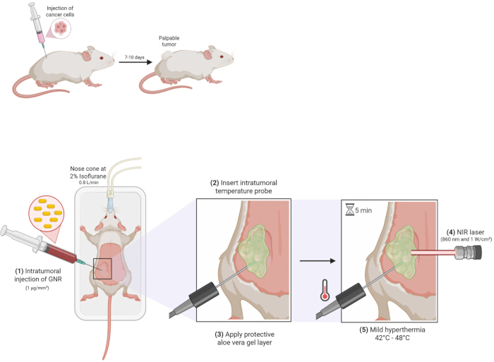
Figure 1: Graphical schematic of GNR-mediated THT procedure. Methodology of gold-nanorod mediated targeted hyperthermic therapy for subcutaneous flank tumors in a murine model. After establishment of a palpable tumor (approximately 50 mm3) and mouse anesthetization, (1) inject GNRs intratumorally at a concentration of 1 µg/mm3, (2) insert intra-tumoral temperature probe in the middle of tumor mass, (3) apply a ~5 mm layer of aloe vera gel on tumor surface, (4) begin NIR laser administration, and (5) maintain internal tumor temperature in mild hyperthermic range (42 °C-48 °C) for 5 min. Abbreviations: GNR = gold nanorod; THT = targeted hyperthermic therapy; NIR = near-infrared. Please click here to view a larger version of this figure.
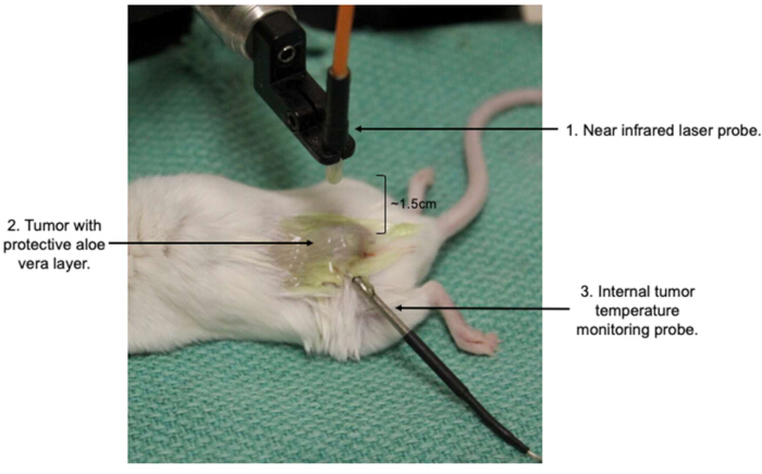
Figure 2: Visual representation of experimental setup. A BALB/c mouse bearing a heterotopic colorectal tumor with a centrally inserted intra-tumoral temperature probe, and topical aloe vera gel layer applied directly below exposure to near-infrared laser beam. Please click here to view a larger version of this figure.
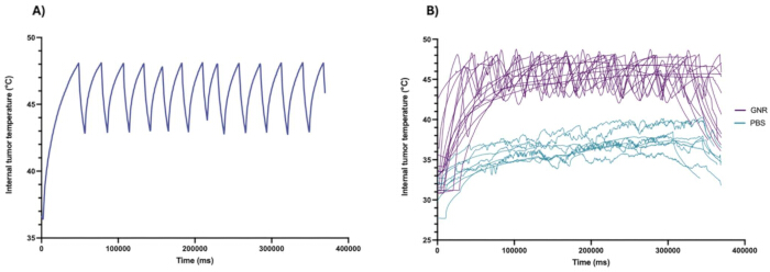
Figure 3: Real-time internal temperature graphs. Representative internal tumor temperature profile(s) over time (in ms) recorded by the internal temperature probe throughout tumor exposure to the NIR laser. (A) On/off NIR laser cycling pattern in a GNR-injected mouse required to maintain internal tumor temperature in a mild hyperthermic range (42 °C-48 °C) for 5 min. This highlights the importance of close monitoring of the internal tumor temperature and cycling of laser administration to maintain the hypothermic range throughout the procedure. (B) Average difference in internal tumor temperature between GNR-injected (n = 15) and control (PBS-injected) mice (n = 8) throughout 5 min NIR laser exposure. Tumors of PBS-injected mice do not get heated into a mild hypothermic range and plateau around 39-40 °C despite receiving the same length of NIR laser exposure as the GNR-injected group, whose tumors reach hyperthermic temperatures of 42-48 °C. Abbreviations: GNR = gold nanorod; NIR = near-infrared. Please click here to view a larger version of this figure.
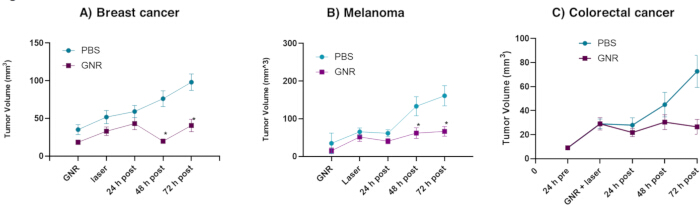
Figure 4: Tumor volume graphs following GNR-mediated THT. GNR-mediated THT induces tumor volume shrinkage across multiple murine cancer models within 48-72 h post treatment. (A) Tumor volume measurements in 4T1 (breast cancer) model following GNR-mediated THT (n= 24 PBS, n = 28 THT). (B) Tumor volume measurements in B16-F10 (Melanoma) model following GNR-mediated THT (n = 19 PBS, n = 23 THT). (C) Tumor volume measurements in CT26 (colorectal cancer) model following GNR-mediated THT (n = 9 PBS, n = 9 THT). A reduction in, or slowing, of tumor volume growth within 48-72 h following treatment is indicative of a positive result. Tumor volume data for CT26 model at '24 h pre' timepoint is not merged-the values are similar and overlap. Error bars represent SEM. * p < 0.05. Please click here to view a larger version of this figure.
Discussion
Critical steps within the protocol
The design and implementation of this GNR-mediated THT technique provides the ability to conduct targeted, accurate, low-resource-intensive, and reproducible heating of solid tumors in vivo. A key advantage of this system lies in its spatial precision and thermal control, which allows for localized treatment without damaging surrounding tissue. One critical step for successful photothermal conversion is ensuring compatibility between the GNRs used and the NIR light source. In this study, we used GNRs optimized for absorption at 860 nm. At this wavelength, light penetrates tissue effectively, and heat is selectively generated in GNR-containing tumors, while surrounding healthy tissue remains near physiological temperature20. Additionally, when performing this protocol, accurate real-time monitoring of the internal tumor temperature is crucial for confirming that a mild hyperthermic range is achieved and that treatment is being delivered consistently. The placement of an intratumor temperature probe, as shown in Figure 2, allows for real-time monitoring of tumor thermal dose, as seen in Figure 3B.
Troubleshooting of the technique
Depending on the in vivo model and tumor type, slight modifications to the protocol may be necessary to optimize results. For example, in the murine melanoma, colorectal cancer, and breast cancer models referenced in Figure 4, we have optimized the GNR-to-tumor volume concentration at 1 µg/mm3. We have also optimized the efficiency of this protocol by conducting NIR laser exposure directly following GNR injection, as no differences in treatment efficacy were observed when comparing this injection and treatment timing to longer intervals of 0.5, 1, 4, 18, or 24 h. For tumor models that vary from our model(s) in terms of tumor pathophysiology or body localization, a different GNR concentration, injected at a different time point, may be optimal for achieving and/or staying within hyperthermic range. Following this procedure, daily tumor volume measurements should be performed to record changes in tumor volume. Tumor regression within 2-3 days is a positive indicator of treatment success.
Due to the nature of the laser protocol, superficial skin burns and ulcerations are a potential risk factor, particularly in pigmented or sensitive skin. Adjustments to laser current output may be necessary for maintaining an appropriate balance between skin surface and internal tumor temperature, depending on the in vivo model used. For example, we have found that because of differences in water content and melanin pigment in the skin21, C57Bl/6 mice require NIR laser administration at a lower current in comparison to BALB/c mice, to keep the skin temperature below 50°C. When modifying laser current output, ensure laser height is adjusted accordingly to keep a target irradiance of 1 W/cm2 ± 0.2 W/cm2 while still covering most of the tumor surface with the laser beam diameter. Alternatively, applying additional aloe vera gel can help lower skin temperature during the procedure without drastically affecting internal tumor temperature. Maintaining appropriate skin surface temperature through necessary modifications is important to minimize the risk of ulceration, which has been observed as a potential side effect of this treatment. When making modifications to the aloe vera gel layer, ensure to momentarily stop administration of the NIR laser beam. In the days following the laser protocol, ensure that the mice are being monitored closely for potential burns and/or ulcerations.
Limitations of GNR-mediated THT
A major safety concern that must be addressed when performing this procedure is the use of certified laser safety glasses for the wavelength of light used during laser operation. The laser system should include built-in safety features, such as a keyed interlock, to prevent accidental activation. However, additional personal protective equipment -- particularly appropriate laser safety eyewear -- is required to protect against potential exposure from reflected or scattered beams. The eye is the most vulnerable to injury from a laser beam, with retina burns being a major concern with NIR lasers22. Lastly, all personnel operating the laser must have appropriate training.
This protocol opted for the intratumoral injection of GNRs over a systemic injection to minimize system toxicity, limit nanoparticle distribution in off-target organs such as the liver, spleen, and kidneys, and enhance the accuracy, efficacy, and safety profile of hyperthermic treatment. However, because this protocol requires intratumoral injections of GNRs, it may be limited in its ability to induce hyperthermia in certain tumor models. So far, we have proven the effectiveness of this method in subcutaneous and mammary fat pad tumors, both of which reside close to the skin surface and are easily accessible in terms of GNR injection and exposure to NIR light. Logistical difficulties may arise in targeting tumors that reside inside body cavities, such as the peritoneum, or when attempting to target metastatic sites of a primary tumor. A potential modification to this protocol could involve systemic GNR injection, provided that the nanoparticles are engineered or modified to ensure efficient accumulation in tumor sites prior to NIR exposure23. Additionally, it is possible that intratumoral GNR injection could result in uneven nanoparticle distribution in the tumor, affecting heating uniformity. However, non-uniform tumor heating is not expected to have a large impact on the degree of treatment effectiveness, given that the primary outcome of this GNR-mediated THT protocol is the induction of ICD to activate anti-cancer immunity. Lastly, the intratumoral temperature readout is dependent on the location of the temperature probe within the tumor. The middle of the tumor is the most optimal location for the probe to accurately reflect the overall average tumor temperature. However, if it is difficult to ensure and/or confirm a central probe insertion because of tumor size and/or shape, it may be necessary to record using two or more temperature probes at different locations.
Significance with respect to existing methods
This manuscript provides a comprehensive, optimized, and reproducible protocol for GNR-mediated THT in solid and superficially accessible tumors. Compared to other methods of inducing hyperthermia, this technique offers distinct advantages, namely, it is low-resource intensive, spatially precise, and associated with minimal systemic toxicity. While the use of GNRs for photothermal therapy is well-documented, there remains significant variability across the literature in how these protocols are implemented. Key procedural elements such as duration of NIR irradiation, choice of thermal monitoring tools (thermocouple probes or thermal camera), GNR solution preparation, and GNR injection methods often vary between studies.
For example, other methods required longer (20 min)24 or multiple doses (4 x 15 min)25 of NIR exposure to achieve similar results as ours. Furthermore, most protocols use either thermocouple probes or thermal cameras but rarely combine both tools26,27. Additionally, GNR solutions are often prepared with CTAB28,29,30, and their method of delivery varies between systemic injections24,30 and intra-tumoral injections31, which can impact their dispersion/location within the TME. As such, a comprehensive and adaptable method for GNR-mediated THT that optimizes and improves upon current protocols has yet to be established.
Our protocol addresses these shortcomings by integrating the most effective and reproducible components from the literature into a single, standardized approach. Specifically, we use dual thermal monitoring (both thermocouple and thermal camera) to ensure precise and safe heat delivery; we employ intra-tumoral injection of CTAB-free GNRs to maximize tumor localization and minimize toxicity; and we incorporate the use of aloe vera gel to protect against superficial skin burns. Additionally, the GNRs used in this protocol are optimized for efficient photothermal conversion at 860 nm NIR, allowing for rapid achievement of mild hyperthermic temperatures within seconds. Once this temperature range is reached, it can be stably maintained for the 5 min treatment duration, which we have determined to be sufficient for inducing immunogenic cell death and immune activation. The 860 nm NIR wavelength itself also provides a favorable balance between tissue penetration depth and safety, further enhancing the therapeutic window of this method.
In summary, our protocol offers a consolidated, validated, and biologically effective alternative to existing GNR-mediated hyperthermia approaches, and fills a critical gap in the field by providing a fully detailed and adaptable method suitable for preclinical cancer research and combination therapy development.
Future applications of the technique
Lastly, although this protocol could be easily combined with various cancer treatments, it is particularly well-suited for enhancing the effectiveness of immunotherapies, given THT's inherent ability to stimulate anti-cancer immunity. By inducing immunogenic cell death, promoting the release of tumor-specific antigens, and increasing the number of innate immune cells recruited to the TME within 48 hours post-treatment, GNR-mediated THT helps 're-sensitize' immunologically cold tumors to detection and attack by the immune system13. As a result, this technique can be used to improve the effectiveness of immunotherapies in treating cancers that may have previously developed resistance, offering a potential strategy for overcoming cancer treatment barriers and disease progression13.
Disclosures
The authors declare that this study received funding from Sona Nanotech Inc. The funder had the following involvement with the study: providing the funding for the materials and resources necessary for the research, as well as supporting the overall project objectives. Carman Giacomantonio is Chief Medical Officer of Sona Nanotech Inc.
Acknowledgements
The authors gratefully acknowledge the financial support from Sona Nanotech Inc. We also wish to thank The Sidney Crosby Foundation, Dr. Mark Johnston, the Department of Surgery at Dalhousie University, the Gibran and Jamile Ramia QEII Health Sciences Centre Chair in Surgical Oncology Research Foundation, and the DMRF Capital Equipment Grant for their generous contributions, all of which supported this research. Thank you to Rick and Lindsay Clark for providing film equipment and assisting with scene direction. Figure 1 was created with https://BioRender.com.
Materials
| Name | Company | Catalog Number | Comments |
|---|---|---|---|
| 0.25% Trypsin | Thermo Fisher Scientific | 25200-056 | |
| 10 mL Stripette | Fisher Scientific | 7200574 | |
| 15 mL Falcon tube | Corning | 352095 | |
| 1 mL syringe | BD Biosciences | 3331431 | |
| 28 G needle | BD Biosciences | 1298811 | |
| 4T1 breast cancer cell line | American Type Culture Collection | CRL-2539 | |
| 5 mL Stripette | Fisher Scientific | 7200573 | |
| 50 mL Falcon tube | Fisher Scientific | 14-432-22 | |
| 70% Ethanol | Commercial Alcohols | 36195 | |
| Aloe vera gel | Life Brand | 57800985495 | |
| B16-F10 melanoma cell line | American Type Culture Collection | CRL-6475 | |
| Biorender | BioRender | N/A | |
| Bisafety cabinet | Nuaire | N/A | |
| Class 4 laser safety goggles | laservision | F22.P5P18.5000 | We utilized a P22 frame with a P5P18 filter |
| CO2 incubator (37 °C, 5% CO2) | Nuaire | N/A | |
| Corning 1-200 µL Universal Fit Racked Pipet Tips | Corning | 4865 | |
| Corning Filtered Pipette Tips, 30 µL | Fisher Scientific | 07-200-265 | |
| CT26 murine colorectal cell line | American Type Culture Collection | CRL-2638 | |
| DPBS | Thermo Fisher Scientific | 10010-049 | |
| Eppendorf tubes | Thermo Fisher Scientific | 3451 | |
| Female BALB/c mice | Charles River Laboratories (Montreal, Canada) | 028 | |
| Female C57Bl/6 | Charles River Laboratories (Montreal, Canada) | 027 | |
| Fetal Bovine Serum | Thermo Fisher scientific invitrogen | 12483020 | |
| GNR | Sona Nanotech Inc | N/A | |
| Intra-tumoral temperature probe | OMEGA | HYP1-30-1/2-T-G-60-SMP-M | Thermocouple Probe Model HYP-1 |
| IsoTip 1000 µL Universal Filtered Pipet Tips | Corning | 4809 | |
| LDX Laser (Model: LDX- 3520-860-HHLFC) | Minnetronix | N/A | Laser current with set point maximum at 4600 mA |
| Optixcare Eye lube | Aventix | N/A | |
| Penicillin-Streptomycin | Thermo Fisher Scientific | 15140148 | |
| Prism 10.6.0 | Graphpad | N/A | |
| RPMI 1640 Medium | Thermo Fisher Scientific | 11875119 | |
| TC-08 Thermocouple Data Logger Software | Pico Technology | PicoLog V5.25.3 | |
| Thermal camera | HIKmicro | EA-1086922 | |
| Tissue Culture Dish 100 x 20 mm | Fisher Scientific | 877222 | |
| Trypan Blue Solution 0.4% | Thermo Fisher Scientific | 15250061 | |
| Tumor measuring caliper | Giles scientificMitutoyo corporation | 500-321 |
References
- Crezee, J., Franken, N. A. P., Oei, A. L. Hyperthermia-based anti-cancer treatments. Cancers (Basel). 13 (6), 1-4 (2021).
- Zhang, Y., Li, Z., Huang, Y., Zou, B., Xu, Y. Amplifying cancer treatment: advances in tumor immunotherapy and nanoparticle-based hyperthermia. Front Immunol. 14, 1258786(2023).
- Huang, X., Lu, Y., Guo, M., Du, S., Han, N. Recent strategies for nano-based PTT combined with immunotherapy: from a biomaterial point of view. Theranostics. 11, 7546-7569 (2021).
- Yi, G. Y., Kim, M. J., Kim, H. I., Park, J., Baek, S. H. Hyperthermia treatment as a promising anti-cancer strategy: therapeutic targets, perspective mechanisms and synergistic combinations in experimental approaches. Antioxidants (Basel). 11, 625(2022).
- Hajebi, S., et al. Advances in stimuli-responsive gold nanorods for drug-delivery and targeted therapy systems. Biomed Pharmacother. 180, 117493(2024).
- Cancino-Bernardi, J., et al. Gold-based nanospheres and nanorods particles used as theranostic agents: an in vitro and in vivo toxicology studies. Chemosphere. 213, 41-52 (2018).
- Cheng, D., et al. Dual-responsive nanohybrid based on degradable silica-coated gold nanorods for triple-combination therapy for breast cancer. Acta Biomater. 128, 435-446 (2021).
- Qin, H., et al. Biosynthesized gold nanoparticles that activate Toll-like receptors and elicit localized light-converting hyperthermia for pleiotropic tumor immunoregulation. Nat Commun. 14, 5178(2023).
- Wu, L., et al. Enzyme-responsive multifunctional peptide coating of gold nanorods improves tumor targeting and photothermal therapy efficacy. Acta Biomater. 86, 363-372 (2019).
- Chatterjee, D. K., Diagaradjane, P., Krishnan, S. Nanoparticle-mediated hyperthermia in cancer therapy. Ther Deliv. 2, 1001-1014 (2011).
- Scutigliani, E. M., Liang, Y., Crezee, H., Kanaar, R., Krawczyk, P. M. Modulating the heat stress response to improve hyperthermia-based anticancer treatments. Cancers (Basel). 13 (6), 1-20 (2021).
- Liu, Y. T., Sun, Z. J. Turning cold tumors into hot tumors by improving T-cell infiltration. Theranostics. 11, 5365-5386 (2021).
- Kennedy, B. E., et al. Targeted intra-tumoral hyperthermia using uniquely biocompatible gold nanorods induces strong immunogenic cell death in two immunogenically 'cold' tumor models. Front Immunol. 15, 1512543(2025).
- Chen, H., et al. Depleting tumor-associated Tregs via nanoparticle-mediated hyperthermia to enhance anti-CTLA-4 immunotherapy. Nanomedicine (Lond). 15, 77-92 (2020).
- Oei, A. L., et al. Enhancing the abscopal effect of radiation and immune checkpoint inhibitor therapies with magnetic nanoparticle hyperthermia in a model of metastatic breast cancer. Int J Hyperthermia. 36, 47-63 (2019).
- Mouratidis, P. X. E., Costa, M., Rivens, I., Repasky, E. E., Ter Haar, G. Pulsed focused ultrasound can improve the anti-cancer effects of immune checkpoint inhibitors in murine pancreatic cancer. J R Soc Interface. 18, 20210266(2021).
- Liu, Y., et al. Plasmonic gold nanostar-mediated photothermal immunotherapy for brain tumor ablation and immunologic memory. Immunotherapy. 11, 1293-1302 (2019).
- Cano-Mejia, J., et al. Prussian blue nanoparticle-based photothermal therapy combined with checkpoint inhibition for photothermal immunotherapy of neuroblastoma. Nanomedicine. 13, 771-781 (2017).
- Okuyama, S., et al. Avoiding thermal injury during near-infrared photoimmunotherapy (NIR-PIT): the importance of NIR light power density. Oncotarget. 8, 113194-113201 (2017).
- Ruggiero, E., Alonso-de Castro, S., Habtemariam, A., Salassa, L. Upconverting nanoparticles for the near infrared photoactivation of transition metal complexes: new opportunities and challenges in medicinal inorganic photochemistry. Dalton Trans. 45, 13012-13020 (2016).
- Sabino, C. P., et al. The optical properties of mouse skin in the visible and near infrared spectral regions. J Photochem Photobiol B. 160, 72-78 (2016).
- Lasers in health care. , Canadian Centre for Occupational Health and Safety. Available: https://www.ccohs.ca/oshanswers/phys_agents/lasers.html#section-6-hdr (2021).
- Kesharwani, P., et al. Gold nanoparticles and gold nanorods in the landscape of cancer therapy. Mol Cancer. 22, 98(2023).
- Zhang, Z., et al. Near infrared laser-induced targeted cancer therapy using thermoresponsive polymer encapsulated gold nanorods. J Am Chem Soc. 136, 7317-7326 (2014).
- Liu, X., et al. antigen-trapping gold nanorods elicit anticancer immunity against abscopal tumors by photothermal therapy-induced in situ vaccination. Biomaterials. 275, 120921(2021).
- Schuh, E. M., Portela, R., Gardner, H. L., Schoen, C., London, C. A. Safety and efficacy of targeted hyperthermia treatment utilizing gold nanorod therapy in spontaneous canine neoplasia. BMC Vet Res. 13, 294(2017).
- Dickerson, E. B., et al. Gold nanorod assisted near-infrared plasmonic photothermal therapy (PPTT) of squamous cell carcinoma in mice. Cancer Lett. 269, 57-66 (2008).
- Huff, T. B., et al. Hyperthermic effects of gold nanorods on tumor cells. Nanomedicine (Lond). 2, 125-132 (2007).
- Ghafarkhani, M., et al. Mild hyperthermia induced by gold nanorods acts as a dual-edge blade in the fate of SH-SY5Y cells via autophagy. Sci Rep. 11, 23984(2021).
- Gong, B., et al. Thermo-responsive polymer encapsulated gold nanorods for single continuous wave laser-induced photodynamic/photothermal tumour therapy. J Nanobiotechnology. 19, 41(2021).
- Hainfeld, J. F., et al. Gold nanoparticle hyperthermia reduces radiotherapy dose. Nanomedicine. 10, 1609-1617 (2014).
Reprints and Permissions
